
Adult Acquired Flatfoot Deformity (AAFD)
Adult acquired flatfoot deformity (AAFD) is a progressive flattening of the arch of the foot that occurs as the posterior tibial tendon wears down. It has many other names such as posterior tibial tendon dysfunction, posterior tibial tendon insufficiency, and dorsolateral peritalar subluxation. This problem may progress from early stages with pain and swelling along the posterior tibial tendon to complete arch collapse and arthritis throughout the hindfoot (back of the foot) and ankle.
Anatomy
The posterior tibial muscle originates on the bones of the lower leg (tibia and fibula). This muscle then turns into the posterior tibial tendon, which passes behind the inside of the ankle and attaches to the navicular bone along the instep of the foot. The posterior tibial tendon plays a central role in maintaining the arch of the foot when you stand and walk.
In addition to tendons running across the ankle and foot joints, a number of ligaments span and stabilize these joints. The ligaments at the inside of the ankle also can become stretched and contribute to the progressive flattening of the arch.
Several muscles and tendons around the ankle and foot act to counter-balance the action of the posterior tibial tendon. Under normal circumstances, the result is a balanced ankle and foot with normal motion. When the posterior tibial tendon fails, the other muscles and tendons become relatively overpowering. These muscles then contribute to the progressive deformity seen with this disorder.
Symptoms
Patients with AAFD often experience pain, deformity, and/or swelling at the ankle or hindfoot. When the posterior tibial tendon does not work properly, a number of changes can occur to the foot and ankle. In early stages, symptoms often include pain and swelling along the posterior tibial tendon behind the inside of the ankle.
As the tendon fails over time, deformity of the foot and ankle may occur. This deformity can include:
- progressive flattening of the arch
- outward shifting of the heel so that it no longer is aligned underneath the rest of the leg
- rotational deformity of the forefoot
- tightening of the heel cord
- development of arthritis
- deformity of the ankle joint
At certain stages of this disorder, pain may shift from the inside to the outside of the ankle as the heel shifts outward and structures are pinched on the outside of the ankle.
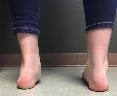
A patient with AAFD of the left foot. You can see that the heel has shifted outward and is no longer aligned under the leg.
Causes
Posterior tibial tendon dysfunction is the most common cause of AAFD. Often there is no specific event or injury that starts the problem. More commonly, the tendon is injured from “wear and tear” over time. Posterior tibial tendon dysfunction occurs more commonly in patients who are born with a flat foot or who develop the condition for other reasons. With a relatively flat arch, more stress is placed on the posterior tibial tendon and also on the ligaments on the inside of the foot and ankle. The result is a progressive disorder. Weight also plays a role in the progression of this disorder. For overweight patients, significant weight loss may lead to some improvement of symptoms.
Diagnosis
The diagnosis of posterior tibial tendon dysfunction and AAFD usually is made from a combination of symptoms, physical exam and X-rays. Your foot and ankle orthopedic surgeon will look at the location of the pain, shape of your foot, flexibility of the hindfoot joints, and how you walk to make the diagnosis and assess how advanced the problem is.