What is triple arthrodesis?

Arthrodesis

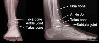
Arthrodesis is a medical term that describes fusion. In a fusion, the bones are locked together by removing the cartilage from the joint surface and creating a “bone bridge” across the joint. A triple arthrodesis is a fusion in the hindfoot (back of the foot) used to treat many types of painful foot deformities. This procedure fuses the joints under the ankle that allow the foot to move from side to side. These joints are the talonavicular, subtalar, and calcaneocuboid.

Surgeons try to avoid fusions, but sometimes pain and deformity are so severe that this procedure offers the best chance of producing a less painful foot with better alignment. Fusions often improve stability and allow for easier standing and walking. The hindfoot fusion is a time-tested, durable procedure.

Three joints in the back of the foot

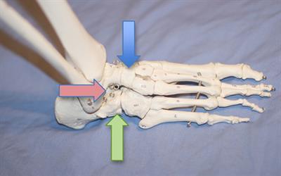
Triple arthrodesis fuses three joints in the back of the foot: the talonavicular (blue arrow), subtalar (red arrow), and calcaneocuboid (green arrow)

Diagnosis

A hindfoot fusion is indicated for severe arthritis, instability, or a deformity that cannot be controlled with non-surgical treatments. Other conditions, such as severe flatfoot, abnormal connections between bones, excessively high arches, and joint instability due to neuromuscular disease, also can indicate the need for a fusion.

Patients who are still growing are not ideal candidates as they may develop additional deformity as they grow. Patients who use tobacco, have an active infection, or have poor healing potential are at higher risk for complications. Non-surgical measures such as bracing and anti-inflammatory medications should be tried first. If another surgical procedure that leaves the joint intact can achieve the same goal, that procedure is preferred over a fusion.

Treatment

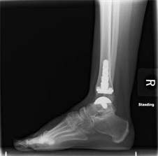
In a triple arthrodesis, your foot and ankle orthopedic surgeon makes one incision on each side of the foot, and works in each joint, removing cartilage, roughening bony surfaces, and filling defects. Once all the desired joints have been prepared, they are put into an appropriate position and hardware is placed to stabilize the reconstruction and promote healthy fusion.

Recovery

The rate of healing is variable and is influenced by many factors. In general, the foot is placed into a splint and kept elevated with no weight on it for the first two weeks to minimize swelling and allow for healing of the skin. Stitches may be removed 2-3 weeks after surgery.

Different weight-bearing protocols may be used. After signs of healing are noted, progressive weight bearing is allowed until full weight bearing is reached. This typically takes three months. During this process, the foot may be placed into a cast. Sometimes, a removable boot may be used rather than a cast.

Risks and Complications

All surgeries come with possible complications, including the risks associated with anesthesia, infection, damage to nerves and blood vessels, and bleeding or blood clots.

Most patients are satisfied with their outcome once they achieve successful fusion. In fact, most feel that the loss of motion is a very acceptable trade-off for pain reduction. In the first two to three weeks, the most likely complication is wound breakdown and/or infection. These are best avoided by not smoking, elevating the foot, avoiding any weight on the foot, and keeping the surgical dressing clean and dry. There also is the risk of the bones not fusing (a non-union). Smoking may place you at increased risk for non-union.

The most common long-term consequence of triple arthrodesis is gradual development of arthritis in other joints of the foot and ankle. These changes can take years or even decades to develop and many never become noticeable to the patient.

FAQs

Will I have stiffness after surgery?

Yes, you can expect some degree of stiffness in your foot as a result of the surgery. You will maintain up-and-down motion, but side-to-side motion will become limited. This will be most noticeable on uneven surfaces but is usually a good trade-off for reasonable pain relief, improved stability and/or deformity correction. Many patients who undergo this surgery have a stiff, painful foot to begin with; after surgery the stiffness may remain but the pain will be relieved.

Will I be able to return to my usual activities?

You can generally expect to return to most activities in life, but the stiffness in side-to-side motion will limit certain activities requiring this motion.

When can I expect to have recovered from a triple arthrodesis?

A significant amount of healing will occur in the first three months. However, it takes about one year for maximum improvement.

Are there side effects to triple arthrodesis?

As one part of the foot is made stiffer, other structures are forced to accept more stress, which increases the chances that they may wear out. This often takes many years or decades to become noticeable.

Will I be able to fit into regular shoes?

Typically patients fit into a shoe after surgery better than before surgery.

When will I be able to bear weight on my foot?

Partial weight bearing typically is allowed once incisions have healed. Full weight bearing generally takes between 2 and 4 months.

What is total ankle replacement?

Total ankle replacement, also known as total ankle arthroplasty, is a surgical procedure that foot and ankle orthopedic surgeons use to treat ankle arthritis. Arthritic changes may be a result of normal wear and tear due to aging or from an injury such as a broken ankle or dislocation. Arthritis eventually leads to loss of cartilage, pain, and/or deformity.

In this procedure, the ankle joint is removed and replaced with an artificial implant made of metal and plastic. The goal of ankle replacement is to provide pain relief while preserving ankle motion so the patient has less pain and better function during activity.

Diagnosis

If you have tried non-surgical treatment including anti-inflammatory medication, bracing, physical therapy, activity modification, and injections, and continue to experience ankle pain and decreased function from arthritis, your foot and ankle orthopedic surgeon may recommend ankle replacement.

Ankle replacement is not recommended if you have severe deformity, dead bone in the talus (the bottom bone of the ankle joint), or bone too soft to support the joint. A history of deep infections of the ankle, significantly abnormal nerve function or sensation (also known as peripheral neuropathy), inadequate or absent leg muscle function, and poor blood flow of the leg also are signs that ankle replacement should be avoided. In these cases, an ankle fusion may be a better option for pain relief.

Post-surgery x-ray of a total ankle replacement

Treatment

Ankle replacement is performed either under general anesthesia or nerve block. Your surgeon will use a tourniquet to control bleeding and improve visualization during the surgery. They will approach the ankle from the front or the side, depending on the type of implant being used. They then cut the bone and place the metal and plastic components that recreate the ankle joint. Sometimes additional procedures will have to be done at the same time to ensure the foot and ankle are properly aligned and the deformity is corrected. Your surgeon then closes the wounds using stitches or staples, and applies a splint.

Recovery

Recovery from a total ankle replacement requires a variable period of non-weightbearing in a cast or boot to allow the implants to heal in place. The procedure is usually performed in an inpatient setting, with the patient spending up to several nights in the hospital. Strict elevation for many days after the procedure is necessary to control swelling and improve wound healing. After the surgical wounds are healed, some foot and ankle orthopedic surgeons will allow the patient to start working on gentle range-of-motion activities even if they are non-weightbearing. Weightbearing usually begins a few weeks after surgery if X-rays show good healing.

Risks and Complications

All surgeries come with possible complications, including the risks associated with anesthesia, infection, damage to nerves and blood vessels, and bleeding or blood clots. A broken bone on the side of the total ankle implant is the most common complication. Patients may also experience an injury to the tendons, nerves, or blood vessels. Wound healing is critical in the early weeks as issues with wound healing can lead to superficial or deep infections. Many of these problems are a greater risk in patients who smoke, have diabetes, or have rheumatoid arthritis. Another possible complication is the failure of the ankle implant to heal into the bone.

Just as with artificial knee and hip replacements, ankle replacements may have problems that happen even years after the initial surgery because there are moving, artificial parts. These issues may require additional surgery and include infection or loosening/wear of the artificial parts.

FAQs

There are two surgical approaches for treatment of end-stage ankle arthritis: ankle fusion and total ankle replacement. Why should I consider an ankle replacement?

There are many factors that go into this decision, so each individual case should be discussed with your foot and ankle orthopedic surgeon. In general, when motion of the ankle is preserved in an ankle replacement, the surrounding joints are protected from increased wear, which is especially important if there is already arthritis in these neighboring joints. The number of ankle replacements being performed is increasing dramatically, as is the amount of published and ongoing research in the area. Newer implant designs and improved surgical techniques constantly are evolving with the promise and goal of helping patients with an ankle replacement get results at least as good as their hip and knee counterparts.

Who should I see to have a total ankle replacement?

A foot and ankle orthopedic surgeon should be seen for management of ankle arthritis. Orthopedic surgeons who specialize in foot and ankle surgery are specifically trained to perform ankle replacements as well as take care of any complications that may arise.

What is the Lapidus procedure?

Lapidus Procedure

The Lapidus procedure is a surgical procedure used to treat a bunion deformity, also known as hallux valgus. It involves fusing the joint between the first metatarsal bone and one of the small bones in your midfoot called the medial cuneiform. Surgery includes removing the cartilage surfaces from both bones, correcting the angular deformity, then placing hardware (screws and often a small plate) to allow the two bones to grow together, or fuse.

Your foot and ankle orthopedic surgeon may perform this procedure to correct a bunion deformity with a very large angle, or when there is increased mobility through the tarsometatarsal (TMT) joint. When the TMT joint has too much looseness or movement, the condition is known as hypermobility or instability. When this joint becomes hypermobile, the first metatarsal moves too far in one direction and the big toe compensates by moving too much in the other direction. When this happens, a bunion can develop.

The goal of the Lapidus procedure is to surgically treat hallux valgus that is caused by first TMT joint hypermobility. When the first TMT joint is fused, the first metatarsal will not move abnormally. This will allow the first toe to stay straight and decrease the risk of the bunion coming back.

Symptoms

Signs surgery may be needed include:

  • A painful bunion on the inner part of the big toe. Typically, this bump causes pain when it rubs the inside of a shoe.
  • Pain and/or hypermobility at the first TMT joint.
  • Difficulties wearing shoes. When patients have a severe enough bunion due to first TMT joint hypermobility, the foot can be so wide that it is difficult to find shoes that fit.
  • Pain that doesn’t improve with non-surgical treatments such as wearing shoes with a wider toe box.

Treatment

The Lapidus procedure is an outpatient procedure, meaning the patient can go home the same day as surgery. Surgery is performed under general anesthesia so the patient is fully asleep or a nerve block is used.

Specific Technique

The Lapidus procedure often is one part of bunion correction surgery. Once the large bony prominence near the big toe is removed, attention is turned to the TMT joint. After the cartilage surfaces of each bone are removed, the alignment is corrected, and the bones are compressed together with hardware. This may be screws or a combination of a plate with screws.

Once the Lapidus is completed, an additional procedure may be necessary to complete the correction of the bunion deformity.

Recovery

Patients typically are immobilized in a splint or boot for the first two weeks after surgery to allow for the incisions to heal. They often are restricted from putting full weight on the foot.

Around six weeks after surgery, patients progress to full weightbearing in either a boot or post-op shoe, then slowly transition to regular shoes a few weeks later.

Some residual swelling and discomfort is normal up to a year after surgery. Most patients are able to return to normal activities with minimal pain and/or problems by four to six months after the surgery.

FAQs

By making the bones grow together, does that affect my ability to walk or run?

A successful Lapidus procedure should allow patients to walk or run with minimal problems or pain once they are fully recovered.

Why do I need to be non-weightbearing for so long?

Patients are asked to limit their weight bearing for several weeks in order to prevent movement between the first metatarsal and medial cuneiform bones that are trying to fuse together. If there is too much motion between the bones, it can take longer for them to heal. Typically, bones take 6-8 weeks to heal, so patients must limit weight bearing during that time.

What if my bones do not heal together?

When bones do not heal together the condition is called a nonunion. Patients who are diabetic or smoke are at higher risk for having this problem. This can also happen if patients put too much weight on the foot before the bones have a chance to fuse together. The most common symptom of a nonunion is continued pain after surgery. X-rays may show broken hardware, which suggests that there is still movement at the fused joint. Most nonunions need further surgery to achieve healing.

What is talar fracture surgery?

Talar Fracture

The talus bone makes up part of the ankle joint and the subtalar joint. The ankle joint allows for up-and-down motion and the subtalar joint supports side-to-side motion. A talar fracture is a break in the talus bone that often involves both of these important joints.

Parts of the ankle

The ankle and foot must be well-aligned for proper function. The goal of surgery is to realign the bone pieces and restore the normal bone shape. The surgery also will restore the function of the ankle and subtalar joints. This surgery should reduce the chances of developing arthritis or losing blood supply to the bone.

Diagnosis

Talar fracture surgery is needed if the bone has shifted or broken through skin, if the nerves or blood vessels are damaged, or if there is an uneven cartilage surface in the ankle or subtalar joints.

Surgery should not be performed in fractures that haven’t shifted, patients who are sick or elderly and cannot risk having anesthesia, or in patients who have very injured or burned skin over the fracture.

Treatment

Talar surgery puts the talus bone back together as best as possible. During surgery you may have a general anesthetic and be completely asleep or have your leg numbed with a nerve block. The bone is exposed with one or two incisions and the broken pieces are realigned. When the fracture is in the appropriate position, your foot and ankle orthopedic surgeon will fix the bone pieces together with plates and/or screws. Then the incisions are closed and the foot is placed into a cast or splint.

Specific Techniques

Every break has a unique fracture pattern, so surgery requires its own tailored approach. Most surgeons will place a tourniquet on the leg above or below the knee. Your surgeon will then make one or two incisions over the bone on either side of the foot. There are important tendons, nerves and blood vessels that are carefully moved out of the way in order to expose the fractured bone.

Your surgeon uses many different tools to move the fractured bone into the appropriate position. The bone pieces are then held in position with temporary pins or clamps and the positioning is checked with an X-ray. When the positioning is correct, the surgeon will place permanent screws and plates across the fracture. The final position of the bone, joints and screws/plates is confirmed on X-ray. The wounds are closed with layers of suture before the foot is placed into a cast or splint below the knee.

Recovery

You may have a short stay in the hospital depending on the severity of the fracture and other injuries. Your surgeon will monitor the incisions and bone healing for the first several weeks after surgery. You should avoid putting weight on the leg until approved by your surgeon. If the fracture was sufficiently stabilized with the plates and screws, you may be placed into a removable boot that will allow you to start moving the ankle to combat stiffness and to bathe.

For certain fractures, your surgeon may decide to place a new cast. You typically will be on crutches, putting no weight on the injured foot for 8-12 weeks after surgery until X-rays show that the fracture has healed sufficiently.

You can expect to have some degree of pain and stiffness after treatment. Some patients will require physical therapy. The complete recovery may take six to 12 months from the time of injury.

Risks and Complications

All surgeries come with possible complications, including the risks associated with anesthesia, infection, damage to nerves and blood vessels, and bleeding or blood clots. Immediate possible complications from talar surgery include wound healing problems, excess swelling, and infection. Patients typically receive intravenous antibiotics prior to surgery, but an infection still may develop in the days and weeks after surgery.

Most wounds will take about 2-4 weeks to heal safely, but this can take much longer if there were traumatic wounds, or if the patient has diabetes or smokes. In the hours to days after surgery, the foot may swell considerably after a talar fracture. If the swelling gets to be too much it may limit blood flow to the foot, resulting in a condition called compartment syndrome. There also is a chance that the bone cannot be put back to its original state, a complication called malunion. Any of these complications may require another procedure to correct.

Some of the most common long-term complications after talar fractures are arthritis and a condition called avascular necrosis (AVN) of the talus. Arthritis can occur after any severe injury to the ankle and is more likely if the fracture has shifted. AVN, which is the death of bone tissue due to a lack of blood supply, also is more frequent with fractures that have shifted.

FAQs

Will I develop arthritis after surgery?

Even if the bones heal well, the talus may develop arthritis at any of three joints: the ankle joint, the talonavicular joint, or the subtalar joint. The subtalar joint is directly below the ankle joint and is responsible for most of the side-to-side motion of our foot. Many patients experience some degree of pain, stiffness, and/or weakness after surgery.

What are the treatment options if I develop arthritis?

If arthritis develops in one or more joints after a talus fracture, it can be treated with medication, braces, injections, and activity modification. If these treatments are unsuccessful, your foot and ankle orthopedic surgeon may discuss additional surgery with you.

What if I develop avascular necrosis of the talus?

Avascular necrosis (AVN) of the talus can be a serious complication of fractures of the talus. AVN can occur with any injury depending on the severity of the fracture. If it does develop, it is not always painful. If it is painful then many of the same treatments for arthritis may be indicated, including fusion surgery.

Does smoking affect my outcome?

Yes. Smoking affects your body’s ability to heal the broken bone as well as the surrounding tissues. Smoking also increases the risk of infection. You should quit smoking immediately in order to minimize these risks.

What is syndesmosis surgery?

Syndesmosis

The two bones in the lower leg are the tibia and the fibula. The point just above the ankle where these two bones meet is called the syndesmosis. While technically a joint, it does not function like most joints as there is very little motion between the two bones. Its main functions are to provide stability to the ankle joint and to allow the joint to move.

The most common way the syndesmosis gets hurt is from a twisting or rotational injury to the ankle. The ligaments that support the syndesmosis are needed to stabilize it, and it is these ligaments that are stretched or torn when this type of injury occurs. Ankle sprains can injure the syndesmosis. The ligaments also can be injured when the ankle is broken. High ankle sprains that are commonly seen in football players are injuries to the syndesmosis.

Surgery of the syndesmosis most often is needed after a traumatic disruption. The goal of surgery is to properly align and stabilize the joint so the ligaments can heal in the correct position.

Diagnosis

Your foot and ankle orthopedic surgeon will examine your ankle. X-rays will be taken and may include a stress X-ray. This is an X-ray that is taken while your doctor carefully twists or stresses your ankle to test the stability of the syndesmosis. If there is an unstable joint, surgery is typically necessary to provide stability.

If the syndesmosis is found to be stable, it usually will not require surgical management. If you have other medical conditions that make surgery too risky for your health, your surgeon may recommend non-surgical treatment. Surgery should also be avoided if you have any active infections or chronic wounds around your ankle.

Treatment

Surgery usually is done on an outpatient basis, but sometimes an overnight stay is required. A general anesthetic typically is used and a nerve block may also be used during surgery or to provide pain relief after surgery. Your surgeon will put the syndesmosis into its proper position and secure it in place with screws or suture implants. A plate also may be used. Some foot and ankle orthopedic surgeons also look inside the ankle joint with an arthroscope to see if the cartilage is injured.

Specific Technique

After making an incision over the outside of the ankle, your surgeon will identify and expose the fibula bone and syndesmosis. Using direct vision and live X-ray techniques, your surgeon will place the syndesmosis into the correct position and set it with an implant. This typically involves one or two screws that go from the fibula bone into the tibia bone. The screws may be placed through a plate that sits on the fibula bone. Alternatively, your surgeon may use a suture device instead of screws.

A stress X-ray is performed to confirm that the syndesmosis is stable. Any additional injuries (e.g., fractures) are repaired if necessary. Your surgeon will close the incision(s) with stitches and then place your let in a splint, cast, or boot.

Recovery

After surgery, you may be immobilized in a splint for the first 10-14 days. You will typically be kept non-weightbearing for 6-8 weeks and then allowed to put weight on your foot in a cast or boot. Swelling persists for many months after this surgery. Stiffness can be problem and physical therapy often is necessary.

Risks and Complications

All surgeries come with possible complications, including the risks associated with anesthesia, infection, damage to nerves and blood vessels, and bleeding or blood clots.

The main complications that can occur after this surgery include irritation or failure of the hardware, the development of arthritis in the syndesmosis, and failure of the syndesmosis to heal properly.

FAQs

Does my hardware need to be removed?

Most of the time, orthopedic hardware does not need to be removed. In the case of syndesmosis surgery, your surgeon may recommend removing the screws that go from the fibula to the tibia. Because there normally is motion between these two bones, the screws may cause pain or limit motion. The hardware is not removed until after the syndesmosis is healed. You and your surgeon will discuss what is best for you.