What is subtalar fusion?

The subtalar joint is located just below the ankle joint between the talus bone and the calcaneus (heel) bone. The main job of the subtalar joint is to allow for side-to-side movement of the foot and ankle. This movement aids in walking, especially on uneven surfaces.
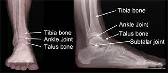
Parts of the ankle
Parts of the Ankle

A fusion surgery locks bones together and is appropriate for diseased joints that can’t be replaced. Once a fusion heals together, it acts as one unit and can restore function and provide significant pain relief. Generally speaking, fusion also is very durable.

Subtalar Fusion

Subtalar fusion is performed to either correct rigid, painful deformities or instability of the subtalar joint, or to remove painful arthritis of this joint. During surgery, this joint between the talus bone above and calcaneus bone below is removed as the joint surfaces are fixed together. The goals of subtalar fusion are to decrease symptoms and allow improved function with less pain.

Diagnosis

Those with subtalar problems typically complain of pain along the outer side of the foot just below the ankle. Subtalar pain may be mistaken for ankle pain. Patients with subtalar joint problems frequently limp, favor the painless other foot, and notice swelling in this region. People commonly have difficulty and pain while walking on uneven surfaces and complain of stiffness in the foot.

Subtalar fusion generally is performed for three reasons: to correct deformity, correct instability, or treat painful arthritis. Causes of arthritis include trauma, degeneration, rheumatologic conditions, and infections.

Treatments

Your foot and ankle orthopedic surgeon will discuss your treatment options with you before proceeding with a subtalar fusion. Skin problems in the area of the surgical site, poor overall health, or active infections may cause your surgeon to delay the procedure.

Smoking increases the risk of blood clots, wound healing problems, and the possibility the fusion won’t heal. You should completely stop nicotine use at least one month before surgery and abstain until the fusion has healed. Inability or unwillingness to follow the treatment plan may mean surgery is not for you.

Specific Technique

Patients are positioned on their back or side to allow exposure of the operative leg. Subtalar fusion is most often performed through an incision on the outer side of the foot. The joint surfaces are prepared by removing all cartilage and correcting all deformity. The bone surfaces are roughened to stimulate bleeding. This bleeding allows the two bones to heal together after the joint is fixed with hardware (screws). X-rays will be used during the surgery to ensure proper alignment and hardware position. Sometimes bone graft is added to help the healing. Once surgery is finished, the foot and ankle are placed in a well-padded splint.

Recovery

After surgery, pain medication will be required for a period of time. Some people may require medication only for a day or two and others for longer. In the first few weeks after surgery you must rest and elevate the operative leg to control swelling and allow the skin incisions to heal. When upright, you may experience throbbing and discoloration in the toes as the blood rushes back to the foot, but typically this resolves with elevation. It is important to keep weight off the foot.

Once stitches are removed, there will be fewer restrictions. A boot or cast usually is placed after the initial surgical bandages are removed. The boot or cast will be in place for 8-12 weeks, sometimes longer for certain patients. Depending on your surgeon’s assessment, weightbearing may be restricted until the X-rays show healing, or weight may slowly be added throughout the period of recovery. If the surgery is on the right foot, do not plan on driving until fully healed. X-rays will be necessary until full healing is seen.

Risks and Complications

All surgeries come with possible complications, including the risks associated with anesthesia, infection, damage to nerves and blood vessels, and bleeding or blood clots.

A potential complication of any fusion procedure is a failure of the fusion to heal, which is called a nonunion. Healing in a bad position also can occur, but this is rare. Following your surgeon’s instructions is very important to avoid complications.

What is sinus tarsi syndrome?

Sinus Tarsi Syndrome

Sinus tarsi syndrome is painful swelling on the outside of the joint below the ankle known as the subtalar joint. This joint allows the foot to move from side to side.

Causes

A common cause of sinus tarsi is flatfoot deformity. With flatfoot deformity, the arch of the foot drops and the two bones on the outside portion of the subtalar joint pinch against each other. This can put increased pressure on the soft tissue in that area, leading to inflammation of the joint lining or the tissue outside the joint.

Sinus tarsi syndrome also can occur due to arthritis in the subtalar joint, scar tissue, joint instability, or as a result of injury.

Symptoms

Sinus tarsi syndrome commonly leads to pain over the outside of the back of the foot. Swelling over the hollow between the ankle bone and the heel bone can develop. The swelling can enlarge so that it can be mistaken for a cyst or tumor.

Diagnosis

This syndrome is usually diagnosed by an exam by a foot and ankle orthopedic surgeon. Your surgeon will see swelling over the outside of the joint below the ankle and tenderness over a specific area of the foot. X-rays can be helpful in diagnosis. On X-rays, your doctor may see collapse of the arch or arthritis.

Treatments

There are non-surgical and surgical treatment options available. In most cases, your doctor will attempt non-surgical treatments first. Anti-inflammatory medications may decrease the swelling in the sinus tarsi. A steroid injection may be tried if other medicines do not relieve the pain. An arch support can be used to relieve the pinching of the subtalar joint. A brace can be applied to the ankle and back of the foot to support and rest the subtalar joint.

Surgical treatments vary depending on the cause of the sinus tarsi pain. Options include removal of inflammation and scarring of the sinus tarsi. This can be done in an open or arthroscopic technique.

If a flatfoot is the cause of the sinus tarsi pain, your surgeon may recommend correction of the flatfoot. If the subtalar joint has advanced arthritis, your doctor may recommend a subtalar fusion (arthrodesis).

Recovery

If surgery is performed, the recovery involves limited weightbearing until the stitches are removed and a fracture boot is placed on the foot. Weightbearing may be allowed at that time depending on the surgery performed. Usually, physical therapy is ordered to help regain range of motion and strength. A boot may be used for several weeks to aid walking.

Risks and Complications

All surgeries come with possible complications, including the risks associated with anesthesia, infection, damage to nerves and blood vessels, and bleeding or blood clots.

FAQs

Are there things that make people more likely to develop sinus tarsi syndrome?

People with flat feet and those who participate in activities that require cutting maneuvers can be more prone to this syndrome. Also, repetitive activities on uneven surfaces can make someone more likely to develop symptoms.

How often is surgery necessary to treat sinus tarsi syndrome?

Surgery usually is not necessary in most patients who develop sinus tarsi syndrome. Non-surgical treatment can be very successful in relieving pain and swelling. Prior to considering surgery, it is important to see a foot and ankle orthopedic surgeon to identify the cause of the sinus tarsi syndrome and the best treatment for that problem.

What is second metatarsal shortening osteotomy?

The metatarsals are the long bones in the foot that connect the toes to the midfoot. The metatarsals are numbered one through five, starting with the big toe. So, the second metatarsal is the long bone of the second toe.

Second Metatarsal Shortening Osteotomy

A second metatarsal shortening osteotomy is a procedure that cuts and shortens the second metatarsal. The goals of shortening the metatarsal are to decrease pain at the base of the second toe (in the ball of the foot) and/or help straighten out the second toe.

Diagnosis

Many patients with problems have pain in the ball of their foot. Typically, they feel like they are walking on a pebble or marble. They often note more discomfort when barefoot. Many patients develop a callus (thickened skin) under the affected metatarsal head. Some patients are at risk for this due to a longer-than-normal second metatarsal.

Before proceeding with this surgery, your foot and ankle orthopedic surgeon typically will try a non-surgical treatment such as a shoe insert with pads to decrease the pressure on the painful area. Note that the pad should not be placed under the painful area, but just toward the heel side of the painful area. A stretching program for the Achilles and calf muscles aimed at decreasing the pressure in the front part of the foot also can be helpful. Steroid injections should never be performed into the fat pad under the ball of the foot, but depending on the diagnosis and thought process of your surgeon, an injection may be done through the top of your foot into the joint or tissue to the side of the joint. Surgery may be an option if these initial treatments do not help. This procedure commonly is performed in combination with other procedures aimed at straightening the foot and toes and decreasing the pressure on the front part of the foot.

If you have not tried conservative treatment, you should do so before considering surgery. You should avoid this surgery if you have an infection in the foot, have poor circulation in the foot, or other medical problems that make the risks of surgery too dangerous for you.

Treatment

This surgery usually is done as an outpatient procedure, meaning the patient can go home the same day. The procedure involves cutting the second metatarsal and removing a small section of the bone. Screws and sometimes a plate are used to hold the metatarsal in the shortened position until it heals.

Specific Technique

The patient receives numbing medication for the foot along with sedation given through an IV. The foot and ankle orthopedic surgeon makes an incision on top of the foot in line with the second toe. The bone is visualized and a saw is used to make a cut in the bone. The bone is shortened to the desired length. It is held in place with some type of implant. The surgeon closes the incision and places a dressing. The surgeon also may place a splint to protect the foot. Sometimes a boot or hard-soled shoe is used.

Recovery

Your foot and ankle orthopedic surgeon will determine if you are allowed to walk or put any weight on your foot. For some patients, weightbearing is allowed the same day, but for others it may not be allowed for 6-8 weeks. The goal is to transition back to supportive shoes at 6-8 weeks based on bone healing. It can take up to 12 weeks to get back to regular shoes, and recovery time is largely determined by other procedures performed at the same time as the shortening osteotomy. The overall goal is to be 75% recovered at three months and 90-100% recovered between six and twelve months after surgery.

Risks and Complications

All surgeries come with possible complications, including the risks associated with anesthesia, infection, damage to nerves and blood vessels, and bleeding or blood clots.

Complications with this specific procedure can include stiffness and numbness near the incision. A delay in bone healing or a deep infection may require additional surgery.

FAQs

Why do I need to be non-weightbearing?

Depending on the specific procedure performed as well as other factors, the patient may be told to remain non-weightbearing to prevent motion between the parts of the bone that are trying to heal together. This means they should not put any weight on the affected foot. If there is too much motion between the bones it can take longer for them to heal or they may not heal at all. Bones typically take six to eight weeks to heal, so being non-weightbearing during this time should allow the bones to heal together.

What if my bones do not heal together?

This complication is more common in patients who have diabetes or smoke. This may cause continued pain after surgery, and is recognized on X-rays or CT scan. A nonunion may require a second procedure. New metal is typically placed during the second surgery and usually some form of bone graft is used to help the bones heal.

What is rheumatoid arthritis?

Rheumatoid Arthritis

Rheumatoid arthritis (RA) is a systemic disease that attacks multiple joints throughout the body. About 90% of people with RA eventually develop symptoms related to the foot or ankle. Usually symptoms appear in the toes and forefeet first, then in the middle and back of the foot, and finally in the ankles. Other inflammatory types of arthritis that affect the foot and ankle include gout, ankylosing spondylitis, psoriatic arthritis, and Reiter’s syndrome.

The exact cause of RA is unknown but there are several theories. Some people may be more likely to develop RA because of their genes. However, it usually takes a chemical or environmental trigger to activate the disease. In RA, the body’s immune system turns against itself. Instead of protecting the joints, the body produces substances that attack and inflame the joints.

Symptoms

The most common symptoms of RA in the foot are pain, swelling, and stiffness. Symptoms usually appear in several joints on both feet. You may feel pain in the joint or in the sole or ball of your foot. The joint may be warm and affect the way you walk. You may develop corns or bunions, and your toes can begin to curl and stiffen in positions called claw toes or hammertoes.

If your hindfoot (back of the foot) and ankle are affected, the bones may shift position. This can cause the arch on the bottom of your foot to collapse (flat foot), resulting in pain and difficulty walking.

Because RA affects your entire system, you may also feel feverish, tire easily, and lose your appetite. You may develop lumps near your joints, particularly around the elbow.

Diagnosis

Sometimes arthritis symptoms in the foot are the first indication that you have RA. Your foot and ankle orthopedic surgeon will ask you about your medical history, occupation and recreational activities, as well as any other persistent or previous conditions in your feet and legs. The appearance of symptoms in the same joint on both feet or in several joints is an indication that RA might be involved.

Your surgeon also will request X-rays to see how much damage there is to the joints. Blood tests will show whether you are anemic or have an antibody called the rheumatoid factor, which often is present with RA. If you’ve already been diagnosed with RA, you and your doctor should be aware that the disease will probably spread to your feet and ankles.

Treatments

Many people with RA can control their pain and the disease with medication and exercise. Some medications, such as aspirin or ibuprofen, help control pain. Others, including methotrexate, prednisone, sulfasalazine, Humira®, Enbrel®, etc., may help slow the spread of the disease itself. In some cases, a steroid injection into the joint can help relieve swelling and inflammation.

Your doctor may prescribe special shoes. If your toes have begun to stiffen or curl, you should wear a shoe with an extra-deep toe box. You also may need to use a soft arch support with a rigid heel. In more severe cases, you may need to use a molded ankle-foot orthotic device, canes, or crutches.

Exercise is very important in the treatment of RA. Your doctor or physical therapist may recommend stretching as well as functional and range-of-motion exercises.

Surgical options for the foot or ankle

Surgery can correct several of the conditions associated with RA of the foot and ankle, including bunions and hammertoes. In many cases, however, the most successful surgical option is fusion (arthrodesis). Fusion is often performed on the big toe, the midfoot, the hindfoot, and in the ankle with RA.

With a fusion, the joint cartilage is removed. The bones are held in place with screws, plates and screws, or a rod through the bone. Eventually, the bones unite to create one solid bone.

There is loss of motion after a fusion, but the foot and ankle remain functional and generally pain-free. Replacing the ankle joint with an artificial joint (known as total ankle replacement or arthroplasty) may be an alternative. Discuss your options with your foot and ankle orthopedic surgeon before proceeding with any surgery.

Recovery

Your doctor will prescribe pain medication for your use after the surgery. Before you leave the hospital, you will be taught how to use crutches. It takes a long time to recover from foot surgery. Here are some things to consider as part of your recovery:

  • Ask friends or family for help in preparing meals and doing other activities of daily living.
  • For the first week or so after surgery, keep your foot elevated above the level of your heart as much as possible.
  • Be sure to do the prescribed physical therapy exercises. They will help you regain strength, motion, and the ability to walk.
  • You won’t be able to put all your weight on your foot for several weeks, and you may need to wear a special shoe or a cast for several months.
  • It may take 6-12 months after surgery to resume regular activities.

RA is a progressive disease that currently has no cure. However, medications, exercises and surgery can help lessen the effects of the disease and may slow its progression.

Risks and Complications

As in all surgeries, there is some risk. Infections, failure for the fusion to heal and loosening of the hardware are the most common problems. Intravenous antibiotics and/or repeat surgery may be needed. Severe complications may require amputation, but this is rare.

What is progressive flatfoot (posterior tibial tendon dysfunction)?

The posterior tibial tendon helps hold up your arch and provides support as you step off on your toes when walking. If this tendon becomes inflamed, overstretched, or torn, you may experience pain on the inner ankle and gradually lose the inner arch on the bottom of your foot, leading to a flatfoot deformity.

Symptoms

The symptoms of progressive flatfoot are gradual and typically include pain and swelling on the inside or outside of the ankle or foot, loss of the arch and the development of a flat foot, weakness and an inability to stand on the toes, and tenderness over the midfoot, especially during physical activity.

Causes

Progressive flatfoot often occurs in women over 50 and may be due to an inherent abnormality of the tendon. But there are several other risk factors, including:

  • Obesity
  • Diabetes
  • Hypertension (high blood pressure)
  • Previous surgery or trauma, such as an ankle fracture, on the inner side of the foot
  • Local steroid injections
  • Inflammatory diseases such as Reiter’s syndrome, rheumatoid arthritis, spondylosing arthropathy, and psoriasis
  • Athletes who are involved in sports such as basketball, tennis, soccer, or hockey may tear the posterior tibial tendon. The tendon may also become inflamed if excessive force is placed on the foot, such as when running on a banked track or road.

Diagnosis

The diagnosis is based on both a history and a physical examination. Your foot and ankle orthopedic surgeon may ask you to stand on your bare feet facing away from him/her to view how your foot functions.

You also may be asked to stand on your toes or to do a single heel raise. You will stand with your hands on the wall, lift the unaffected foot off the ground, and raise up on the toes of the other foot. Normally, the heel will rotate inward; if not, this indicates posterior tibial tendon dysfunction. Your surgeon may request X-rays or an MRI of the foot.

Treatments

Without treatment, the flatfoot that develops from posterior tibial tendon dysfunction eventually becomes arthritic and rigid. Pain increases and spreads to the outer side of the ankle. The way you walk may be affected and wearing shoes may be difficult.

The treatment your surgeon recommends will depend on how far the condition has progressed. In the early stages, posterior tibial tendon dysfunction can be treated with rest, non-steroidal anti-inflammatory drugs such as aspirin or ibuprofen, and immobilization of the foot for 6-8 weeks with a boot to prevent overuse. After the boot is removed, shoe inserts such as a heel wedge or arch support may be helpful. If the condition is advanced, your doctor may recommend that you use a custom-made ankle-foot orthosis or support.

If conservative treatments don’t work, your doctor may recommend surgery. Several procedures can be used to treat progressive flatfoot; often more than one procedure is performed at the same time. Your doctor will recommend a specific course of treatment based on your foot. Surgical options include:

Osteotomy: This procedure changes the alignment of the calcaneus (heel bone). The surgeon may sometimes have to remove a portion of the bone.

Tendon transfer: This procedure uses some fibers from another tendon (the flexor digitorum longus, which helps bend the toes) to repair the damaged posterior tibial tendon.

Lateral column lengthening: In this procedure, the surgeon removes a small wedge-shaped piece of bone from either your hip or that of a cadaver and places it into the outside of the heel bone. This helps realign the bones and recreate the arch.

Arthrodesis: This procedure fuses one or more bones together, eliminating movement in the joint. This stabilizes the hindfoot and keeps the condition from progressing further.