What is posterior ankle endoscopy or arthroscopy?

Posterior Ankle Endoscopy/Arthroscopy

Posterior ankle endoscopy/arthroscopy is a technique used to look at and treat problems in the back of the ankle.

First, it’s important to understand ankle anatomy. The ankle joint is the joint between the lower leg bones (tibia and fibula) and the ankle bone (talus). The joint below the ankle joint is called the subtalar joint; it lies between the ankle bone and the heel bone (calcaneus). The talus has a bony prominence in the back next to the flexor hallucis longus (FHL) tendon. This is the tendon that moves the big toe downward toward the floor.

The bony posterior prominence might be the cause of ankle pain in some people if it is large (called a trigonal process) or it is not completely fused with the talar bone (called an os trigonum).

Pain might also occur if the FHL tendon gets irritated. This can happen if the tendon doesn’t fit well because the tunnel is too tight or the tendon is too big, or if the tendon is inflamed and swollen (called tenosynovitis).

An ankle sprain may cause a tear of the posterior ankle ligaments. The torn pieces can flip inside the joint. They can get pinched between the joint surfaces and cause pain. This problem is called posterior soft tissue impingement.

The Achilles tendon attaches to the back of the heel bone. It can get pinched by a prominent piece of bone at the top of the heel (called a Haglund’s deformity). This can lead to wear of the Achilles tendon and calcium deposits in the tendon (called insertional Achilles tendinitis).

Diagnosis

Patients typically experience pain in the back of the ankle. The precise location of the pain may differ depending on the cause. The pain from Achilles tendinitis is typically at the surface in back of the heel. This pain often increases when wearing closed shoes and improves with shoes that have open heels (e.g. clogs).

The pain from an os trigonum, an FHL problem or posterior soft tissue impingement tends to be deeper. It typically increases with downward motion of the ankle (pointing the toes). Soccer players and ballet dancers tend to be at higher risk for these problems.

You should avoid a posterior ankle endoscopy or arthroscopy if you have Infection in the skin or soft tissue of the posterior ankle area. You should discuss all of your medical conditions with your surgeon before you have this procedure.

Surgery should be considered after at least three months or non-surgical treatment has failed. Non-surgical approaches include rest, anti-inflammatory medications, a cast or walking boot for a short period of time, physical therapy and local steroid injection.

An X-ray can diagnose an os trigonum or enlarged trigonal process and can reveal other bony problems. MRI can be beneficial in evaluating soft tissues such as ligaments and tendons. In some cases, MRI can provide a better understanding of the problem.

Treatment

With the patient lying face-down or on the side, the foot and ankle orthopedic surgeon makes incisions at the back of the ankle. Typically, two incisions are made on either side of the Achilles tendon. An arthroscope (a tube-shaped device with a camera at the tip) is inserted and allows the surgeon to see the area. Fatty tissue at the back of the ankle is removed to create a workspace. The FHL tendon is identified and the blood vessels and nerves are protected. A small part of the posterior ankle capsule might need to be removed in order to enter the ankle joint. A device that “stretches” the ankle joint is often used to help with visualization.

The problem causing the pain is identified and treated accordingly using various small instruments:

  • The os trigonum is freed from the surrounding soft tissues then removed.
  • The FHL tenosynovitis is cleaned up using a shaver and the tunnel is released if necessary.
  • The torn ligaments causing posterior soft tissue impingement are cleaned up with the shaver.
  • The Haglund’s deformity is removed using a burr.

Recovery

The post-operative dressing is usually a splint or bulky soft dressing. A post-op shoe or boot may be added for protection. Weight bearing may be restricted depending on the surgery that is done.

Foot elevation is encouraged in the first 48 hours after the procedure. The stitches are removed in 10 to 14 days and more aggressive exercises can be started thereafter. Early motion of the ankle and foot joints usually is recommended. Formal physical therapy may be ordered. A night splint to keep the ankle at 90 degrees may be used to prevent tightening of the posterior ankle soft tissue.

Risks and Complications

Injury to blood vessels and nerves is uncommon but remains a complication of this procedure. Other complications include numbness on the bottom of the foot, very sensitive skin on the outside part of the foot, Achilles tendon tightness, chronic pain syndrome, infection, and the formation of a cyst at the incision site.

FAQs

What are the advantages of arthroscopic surgery over open surgery?

Arthroscopic surgery for posterior ankle and subtalar joint problems is much less invasive and produces less scar tissue in most cases. The magnification of the arthroscope and the nature of arthroscopy often allow for the evaluation of the tissues and pathologic problems in a more natural state with less injury to the surrounding tissues. This may provide advantages over traditional open surgery.

How much time it will take an athlete or ballet dancer to return to play or performance after this procedure?

It usually takes 8-12 weeks for a ballet dancer to return to performing after posterior ankle arthroscopy and os trigonum excision, but this time can vary. Always check with your surgeon about the anticipated timeline for recovery. Some swelling and discomfort can continue for several months after surgery.

What is plantar fasciitis?

Plantar Fasciitis

If your first few steps out of bed in the morning cause severe pain in the heel of your foot, you may have plantar fasciitis, an overuse injury that affects the sole of the foot. A diagnosis of plantar fasciitis means you have inflamed the tough, fibrous band of tissue (fascia) connecting your heel bone to the base of your toes.

Causes

You’re more likely to develop the condition if you’re female, overweight, or have a job that requires a lot of walking or standing on hard surfaces. You’re also at risk if you walk or run for exercise, especially if you have tight calf muscles that limit how far you can flex your ankles. People with very flat feet or very high arches also are more prone to plantar fasciitis.

Symptoms

Plantar fasciitis typically starts gradually with mild pain at the heel bone often referred to as a stone bruise. You’re more likely to feel it after (not during) exercise. The pain classically occurs right after getting up in the morning and after a period of sitting. If you don’t treat plantar fasciitis, it may become a chronic condition.

Treatments

Stretching is the best treatment for plantar fasciitis. It may help to try to keep weight off your foot until the initial inflammation goes away. You can also apply ice to the sore area for 20 minutes 3-4 times a day to relieve your symptoms. Rolling a frozen water bottle on the bottom of your foot can be beneficial.

Your foot and ankle orthopedic surgeon may prescribe a nonsteroidal anti-inflammatory medication such as ibuprofen or naproxen. Home exercises to stretch your Achilles tendon and plantar fascia are the mainstay of treatment and reduce the chance of recurrence.

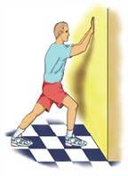
In one exercise, you lean forward against a wall with one knee straight and heel on the ground. Your other knee is bent. Your heel cord and foot arch stretch as you lean. Hold for 10 seconds, relax and straighten up. Repeat 20 times for each sore heel. It is important to keep the knee fully extended on the side being stretched.

Stretch for Plantar Fasciitis

What is peroneal tendinosis?

Peroneal Tendons

The peroneal tendons are on the outside of the ankle just behind the bone called the fibula. Peroneal tendinosis is the name for the enlargement, thickening, and swelling of these tendons. This usually occurs with overuse, such as a repetitive activity that irritates the tendon over long periods of time.

Symptoms

People with peroneal tendinosis typically have tried a new exercise or markedly increased their activities. Characteristic activities include marathon running or others that require repetitive use of the ankle. Patients usually have pain around the back and outside of the ankle. There often is no history of a specific injury.

Causes

Improper training or rapid increases in training and poorly fitting shoes can lead to peroneal tendinosis. Also, patients who have high arches may be more susceptible because their heel is turned inwards slightly, which requires the peroneal tendons to work harder to turn the ankle to the outside. The harder the tendons work, the more likely patients are to develop tendinosis.

Anatomy

Tendons connect muscle to bone and allow them to exert their force across the joints that separate bones. Ligaments, on the other hand, connect bone to bone. There are two peroneal tendons that run along the back of the fibula. The first is called the peroneus brevis. It runs down around the back of the bone called the fibula on the outside of the leg and connects to the fifth metatarsal on the side of the foot.

The peroneus longus takes its name because it has a longer course. It runs all the way underneath the foot to connect to the first metatarsal on the other side. Both tendons share the major job of turning the ankle to the outside. The tendons are held in a groove behind the back of the fibula and are covered by a ligament-type tissue called a retinaculum.

What is percutaneous Achilles tendon lengthening?

Percutaneous Achilles Tendon Lengthening

Percutaneous Achilles tendon lengthening in a procedure used to stretch a tight Achilles tendon and increase motion at the ankle joint.

Diagnosis

People with a tight Achilles tendon tend to walk on their toes. The tight tendon prevents the foot from sitting flat on the floor, and ankle motion can be limited. When this tightness cannot be treated with non-surgical stretching or physical therapy, surgery is needed.

This procedure may be only one part of a surgery to help correct the position of the foot. Patients that develop ulcers in the front part of the foot may require an Achilles lengthening to decrease pressure on the front part of the foot and allow the ulcers to heal.

This procedure is not recommended when there is active infection or redness around the Achilles tendon. A very tight tendon may require a more complex lengthening surgery.

Treatment

The procedure is minimally invasive. It may be done alone or with other procedures in order to improve the overall position of the foot. It is typically an outpatient procedure, and general or regional anesthesia is typically used. It only takes a few minutes to perform this procedure.

The foot and ankle orthopedic surgeon makes three small incisions at the back of the ankle along the Achilles tendon. The tendon is cut approximately 50 percent at each of these three sites in an alternating pattern. The surgeon does this while an assistant is holding the ankle and stretching the tendon. The tendon stretches as the fibers are cut.

Recovery

Healing time for tendons is approximately 6-8 weeks. You may be in a protective cast, splint, or walking boot initially while the tendon heals. Physical therapy and rehabilitation often are needed after the initial healing period to help with strength and range of motion.

Risks and Complications

All surgeries come with possible complications, including the risks associated with anesthesia, infection, damage to nerves and blood vessels, and bleeding or blood clots. With a percutaneous Achilles tendon lengthening, specific complications are rare. Wound problems such as nonhealing incisions or infection can occur. The Achilles tendon can remain tight after surgery. The Achilles tendon also can completely rupture during surgery or recovery.

FAQs

When will I be able to walk again after this procedure?

Generally, the Achilles tendon will take about 6-8 weeks to heal, so weight bearing and therapy will begin at that point. It may take several months to be able to walk normally. The routine post-operative course may be altered if other procedures are performed.

What is os trigonum syndrome?

Os Trigonum

An os trigonum is a small extra (accessory) piece of bone in the back of the ankle. While up to 15% of people have this extra bone, it usually does not cause any symptoms. It may be present only on one side but can be found in both feet. An os trigonum does not move as it attached by thick tissue to the talus bone just behind the ankle joint.

Os trigonum syndrome is the term used when it becomes painful. This is due to the piece of bone and surrounding tissue becoming irritated and inflamed. It may happen as a result of a single injury or due to repetitive/overuse injury such as pointing the toes in dancers, downhill running, or frequent kicking seen in soccer players. It has been termed a “nutcracker injury” as the os trigonum is compressed like a nut when the toes are pointed down.

Symptoms

Frequently, people experience a deep achy pain at the back of the ankle and pain when pointing the toes. It also can be associated with pain when simply moving the big toe because the big toe tendon runs close to the os trigonum at the back of the ankle. Often symptoms are worse when patients are active and improve with rest. As it is quite deep at the back of the ankle, swelling is not common.

Treatments

Patients are usually successfully treated with rest, ice, anti-inflammatory medication, and occasionally immobilization in a walking boot. Sometimes an injection of steroid medications will be considered to relieve the pain and settle the inflammation. If pain persists despite these therapies, surgery can be considered. This simply involves removal of the bone and inflamed tissue which can be performed either by an open procedure or arthroscopically using two small incisions.

Recovery

Complete healing usually takes 4-6 weeks but depending on their activity level some people find they can have discomfort for several months. If surgery is performed, patients usually wear a surgical boot for 2-4 weeks to let the tissues heal. Activities can be resumed as symptoms allow, although the ability to run and perform jumping activities comfortably may take 3-6 months.

Risks and Complications

All surgeries come with possible complications, including the risks associated with anesthesia, infection, damage to nerves and blood vessels, and bleeding or blood clots. Also, there are two main sensory nerves on either side at the back of the ankle which are at slight risk, but the surgical approach generally avoids them.

FAQs

Did this occur because I broke a bone in my ankle?

No. The os trigonum is a naturally occurring bone in up to 15 percent of people who are walking around with no symptoms in this area. It is joined to the rest of the ankle bone by a thick but slightly flexible cartilage. Some people can develop pain if they damage this attachment. As noted above, this damage can occur either with repetitive small injuries or by one big injury. There can be a broken bone in this same area, but generally it looks quite different on X-ray.

Does the removal of this bone affect my athletic ability?

No studies to date show an impact on athletic ability. Most people improve because they no longer have the pain, and most people get back to full athletic activities. It can take several months to get there to allow for slow healing.