What is naviculocuneiform fusion?

Naviculocuneiform Joint

The naviculocuneiform (NC) joint is located in the middle of the foot. It consists of four bones: the tarsal navicular and the medial, middle, and lateral cuneiforms.

The main reason to perform NC joint fusion is to relieve pain related to arthritis. Arthritic joints in the midfoot typically occur after trauma to that region or as part of a collapsing foot arch. When a joint is arthritic, the cartilage has worn away and the bony surfaces rub together, which causes pain. The goal of the fusion is to get the bones to heal together so the pain goes away. Sometimes patients whose arches have collapsed will have a deformity that requires NC fusion to correct it.

Diagnosis

Patients will notice pain on top of the foot with tight shoes or weightbearing activities. This pain can prevent them from being able to walk for any length of time. The pain usually is worst after the patient gets up from resting or sitting. It can improve slightly with walking but then gets worse with continued walking.

Surgery should be avoided if you have any signs or symptoms of a bone or skin infection to the same foot or ankle as the planned procedure. An infection can prevent the bones from healing together and lead to more surgery in the future.

Smoking increases the risk of blood clots, wound healing problems, and the possibility the fusion won’t heal. You should completely stop nicotine use at least one month before surgery and abstain until the fusion has healed. Inability or unwillingness to follow the treatment plan may mean surgery is not for you.

Treatment

Depending on the general health of the patient and what other procedures are planned to take place with the NC fusion, the procedure often is done on an outpatient basis. Patients are put to sleep with general anesthesia. A nerve block may also be offered. This will make the operative foot and ankle numb and help with pain control after surgery.

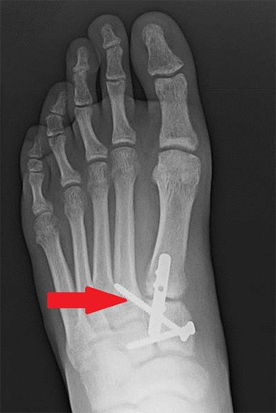
Generally, a single incision along the inside of the foot is used to gain access to the joint. After the joint is identified, the cartilage that remains on the bones is removed and the bones are then positioned back in their correct location and held in place with screws or a combination of plates and screws. The wounds are then closed with stitches or staples and the patient is placed into a well-padded splint. Patients stay off the operative foot for six to eight weeks.

Specific Techniques

During surgery, the lining of the joint is opened and the joint surfaces are evaluated. More often than not, the top of the joint surface has been worn away while the bottom of the joint still has cartilage in place. After all of the cartilage has been removed, holes are placed in the underlying bone to allow bleeding at the joint, which helps the bones heal together. Bone graft also may be used to help the bones heal together.

Recovery

Normally, patients are placed into well-padded splints while in the operating room. Patients are asked to keep their dressings clean and dry at all times and to not remove the dressing unless instructed to do so by their foot and ankle orthopedic surgeon.

The first two weeks after surgery usually are spent with the foot elevated to help decrease swelling. At around two weeks the stitches/staples are removed and X-rays are taken. The patient typically will be placed into a cast or boot and remain non-weightbearing for another 4-6 weeks.

Six to eight weeks after surgery, the patient will come out of the cast or boot and X-rays will be taken to assess healing. If all looks good the patient can begin weightbearing slowly. Physical therapy may be recommended to improve strength and range of motion of the foot and ankle. Some patients may have residual swelling and periodic discomfort in the year following surgery, but the majority of patients are back to normal activities in four to six months.

Risks and Complications

All surgeries come with possible complications, including the risks associated with anesthesia, infection, damage to nerves and blood vessels, and bleeding or blood clots.

Complications from this procedure can include delayed wound healing, infection and delayed bone healing or no bone healing at all. All of these complications generally require further surgery to try and correct the problems. These complications are rare but happen more frequently in diabetic patients and those who smoke.

FAQs

After the bones grow together, will I still be able to walk or run?

In a normal foot, there is limited motion at these joints, so removing painful motion generally will not have any negative effect on your ability to walk or run. A successful naviculocuneiform fusion should allow you to walk or run pain-free once you have recovered fully from the surgery.

Why do I need to be non-weightbearing for so long?

Typically, in a healthy non-smoking patient without diabetes, bones take six to eight weeks to heal. Patients are asked to remain non-weightbearing for that period to prevent motion between the bones that are trying to heal together. If there is too much motion between the bones, it can take longer for them to heal or they may not heal at all.

What if my bones do not heal together?

The term for this is nonunion. It is more common in patients who are diabetic or who smoke. Additionally, patients who put weight on the foot prior to the bones healing can cause this. If this happens the patient will continue to have pain just like before surgery. A nonunion requires another surgery. More or bigger plates and screws can be tried the second time, and usually some form of bone graft is used to try and help the bones heal.

What is midfoot fusion?

The midfoot is the middle of the foot. It refers to the bones and joints that make up the arch and connect the forefoot (front of the foot including the bones of the toes) to the hindfoot (back of the foot including the ankle bone and the heel bone).

Midfoot Fusion

In a midfoot fusion, your foot and ankle orthopedic surgeon fuses together the different bones that make up the arch of the foot. Fusion eliminates the normal motion that occurs between two bones. Since there is very little movement in the small joints of the midfoot, the function of the foot can be preserved.

Midfoot fusion can involve all of the midfoot joints, but in most cases just one or a few of the joints are fused. The joints of the midfoot do not bend and move like your knee or elbow. They are designed to be relatively stiff to give your foot strength and support your body. Midfoot fusion generally does not produce much noticeable loss of motion because there is fairly little motion to begin with.

The primary goals of midfoot fusion are to decrease pain and improve function. This is achieved by eliminating the painful motion between arthritic joint surfaces and restoring the bones to their normal positions. Other goals include the correction of deformity, returned stability to the arch of the foot, and restored normal walking ability.

Diagnosis

The most common reason for midfoot fusion is painful arthritis in the midfoot joints that has not improved with non-surgical treatment. Other common reasons to do a midfoot fusion include too much motion of one or more of the midfoot joints or deformity of the midfoot. Examples of conditions that may result in midfoot deformity include severe bunions, flatfoot deformity, and arthritis. Midfoot fusion also is indicated for certain acute fractures and joint displacement involving the midfoot.

Midfoot fusion should not be performed if there is active infection or if the patient’s health is poor enough that the risk of surgery is too high. Conditions such as uncontrolled diabetes and blood circulation problems may make a patient a poor candidate for surgery. Other reasons to not perform midfoot fusion include osteoporosis and poor skin quality. Smoking significantly increases the risk that bones will not fuse.

Treatment

Successful midfoot fusion depends on complete removal of all joint surfaces (cartilage) and stable fixation of the joints being fused. Residual cartilage can prevent the bones from fusing together. Failure to achieve adequate stability may allow too much motion for fusion to occur.

Typically, your foot and ankle orthopedic surgeon will make one or two incisions on the top of the foot. The number and length of the incisions is determined by the number of joints to be fused. Your surgeon will pay careful attention to protecting tendons and nerves.

Stability is achieved during midfoot fusion using metal implants such as screws and plates. These are designed to immobilize the joints and allow for the formation of bone across the joint space. Your surgeon may add bone graft material to fill any gaps that might exist between the bones after the cartilage has been removed. This bone graft material may be taken from another location in the patient’s body (autograft). It may also come from donated bone (allograft) or from a synthetic material. A combination of these materials may be used.

Recovery

After surgery a period of protection and immobilization is required for successful fusion to occur. A cast is typically placed for the first 6-10 weeks. You will not be able to put weight on the affected foot for 6-12 weeks after surgery. X-rays usually are obtained every four weeks to assess progress of the fusion.

Gradually increased weightbearing is allowed as healing progresses. Initial weightbearing is protected in a prefabricated boot with gradual transition to supportive shoes. Physical therapy may be prescribed on a case-by-case basis to help the patient’s walking and balance.

Risks and Complications

All surgeries come with possible complications, including the risks associated with anesthesia, infection, damage to nerves and blood vessels, and bleeding or blood clots.

A major potential complication after midfoot fusion is failure of the bones to fuse (nonunion). Other complications can include over-correction or under-correction of deformity (malunion). There can be problems with wound healing. Prominent plates and screws can be painful and may require removal of the hardware. Injury to nerves on the top of the foot can occur.

Smoking is one of the leading risks for nonunion. Premature weightbearing can also result in failure of the bones to fuse.

FAQs

How much motion in my foot will I lose after midfoot fusion?

Motion of the midfoot joints is normally somewhat limited. Loss of that motion after fusion surgery tends to be well-tolerated by patients. The more mobile joints of the ankle, hindfoot, and forefoot are unaffected by midfoot fusion and thus continue to provide motion to the foot.

Will I set off an airport metal detector after midfoot fusion?

The strength of the metal detector and the amount of metal implants used determine whether hardware from a midfoot fusion will be detected. It is uncommon for the metal implants to be detectable by airport screening methods.

How will I get around after surgery before I am allowed to put any weight on the foot?

A combination of devices can be used, including crutches, walkers, knee-rollers, scooters, and wheelchairs. Physical therapy is used to help assess patient needs and improve mobility and safety. Certain patients may benefit from the assistance provided by a skilled nursing facility or post-operative rehabilitation unit.

Will the plates and screws have to be removed after midfoot fusion?

Metal implants used for midfoot fusion are not routinely removed. Hardware may need to be removed if there is a failure of the fusion or if infection develops. Painful hardware can be removed once the fusion is healed.

What is metatarsalgia?

Metatarsalgia

Pain in the ball of your foot, the area between your arch and the toes, is called metatarsalgia (MET-ah-tar-SAL-gee-ah). The pain usually centers on one or more of the five bones (metatarsals) in this area under the toes.

Causes

Calluses: If one of your metatarsal bones is long, the bone may take on extra weight. This extra pressure on the metatarsal bone can cause a callus, or buildup of skin, to form. The combination of the extra weight on the bone and the callus can cause pain; however, a callus does not need to be present for the patient to have pain.

Bunions: Metatarsalgia often is associated with a bunion deformity of the big toe. In this case, the big toe metatarsal bone cannot support the weight it needs to, and the weight shifts to the smaller metatarsal bones.

Hammertoes: A hammertoe is an imbalance of the tendons that support the toes. Hammertoes can push the metatarsal bone into the ground, which increases the pressure on the bone and may cause metatarsalgia pain.

Another cause can be shoes that don’t fit properly. Tight shoes squeeze the foot and increase pressure, while loose shoes let the foot slide and rub, which creates friction. Torn ligaments or inflammation also can cause metatarsalgia. Pain on the underside of the foot may indicate a torn ligament or inflammation of the joint. Your foot and ankle orthopedic surgeon can do some simple tests to assess joint stability.

Treatment

Most of the time, practical measures can help ease foot pain. Your doctor may recommend that you use a shoe insert (arch support) as a kind of shock absorber, or that you wear a different kind of shoe. Routinely, a pad is added to the orthotic to shift the weight off the painful metatarsal bones. Sometimes, simply buying shoes that fit properly can solve the problem. Shoes should have a wide toe box that doesn’t cramp your toes. Heels should never be higher than 2 1/4″ high.

Soaking your feet to soften calluses and then removing some of the dead skin with a pumice stone or callus file will relieve pressure. Note, however, that diabetics should not do this themselves. Calluses should be taken care of by your physician or someone your physician recommends for treating diabetic feet. This is a short-term solution that temporarily removes the callus but does not fix the problem.

Occasionally, surgery may be necessary to remove a bony prominence or correct a deformity.

What is Lisfranc surgery?

Lisfranc Ligament and Joint

The Lisfranc ligament runs between two bones in the midfoot (middle of the foot) called the medial cuneiform and the second metatarsal. The place where these two bones meet is called the Lisfranc joint. The name comes from French surgeon Jacques Lisfranc de St. Martin (1790-1847), who was the first physician to describe injuries to this ligament.

Tearing of the Lisfranc ligament and other ligaments around the Lisfranc joint can lead to instability and disruption of the joints in the middle of the foot. The goals of Lisfranc surgery are to put the bones back into their original position and restore the foot’s normal alignment.

Diagnosis

Your foot and ankle orthopedic surgeon may recommend surgery for a Lisfranc injury if your midfoot joints are not lined up anatomically. Most commonly this misalignment is identified on X-ray; however, CT and MRI scans also can be helpful in diagnosis. Surgery will realign and stabilize the misaligned joints. Some injuries with noticeable cartilage damage may require fusion of the joints.

You do not need surgery for a Lisfranc injury if you have a sprain that does not create instability. Such injuries typically require you to restrict activity and use a boot or cast for 6-8 weeks. Surgery also should be avoided if you have significant soft tissue swelling, severe peripheral vascular disease, or fracture due to nerve dysfunction, which can be seen with diabetic neuropathy. You should speak with your surgeon prior to Lisfranc surgery if you have these conditions.

Treatment

Lisfranc surgery is usually an outpatient procedure, meaning you can go home the same day as surgery. You will receive anesthesia, such as general anesthesia, spinal anesthesia, an ankle block, or popliteal with sedation. A nerve block may be used to help control pain after the surgery. A tourniquet usually is used to reduce bleeding. Most patients will require at least one incision on the top of the foot, and a second incision may be needed if the injury is severe.

Specific Technique

Your foot and ankle orthopedic surgeon will make the first incision on the top of the foot, making a line between the big toe and second toe at the middle of the foot. They will carefully protect the tissues to minimize risk of injury to tendons or nerve structures.

Your surgeon will realign the medial cuneiform bone to the base of second metatarsal bone and then realign the other joints around this joint. An X-ray will verify that the joints are aligned. A second incision often is necessary for more severe injuries. This second incision is typically made on the top of the foot but more toward the little toe side.

A series of screws or plates placed beneath the skin will help hold the bones in place. One of the screws often placed is known as a “home run” screw. It runs between the medial cuneiform and the second metatarsal bones. This screw mimics the path of the injured Lisfranc ligaments. Some injuries require wires to be left in place. These wires are left partially exposed outside of the skin.

A fusion surgery involves a similar technique. The main difference is that the cartilage is removed from the joint surfaces prior to inserting plates or screws. The goal is to make the bones grow together to eliminate arthritis.

x-ray after Lisfranc surgery
X-ray after Lisfranc surgery

This X-ray shows a patient who had surgery to realign the bones of the foot. A plate and several screws are holding the bones in place. The arrow points to the “home run” screw that mimics the injured Lisfranc ligament.

Recovery

You will be placed into a non-weightbearing splint immediately after surgery. This protects the bones and incisions. You should elevate the foot as much as possible to help reduce swelling and pain. Pain will typically be controlled with pain pills.

Sutures will be removed about two weeks after surgery and you will have a cast or CAM boot. No weightbearing is allowed for 6-8 weeks after surgery. A walking cast or boot is then used for another 4-6 weeks. If pins were used to hold the fourth and fifth metatarsals in place, they are removed 6-8 weeks after surgery.

Patients usually are able to wean out of the boot and into an athletic shoe in 10-12 weeks. A more rigid shoe with an arch support insert will help reduce stresses through the middle of the foot. Physical therapy may be prescribed for strengthening and improvement in function. It can take longer than one year for full recovery.

Risks and Complications

All surgeries come with possible complications, including the risks associated with anesthesia, infection, damage to nerves and blood vessels, and bleeding or blood clots.

With Lisfranc surgery, there is a nerve that runs very close to the site of the incision. Injury of this nerve can result in numbness. If numbness occurs it typically is not painful and the foot recovers with time. Another common problem after a Lisfranc injury is the development of post-traumatic arthritis in the joints of the middle of the foot. This is due to degeneration of cartilage in the area of the injured joints. This can lead to pain and stiffness in the middle part of the foot.

FAQs

Will the plates and/or screws stay in my foot forever?

The hardware that is placed during surgery is sometimes removed 4-6 months after surgery. Hardware placed for a fusion typically is not removed unless it becomes bothersome.

Should I have my injured foot realigned or fused?

This is a debated topic. For simple Lisfranc injuries, a patient typically will do well with realignment of the bones. More substantial injuries that result in obvious displacement of the joints or a fracture involving the joint surfaces may be better treated with a fusion. Other factors to consider include your age and any existing foot arthritis. Discuss your treatment options with your foot and ankle orthopedic surgeon to find the best solution for your problem.

What is lesser metatarsal shortening osteotomy?

Metatarsals

Each foot has five metatarsals. These are the long bones of the foot. They connect the toes to the rest of the foot. They also make the ball of the foot. The lesser metatarsals are the bones that connect to the second through fifth toes (not the first, or big toe).

An osteotomy is a cut made in the bone. It is similar to breaking the bone but in a very controlled manner. A lesser metatarsal shortening osteotomy changes the pressure distribution under the ball of the foot, relieving pain. It also can be used to put a chronically dislocated toe back in position.

Diagnosis

Surgery is appropriate if non-surgical methods have not adequately relieved pain. This procedure is used to alter pressure across the ball of the foot as well as correct toe angulation or dislocation. This is most commonly seen when the toes are abnormally bent (hammertoe or claw toe deformities). Other less involved options may be more appropriate depending on the deformity and symptoms of the patient. However, when the toe is dislocated, the deformity usually is addressed with a metatarsal shortening osteotomy, as just straightening the toe (without shortening) can cause loss of adequate blood flow to the toe which could cause gangrene. Another reason to perform the procedure is to relieve pressure under a metatarsal head. In this case, there is usually a callus and pain in the ball of the foot.

Reasons to avoid this surgery include lack of attempted non-surgical treatment, active infection around the surgical site, lack of adequate blood supply to allow healing, or arthritis at the joint next to the osteotomy. The shortening osteotomy would correct the toe’s position, but the patient would likely have continued pain due to the arthritis.

Treatment

This procedure typically is performed with the patient going home the same day. General and/or regional anesthesia (a block) can be utilized. The surgeon uses a saw to cut the bone. The two bone ends are shifted so that the bone shortens. This shortening gives “slack” to the toe and allows the toe to be placed in the correct position. Other soft tissue procedures often are performed at the same time to correct toe deformities.

Specific Technique

A one to two inch incision is made over the top of the foot. There are many types of metatarsal shortening osteotomies. Where the incision is made depends on the location of the osteotomy. A bone cut is made using a saw at the end of the metatarsal. The cut is made completely through the bone. After the appropriate amount of sliding has occurred, the osteotomy is held in that position with either pins or screws. The surgeon may also place a temporary wire through the toe itself. The skin is then closed.

Recovery

Immediately after surgery, a padded soft dressing is placed around the foot. The patient is given a hard-soled shoe or boot and allowed to place weight only on the heel.

Instructions are given to keep the dressing clean. Showers/bathing can be done with either a plastic bag or commercially available cover to keep the dressings dry. The patient should keep the foot elevated as much as possible to avoid swelling. Excess swelling can lead to problems with wound healing.

The patient usually is seen in the office one to two weeks after surgery. The dressing is changed and when skin has adequately healed, the stitches are removed. Range-of-motion exercises with the toe may be started. If a wire was placed in the toe, it typically is removed in the office four to six weeks after surgery. At 4-6 weeks, weight bearing usually is allowed as tolerated.

Risks and Complications

All surgeries come with possible complications, including the risks associated with anesthesia, infection, damage to nerves and blood vessels, and bleeding or blood clots. There also are complications specific to metatarsal osteotomies. Failure of the bone to heal after it has been cut may result in continued pain. This complication typically requires another surgery to get the bone to heal in order to alleviate the pain. Excessive scaring can occur as can recurrence of the deformity.

FAQs

Will I have normal function of my toes after this surgery?

You should expect some degree of stiffness of the operative toes after this procedure. The amount of motion regained depends on the severity of the deformity before surgery and any other procedures done. Range-of-motion exercises after surgery can help to decrease stiffness.

How long will my toe be swollen after surgery?

Even with appropriate healing, toes may remain swollen for months after surgery.