Anterior Cruciate Ligament Injuries

anterior-240x300Anatomy
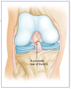
The anterior cruciate ligament (ACL) runs from the front of the tibia (lower leg bone) to the back of the femur (thigh bone). It prevents the tibia from sliding forward and keeps the knee from pivoting (instability). The ACL sits in the center of the joint and has poor blood supply that contributes to its poor healing potential.

Injury Mechanism
The ACL is often injured in sports with twisting type injuries or hyperextension injuries of the knee. ACL injuries can occur with rapid stopping while running and often as contact injuries. ACLs can be an isolated injury, but often occur with associated injuries to the meniscus (cartilage pads) or other ligament combinations.

Females are known to have a higher rate of ACL injuries than males in the same sports.

Arthroscopic view of the normal ACL

Symptoms
The “classic” ACL injury is described as a sudden “giving way” and hearing a “pop” at the time of the injury. Rapid swelling occurs, usually within the first 24 hours.

The following day, one typically describes stiffness, swelling, and pain with weight bearing.

Over the next 1 to 2 weeks, the swelling starts to subside and the range of motion of the knee improves, but patients may start to experience “giving way” or a sense that they cannot trust the knee.

Diagnosis
The physician’s work-up will start with a careful history and exam. Often the description of a sudden “giving way” episode and “pop” can lead the physician to the suspected injury to the ACL. A history of immediate swelling of the knee (effusion) will further lead to this diagnosis.

Once the patient can relax the muscles around the knee, the physician can feel the instability on the exam. X-rays are often obtained to see that no fractures have occurred with the injury and to help assess the overall condition of the knee joint.

An MRI scan is often obtained to confirm the diagnosis and to evaluate any associated injuries to the menisci, other ligaments, and damage to the joint surfaces. Plain x-rays show the bones of the knee, while MRI scans reveal the soft tissues around the knee including the ligaments, menisci, muscles and tendons.

Treatment
Your physician will discuss treatment options with you. Treatment decisions are based on age, activity level, degree of instability, and associated injuries to other structures about the knee. If a patient is older and does not participate in ACL dependent activities (soccer, basketball, court sports, or other twisting and pivoting sports) the patient may choose nonoperative treatment.

Nonoperative treatment does not mean “no treatment.” The patient is educated about the ACL injury and educated about high-risk activities. Often physical therapy is initiated to help regain full range of motion and strengthen the surrounding muscles that can help stabilize the knee. Occasionally, bracing can help for high-risk activities and sports. If patients in this group have “giving way” episodes after therapy or bracing, they may need to be considered for ACL surgery.

In the younger, more active patients, surgery is undertaken to help stabilize the joint to allow the patient to return to full activities. Surgery involves replacing the torn ACL with another tissue (a graft). Timing of surgery is important.

It has been found best to wait for at least 2 or 3 weeks after the injury before undertaking surgery to give the swelling time to resolve and to allow the patient to recover most of their range of motion before surgery. The chance of developing stiffness following surgery is decreased with better motion going into surgery.

Repairing the native ACL does not work, because of the poor blood supply mentioned above to the ACL, so a substitute tissue must be used for the graft. Tunnels are drilled in the femur and tibia at the attachment site of the normal ACL. The graft can then be passed through the tunnels to replicate the course of the normal ACL. The graft is fixed at both ends until the graft eventually heals into the tunnels and re-establishes a blood supply.

Graft options will be discussed with one’s surgeon, but usually involves taking another tissue from elsewhere around the knee (such as the hamstrings or a portion of the patellar tendon) or taking tissue from a cadaver (another human being). Risks and benefits of each option will be discussed so a proper choice can be made for each patient.

What to expect after surgery
ACL surgery is now performed as an outpatient procedure. Most patients find that they do best by resting their knee for 3 or 4 days following surgery with protected weight bearing with crutches. As swelling and pain subside, most patients are able to progress their weight bearing rapidly.

Once fully weight bearing and not requiring pain medication, the patient can resume driving and returning to office work. Formal physical therapy begins at one week following surgery and continues once or twice a week for six or eight weeks, depending on each individual’s progress.

Patients are using a stationary bike by 3 weeks, and an elliptical or stair climber shortly thereafter. Jogging is restricted until 4 months following surgery and full sports activities are not resumed until 6 months after surgery. Most patients can return to full activities, with no restrictions and no bracing at the 6th month point. Please see the complete ACL physical therapy protocol.

Cubital Tunnel

Cubital Tunnel – Numb hand from nerve pinched at the Elbow

Facts about Cubital Tunnel Syndrome / Ulnar Nerve Compression

6990160What is Cubital Tunnel Syndrome?
Cubital tunnel syndrome is a condition brought on by increased pressure on the ulnar nerve at the elbow. The ulnar nerve controls muscles used for gripping and the coordination of fine movements. The nerve passes through the cubital tunnel, a bony passageway. Cubital tunnel syndrome occurs when the ulnar nerve is compressed as it passes through this tunnel behind the elbow. When compressed, the ulnar nerve causes the sharp, tingling sensation that you feel when you hit your “funny bone.”

Cubital Tunnel Syndrome is the second most common peripheral nerve compression syndrome (after carpal tunnel syndrome).

What are the symptoms?
Common symptoms include pain and numbness in the hand, including the ring and small fingers. More severe cases may also lead to a weak grip and pain at the elbow. Often patients suffering from cubital tunnel syndrome have difficulty sleeping at night due to the pain and numbness.

What causes Cubital Tunnel Syndrome?
While the majority of cases of cubital tunnel syndrome are idiopathic, the syndrome has been termed “cell phone elbow,” as it can result from prolonged hyperflexion of the elbow (holding the elbow in a bent position for a long time). Bending the elbow increases the pressure on the ulnar nerve. Sustained bending of the elbow also tends to occur during sleep.

What are the treatment options?
Treatment usually begins with splinting the elbow, especially at night, and anti-inflammatory medications. Surgery may be necessary. Surgery involves either releasing the ulnar nerve from the compression, or actually moving the nerve (an ulnar nerve transposition) to allow more room for the nerve to move behind the elbow.

Carpal Tunnel Surgery

Carpal Tunnel Syndrome

Carpal Wrist

The Carpal Tunnel – The Median Nerve’s Path to the Hand

What is Carpal Tunnel Syndrome?

Carpal tunnel syndrome is a common condition in the adult hand, affecting nearly 3% of the population and is the most common peripheral nerve compression syndrome. The carpal tunnel is a bony passageway in the wrist, housing both the median nerve along with nine wrist flexor tendons.

Due to the nonconforming shape of the tunnel, any increase in the pressure of the tunnel causes compression on the median nerve, thereby decreasing its ability to function properly. The median nerve controls important muscles in the hand, giving sensation to many of the digits.

Carpal tunnel syndrome occurs when the median nerve is compressed as it passes through this tunnel in the hand. When compressed, the median nerve causes numbness, tingling, weakness and pain in the hand and wrist. The pain is often positional, meaning that extreme flexion or extension of the wrist exacerbate the symptoms.

What are the symptoms?
Compression of the median nerve reliably causes numbness in the thumb, index finger, middle finger and half of the ring finger. In addition, if the median nerve is compressed for a critical period of time, the nerve loses its ability to power key muscles of the thumb and hand, causing a loss of strength and dexterity for fine movements.
Other common symptoms include hand pain which is often worse at night or while driving, and a need to shake the hand to allow the hand to “wake up”.

What causes Carpal Tunnel Syndrome?

Contrary to much of the information readily available for patients, it is important to emphasize that the vast majority of patients have no known cause for their carpal tunnel syndrome. While it is tempting to attach blame to repetitive activity such as typing, or blame a particular injury, many well designed objective studies have consistently failed to show any effect of repetitive activity on the development or exacerbation of carpal tunnel syndrome.  

The best evidence suggests that some patients likely have a genetic predisposition to the development of carpal tunnel syndrome, despite intuitive interventions such as ergonomic workplace environments or other similar modifications.

Caveat: This is not to say that ergonomic improvements in the way we live and work are not positive improvements. Many significant advances have been made in workplace safety, and certainly in a patient with known carpal tunnel syndrome, minor modifications can be made which can decrease one’s symptoms. However it is important to underscore the fact that though these interventions may help symptomatic management, the lack of such interventions are not the cause of carpal tunnel syndrome.

What are the treatment options?
Treatment usually begins with wrist splints, especially at night, anti-inflammatory medications, and activity modifications.  Steroid injections into the carpal tunnel may also provide relief of symptoms, but unfortunately no interventions short of surgery give adequate space for the nerve in the carpal tunnel once there is too much pressure.  Ultimately, surgery may be necessary to relieve the pressure on the median nerve in the carpal tunnel.

What is involved in surgery?
Carpal tunnel surgery involves an incision in the base of the palm to gain access to the carpal tunnel. The goal of surgery is to provide more space to for the constricted nerve, which the 30 minute procedure reliably accomplishes. I perform the procedure typically in an outpatient setting and most patients have only a few days of discomfort from the incision are able to use their hand fully by 2 weeks after the sutures are removed. Many patients are able to return to work within 1-3 days, and the dressing stays in place until the sutures are removed.

It is important to note that in severe cases, the nerve has sustained permanent injury from years of compression. Though the surgery reliably relieves pressure on the nerve, sometimes the damage is irreversible and persistent numbness or weakness remain. Interested in learning more about your symptoms?  Contact Dr. Scott Ruhlman’s office at 206-633-8100.

Dupuytren’s Contracture

What is Dupuytren’s disease?

Dupuytren’s (pronounced “duh-pwee-trahns”) disease changes the way your hand looks and makes it impossible to use one or more of your fingers. With Dupuytren’s contracture the tissue under the skin of the palm of your hand thickens and becomes shorter.

This leads to the fingers bending in toward the palm and it becomes difficult to straighten them. This condition occurs more in people who are older than fifty years and is more common in men.
Dupuytren’s disease, sometimes referred to as Viking’s disease often affects both hands, too. Our orthopedic specialists can treat this condition but there is no cure for it. … read more