mm

About Orthopedic Specialists

Orthopedic Specialists of Seattle provides new and advanced procedures including endoscopic carpel tunnel release surgery for carpal tunnel syrome, complex joint restoration procedures, anterior approach hip replacement surgery, and more.

What is calcaneus fracture surgery?

The calcaneus is the heel bone. Fractures or breaks of the calcaneus commonly occur after a fall from a height or car accident. Treatment of these fractures may require surgery.

Calcaneus Fracture Surgery

The goal of heel fracture surgery is to restore the shape of the heel bone as close to normal as possible. Restoration of normal alignment and contour is considered the best way to restore function and minimize pain.

Diagnosis

Surgery is recommended when a broken heel bone has lost its alignment and contour. Identification of the fracture typically is made after a physical examination by obtaining standard foot and ankle X-rays. The specific type, pattern and classification of the fracture is best made by obtaining a CT scan. Your surgeon may require both X-rays and a CT scan to determine if surgery is your best option.

Not all heel fractures require surgery. If the shape of the calcaneus is generally maintained, surgery may not be needed. Patients with diabetes may be at increased risk for infection or wound healing problems. Patients with poor blood flow may also have difficulty healing properly. Elderly individuals may have difficulty with surgical rehabilitation.

Heel surgery often is delayed due to the swelling that typically accompanies these injuries. It may be severe enough to delay surgery for weeks or preclude it altogether. Surgery can safely proceed when the skin at the surgical site at the lateral heel wrinkles, indicating the dangerous swelling has gone away.

Medications such as immuno-suppressants or steroids may slow healing and delay or preclude surgery. Smoking is considered harmful for wound and fracture healing and smokers should quit before any planned calcaneus surgery.

Treatment

The most common surgical techniques utilized to treat a broken heel bone involve cutting through the skin to place the bone back together and using plates and screws to hold the alignment until the bones heal. A classic “open” procedure involves an incision over the lateral aspect of the heel. The incision is likened to a hockey stick or large “L” where the overlying nerve and tendons are moved out of the way. The fracture fragments are restored to the best possible position and a plate and screws hold the fragments in place.

The technique of “closed” reduction and percutaneous fixation can sometimes be utilized. Multiple small incisions are placed in critical areas around the heel. The broken fragments can be realigned with the help of X-rays. Screws are then placed through the skin to hold the position.

The size and location of the incision and the type of screws and plates used are based on skin quality and the surgeon’s judgment on how to best access and fix the broken fragments of bone.

Specific Technique

General anesthesia, used to put a patient to sleep during surgery, commonly is used along with a regional nerve block, which involves a local injection to help with pain control. The addition of a regional block can provide 12 to 24 hours of pain control after surgery. Surgery can be a same-day procedure or planned with a hospital stay.

A tourniquet is used to minimize bleeding and to ensure proper visualization of critical structures that are protected during the surgery. For the standard open approach, a hockey stick or “L” incision is made on the outside of the heel. The sural nerve and the peroneal tendons are moved out of the way and the skin is held back by placing wires in key positions. The bony fragments are then visualized. The general alignment of the heel is restored. The fragments are then placed into position.

All fragments are temporarily held in position with small removable wires. The wires are then removed, and a plate and screws are placed. The skin is then closed. Post-surgical dressings and a splint are applied.

Recovery

Expect a lengthy recovery after calcaneus fracture surgery. You will be given a splint or cast. You should not put weight on your foot for at least 6-8 weeks until there is sufficient healing of the fracture. The foot remains very stiff and some permanent loss of motion should be expected. Most patients have at least some residual pain despite complete healing.

Everyone who sustains a malaligned break of the calcaneus, particularly involving the joint, should expect to develop some arthritis despite having surgery. If arthritis pain and dysfunction of the foot become severe, then further surgery may be required. Heel bone fractures often are severe and can be life-changing.

Risks and Complications

All surgeries come with possible complications, including the risks associated with anesthesia, infection, damage to nerves and blood vessels, and bleeding or blood clots.

Complications from treatments for displaced calcaneus fractures can be severe. The most common early complications are in skin healing and nerve stretch. Most wound healing complications can be treated with wound care. Occasionally further surgical treatment may be required. The development of a deep wound infection often requires surgery and antibiotics. Nearly all nerve stretch complications will resolve over time.

FAQs

Do the plates and screws need to be removed?

No, plates and screws do not need to be removed. If they are causing pain or irritation, your surgeon may consider removing them, but he or she will make sure there is enough fracture healing before proceeding.

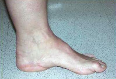
What is arthritis of the foot and ankle?

Arthritis of the Foot and Ankle

The pain and stiffness you feel in your feet and ankles as you age could be arthritis. If left untreated, this nagging pain can get worse over time, eventually making it difficult to walk even short distances. Severe arthritis can restrict your mobility, but with proper treatment, you can minimize the pain and maximize your quality of life.

Arthritis is a broad term for a number of conditions that destroy the workings of a normal joint. Arthritis may occur in your back, neck, hips, knees, shoulders or hands, as well as your feet and ankles. Almost half of people in their 60s and 70s have arthritis of the foot and/or ankle, but not all of them have symptoms.

There are many different types of arthritis. The most common type, osteoarthritis, results from wear and tear to joint cartilage, the cushioning between joints that allows them to glide smoothly. The damage can cause inflammation, redness, swelling, and pain in the joint.

Also, a sudden and traumatic injury such as a broken bone, torn ligament, or moderate ankle sprain can cause the injured joint to become arthritic in the future. Sometimes a traumatic injury will result in arthritis in the injured joint even though the joint received proper medical care at the time of injury. This type of arthritis, called post-traumatic arthritis, is the most common type that affects the ankle.

Another less common type, rheumatoid arthritis, is an inflammatory condition caused by an irritation of the joint lining. Other types of inflammatory arthritis include gout, lupus, ankylosing spondylitis, and psoriatic arthritis.

Diagnosis

The foot has 26 bones and more than 30 joints. Tough bands of tissue called ligaments hold these together. The muscles, tendons, and ligaments work together with the many joints of the foot to control motion and weight bearing position. This smooth motion makes it possible for a person to walk well. When you get arthritis in the foot, you develop pain and limited motion so that you cannot walk as well.

Arthritis can develop in any of these joints but some are more commonly affected than others. A foot and ankle orthopedic surgeon can help you locate the source of your pain and determine the best course of treatment.

If your foot and ankle orthopedic surgeon suspects you have arthritis, he or she will ask you to have a complete medical history and physical examination. X-rays and laboratory tests often can confirm the type and extent of the arthritis. Other tests such as a bone scan, CT scan, or MRI may be used to evaluate your condition.

What is ankle fracture surgery?

The ankle is made up of three bones:

  • the tibia (shin bone), which forms the inside, front, and back of the ankle
  • the fibula, which forms the outside of the ankle
  • the talus, a small bone that sits between the tibia and fibula and the heel bone

The ends of these bones are called malleoli. The tibia has a medial (inside) malleoli and a posterior malleoli. The fibula forms the lateral (outside) malleoli.

A broken ankle (ankle fracture) occurs when the malleoli are broken. These fractures are very common. Ankle fractures happen with twisting of the ankle, falls, car accidents, or other injury. One, two, or all three malleoli can be broken. Ankle fractures can be displaced (out of place) or non-displaced. Symptoms of an ankle fracture include pain especially with weight bearing, swelling, bruising, and problems with ankle motion. X-rays help determine if treatment from a foot and ankle orthopedic surgeon is needed.

Ankle Fracture Surgery

The main goal of ankle fracture surgery is to put the ankle joint back in place and to stabilize the bones to heal. Getting the ankle joint back in place helps to decrease the risk of developing arthritis of the ankle. Stabilizing the ankle with plates and screws may allow earlier motion.

Diagnosis

When the ankle fracture is unstable or in bad position, surgery is needed to repair the ankle. In some cases, the bones of the ankle may poke through the skin. These are called open ankle fractures and require surgery.

Ankle fracture surgery is not needed if the ankle is in position and stable despite the fracture. Surgery may be too risky when patients have a severe medical condition.

Treatment

Antibiotics are given shortly before the start of the surgery. Once the patient receives antibiotics and anesthesia, the surgery can safely begin. Incisions are made through the skin of the ankle where the bones are broken. The breaks in the bone are then repositioned and held in place with implants (screws and plates). After the ankle fracture is repaired, the patient’s leg is placed in either a protective splint, cast or boot. Some patients go home after surgery the same day while others may stay overnight in the hospital.

Specific Technique

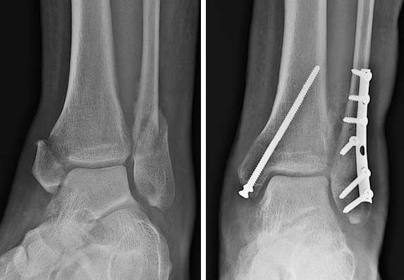
Most ankle fracture surgery involves open reduction and internal fixation (ORIF). An incision is made over the ankle to see the fractured bones. Like a jigsaw puzzle, the pieces of the broken bones are placed back together (open reduction). The broken bones are then held together (internal fixation) in this correct position with metal plates and/or screws. This internal fixation provides stability so movement can begin shortly after surgery as the ankle fracture heals.

ankle fracture surgery
Left, a bimalleolar ankle fracture before surgery. Right, the ankle fracture after it has been fixed.

Recovery

Surgical treatment of ankle fractures allows patients to regain ankle function while the bones and joint heal. Immediately after surgery, your ankle will be immobilized for a few weeks. Once the wounds are healed, you will be fitted for either a cast or a boot. Many surgeons use a removable boot. The boot protects the ankle as it heals but can be removed to allow washing, wound checks, and icing.

Once the broken bones start to heal, you will be told when you can put weight on the ankle in your cast or boot. You are then allowed to wean out of the cast or boot. You can progress to normal activities once the bones are fully healed. Some patients may need physical therapy for their ankle to regain full movement and strength. While everyone is different, it may take up to a year after surgery for some patients to regain ankle function.

Risks and Complications

All surgeries come with possible complications, including the risks associated with anesthesia, infection, damage to nerves and blood vessels, and bleeding or blood clots. Potential long-term problems after ankle fracture surgery include ankle joint stiffness, weakness, and arthritis. Possible complications include infection and problems with healing. Factors that increase risks after ankle fracture surgery include diabetes, steroid use, and cigarette smoking.

FAQs

Will my ankle hardware (plate and/or screws) have to be removed at some point?

Plates and screws used to fix an ankle fracture are not removed if they are not causing problems. Most people do not have problems with the plate and screws. In rare cases, the plate and screws can cause some pain or irritation. When this happens, the hardware may be removed after the fracture is healed, about a year from the original surgery.

What is ankle arthroscopy?

Ankle Arthroscopy

Ankle arthroscopy is a minimally invasive surgical procedure that orthopedic surgeons use to treat problems in the ankle joint. Ankle arthroscopy uses a thin fiber-optic camera (arthroscope) that can magnify and transmit images of the ankle to a video screen. Ankle arthroscopies can reduce ankle pain and improve overall function.

Arthroscopy can be used to diagnose and treat different disorders of the ankle joint. The list of problems that can sometimes be treated with this technology is constantly evolving and includes:

Ankle arthritis: Ankle fusion is a treatment option for many patients with end-stage ankle arthritis. Ankle arthroscopy offers a minimally invasive way to perform ankle fusion. Results can be equal to or better than open techniques.

Ankle fractures: Ankle arthroscopy may be used along with open techniques of fracture repair. This can help to ensure normal alignment of bone and cartilage. It also may be used during ankle fracture repair to look for cartilage injuries inside the ankle.

Ankle instability: Ligaments of the ankle can become stretched out, which can lead to a feeling that the ankle gives way. These ligaments can be tightened with surgery. Arthroscopic techniques may be an option for treating moderate instability.

What is ankle arthrodesis?

Ankle Arthrodesis or Ankle Fusion

The goal of ankle arthrodesis (also known as ankle fusion) is to relieve pain and maintain or improve function for patients with ankle arthritis. Ankle arthritis is degeneration of the cartilage that covers the ends of the bones that form the ankle joint. These bones are the tibia, the fibula, and the talus. Pain typically is made worse with movement of the arthritic ankle. In ankle arthrodesis the ankle bones are fused into one bone. This eliminates the joint motion and reduces pain coming from the arthritic joint.

Diagnosis

Patients may be candidates for ankle arthrodesis if they have severe ankle arthritis and non-surgical treatments have failed. Many patients may find relief from the pain associated with ankle arthritis using:

  • Anti-inflammatory medication (such as ibuprofen)
  • Injections of steroids into the ankle joint
  • Modification or limitations of activity
  • Walking aids (such as canes)
  • Specialty braces that stabilize the ankle and restrict its movement
  • Cushioned and specially contoured shoes

These treatments do not reverse ankle arthritis. In many patients they may temporarily or permanently provide relief from pain. If these measures fail to provide adequate pain relief or maintain function, a patient may be a candidate for ankle fusion. You should discuss your options with your foot and ankle orthopedic surgeon.

Patients should avoid ankle arthrodesis if they have:

  • Insufficient quantity or quality of bone for fusion
  • Poor blood supply to the ankle
  • Severely impaired nerve function
  • Medical conditions that increase the risk of anesthetic
  • Severe deformity of the limb

Treatment

Patients are asleep or sedated in the operating room during the procedure. Incisions are made in and around the ankle to access the joint. Any remaining cartilage within the ankle joint is removed so there is contact between the bony surfaces. The ankle is held in the most functional position with metal hardware. This allows the bones to heal together.