mm

About Orthopedic Specialists

Orthopedic Specialists of Seattle provides new and advanced procedures including endoscopic carpel tunnel release surgery for carpal tunnel syrome, complex joint restoration procedures, anterior approach hip replacement surgery, and more.

What is ankle arthroscopy?

Ankle Arthroscopy

Ankle arthroscopy is a minimally invasive surgical procedure that orthopedic surgeons use to treat problems in the ankle joint. Ankle arthroscopy uses a thin fiber-optic camera (arthroscope) that can magnify and transmit images of the ankle to a video screen. Ankle arthroscopies can reduce ankle pain and improve overall function.

Arthroscopy can be used to diagnose and treat different disorders of the ankle joint. The list of problems that can sometimes be treated with this technology is constantly evolving and includes:

Ankle arthritis: Ankle fusion is a treatment option for many patients with end-stage ankle arthritis. Ankle arthroscopy offers a minimally invasive way to perform ankle fusion. Results can be equal to or better than open techniques.

Ankle fractures: Ankle arthroscopy may be used along with open techniques of fracture repair. This can help to ensure normal alignment of bone and cartilage. It also may be used during ankle fracture repair to look for cartilage injuries inside the ankle.

Ankle instability: Ligaments of the ankle can become stretched out, which can lead to a feeling that the ankle gives way. These ligaments can be tightened with surgery. Arthroscopic techniques may be an option for treating moderate instability.

What is ankle arthrodesis?

Ankle Arthrodesis or Ankle Fusion

The goal of ankle arthrodesis (also known as ankle fusion) is to relieve pain and maintain or improve function for patients with ankle arthritis. Ankle arthritis is degeneration of the cartilage that covers the ends of the bones that form the ankle joint. These bones are the tibia, the fibula, and the talus. Pain typically is made worse with movement of the arthritic ankle. In ankle arthrodesis the ankle bones are fused into one bone. This eliminates the joint motion and reduces pain coming from the arthritic joint.

Diagnosis

Patients may be candidates for ankle arthrodesis if they have severe ankle arthritis and non-surgical treatments have failed. Many patients may find relief from the pain associated with ankle arthritis using:

  • Anti-inflammatory medication (such as ibuprofen)
  • Injections of steroids into the ankle joint
  • Modification or limitations of activity
  • Walking aids (such as canes)
  • Specialty braces that stabilize the ankle and restrict its movement
  • Cushioned and specially contoured shoes

These treatments do not reverse ankle arthritis. In many patients they may temporarily or permanently provide relief from pain. If these measures fail to provide adequate pain relief or maintain function, a patient may be a candidate for ankle fusion. You should discuss your options with your foot and ankle orthopedic surgeon.

Patients should avoid ankle arthrodesis if they have:

  • Insufficient quantity or quality of bone for fusion
  • Poor blood supply to the ankle
  • Severely impaired nerve function
  • Medical conditions that increase the risk of anesthetic
  • Severe deformity of the limb

Treatment

Patients are asleep or sedated in the operating room during the procedure. Incisions are made in and around the ankle to access the joint. Any remaining cartilage within the ankle joint is removed so there is contact between the bony surfaces. The ankle is held in the most functional position with metal hardware. This allows the bones to heal together.

What is an flexor digitorum longus tendon transfer to posterior tibial tendon?

Flexor Digitorum Longus

The flexor digitorum longus (FDL) is one of the tendons responsible for bending the toes down to the floor. The goals of a FDL tendon transfer surgery are to relieve pain and to help restore the arch in patients with painful fallen arches. A fallen arch occurs when the foot loses its support and flattens out, generally due to weakening of tendons and ligaments in the foot.

Diagnosis

Tendon transfer surgery is indicated for people with a flexible flatfoot that can be moved into a more normal position. The posterior tibial tendon (PTT) is a main support for the arch of the foot. If it becomes diseased, it no longer functions properly and the arch begins to fall. The patient begins to walk on the inside of the foot as it flattens. The toes may begin to turn outward resulting in a flatfoot deformity. When non-surgical treatment such as arch supports fail to provide relief, surgery may be necessary.

If the deformity becomes stiff or arthritis develops, more advanced surgery is needed. This typically includes re-making the arch by fusing bones of the foot together. Patients with other medical problems may be too sick to safely undergo surgery.

Treatment

The PTT connects to the navicular bone near the middle of the foot at the instep. In this surgery, the FDL tendon is moved from its usual position and transferred to the navicular bone. This helps support or replace the diseased PTT to improve function. The diseased PTT is cleaned up or removed to eliminate it as a source of pain.

Specific Technique

The surgery is done through an incision on the inside of the ankle and foot. The initial step is to remove the scarred or inflamed tissue of the PTT. The tendon may be completely removed if it is severely damaged. Just below the PTT is the tendon of the FDL. The FDL is cut so that it is as long as possible. A hole is drilled in the navicular bone. The end of the FDL is placed through the bone. While the foot is held in the corrected position, the tendon is attached to the bone. It may be held in place with stitches and/or an anchor or screw. The incision is then closed. Other procedures may be performed with the tendon transfer to improve the arch. These can include moving or shifting of bones and stretching of the calf muscles or Achilles tendon.

Recovery

Patients usually are placed in a well-padded dressing with a splint or split cast. No weight is allowed on the ankle and foot. Patients are given crutches, a walker, or a knee walker/scooter. This procedure may be done as an outpatient or may require an overnight stay.

At two weeks the sutures are removed and a new cast or removable brace is applied. At six weeks most patients transition into a walking cast or boot with a well-molded arch. At three months, patients return to a shoe with an arch support. Elastic hose may be used for swelling and physical therapy may be prescribed to help with walking and to restore muscle strength and joint flexibility. It may take up to one year for patients to fully recover.

Risks and Complications

All surgeries come with possible complications, including the risks associated with anesthesia, infection, damage to nerves and blood vessels, and bleeding or blood clots. Standard wound complications can occur and may include delayed healing and infection. Irritation of an adjacent nerve can occur and may cause numbness or burning. These symptoms typically resolve with time. Rarely the repositioned tendon may pull out of the navicular bone and require re-placement. A more common problem is failure to restore the arch and a residual flat foot.

FAQs

Can I expect pain relief with this surgery?

Typically, improvement in pain control is achieved with the procedure. The use of arch supports often is recommended even after successful surgery. Ankle bracing may also be helpful.

What alternatives do I have if my arch is not restored or falls again?

If the arch is not restored or falls further, and arch supports and/or bracing are not helpful, additional surgery may be considered.

What is an ankle cheilectomy?

Ankle Cheilectomy

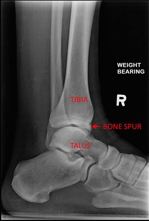
An ankle cheilectomy is a surgery that removes a bone spur from the talus or tibia, which are the lower and upper bones of the ankle joint.

The goal of an ankle cheilectomy is to relieve ankle pain caused by bone impingement or pinching at the front of the ankle. While this may be a sign of early ankle arthritis, an ankle cheilectomy is a joint preserving option that may help with pain and function and buy time before additional treatment is needed.

Diagnosis

If you have pain at the front of the ankle that does not improve with non-surgical treatment, your foot and ankle orthopedic surgeon may consider an ankle cheilectomy. The pain is usually worse with upward bending (dorsiflexion) of the ankle. The pain typically is caused by a bone spur.

An ankle cheilectomy is not recommended if you have severe ankle arthritis. Removal of bone spurs in arthritic ankles typically does not improve pain.

Treatment

An ankle cheilectomy can be performed arthroscopically (minimally invasive) or with an open procedure. The choice for open or arthroscopic procedure is made based on the size of the spur and the preference of the surgeon.

  • Arthroscopic: When an ankle arthroscopy is performed, a camera is placed into the ankle joint through a small incision. The cartilage and soft tissues inside the ankle joint are examined and the bone spur is seen with the camera. It is removed with an instrument such as a burr or chisel placed into the ankle through a separate small incision.
  • Open: An incision is made at the front of the ankle. The ankle joint is opened, and the bone spur is identified and removed.

ankle cheilectomy bone spur

Recovery

Activity after surgery may depend on the size of the bone spur and the degree of swelling or bleeding that occurs during surgery. There may be a period where you may not be allowed to put any weight, or only partial weight, on the ankle. This usually lasts for 1-3 weeks. Physical therapy is then typically started, and weight bearing and activity generally are increased as tolerated.

Risks and Complications

All surgeries come with possible complications, including the risks associated with anesthesia, infection, damage to nerves and blood vessels, and bleeding or blood clots. Patients may experience loss of feeling at the top of the foot after this procedure. Pain may not improve after an ankle cheilectomy, and an increase in ankle pain can occur. While many patients do experience a significant reduction of pain, there is a risk of recurrence of the pain after surgery as underlying arthritis progresses. However, the overall complication rate for this surgery is low.

FAQs

Will I regain range of motion in my ankle if the bone spur is removed?

Range of motion may increase after removal of bone spurs. This does not always occur as soft tissues such as tendons and ligaments around the ankle may still be tight, and these tissues also affect ankle motion. Often, even if motion is not improved, the pain from the bony impingement improves.

What is an Achilles tendon tear?

The Achilles tendon is a large tendon at the back of the lower leg and ankle that connects the calf muscles to the heel. It is the largest tendon in the body, and its strength allows us to push off with forces up to 10 times our body weight.

Achilles Tendon Tear

An Achilles tendon tear, or rupture, usually is a complete gap between the upper and lower portions of this tendon. It most often occurs 2-3 inches above the heel bone but can be directly at the attachment or higher in the leg.

Symptoms

A tear usually occurs during activities such as running and jumping, or trauma such as a slip and fall. Most patients report their first feeling was a “pop” or like they were struck in the back of the ankle, followed by some pain. There often is no pain in this area before the actual tear. After the tear, patients complain of weakness when pushing off of their foot during walking or when trying to stand on tiptoe.

Diagnosis

Your foot and ankle orthopedic surgeon often performs an examination to make a diagnosis. You will lie in a facedown position with your ankles off the edge of the exam table. When the Achilles tendon is not torn, it is taut and the ankle lies in a toe down position of approximately 20 degrees. Squeezing the upper calf will cause the toes to point down even further.

If the tendon is torn, it does not have this tension and the ankle usually will hang at about a 90-degree angle. Squeezing the upper calf will cause weak or no movement. Special tests such as ultrasound, X-rays, and MRI scans typically are not required but can be in some instances.

Treatments

At first, the leg is placed at rest in a splint or special boot. It is important to see a foot and ankle orthopedic surgeon soon after the injury so the best treatment can be started within a few days.

Both non-surgical and surgical treatments have been well studied but remain debated. In general, surgical treatment is thought to give greater strength and result in a lower risk of repeat tear. However, surgery has an increased risk of wound healing problems, nerve damage, and infection plus the usual risks associated with surgery. Blood clots are a concern with both types of treatment. Your foot and ankle orthopedic surgeon will be the best person to guide you through the pros and cons and help you to select the best option.

Non-surgical Treatment

Non-surgical treatment starts with a period of rest in a boot for the injured leg. You will need to use crutches, a walker, or wheelchair in order to not put weight on the leg. Within the first few weeks you will start doing active motion. Gradually, you will be allowed to put weight on the leg and start specific strengthening exercises. The length of treatment may be different for each patient but usually takes about three months. It is often guided by a physical therapist along with the orthopedic surgeon.

Surgical Treatment

In this surgery, your foot and ankle orthopedic surgeon will place stitches into the tendon above and below the area of the tear and then pull the ends together. The surgery often is performed through a very small incision to minimize the risk of wound complications. The recovery after surgery is similar to the non-surgical treatment but can be slightly shorter.

Recovery

Since tendons do not have a great blood supply, healing is a slow process. Patients usually can start light jogging in 3-6 months with return to sports involving cutting and jumping in 6-9 months. Full return of strength and the feeling of being normal may take more than a year.

FAQs

After an Achilles tendon tear, how likely am I to tear the other side?

About 6% of patients with an Achilles tendon tear will have the same injury in the other foot.

Is there anything I can do to make the tendon heal faster?

Starting range-of-motion exercises and putting weight on the injured leg early have shown better results than long periods of immobilization on crutches. However, it has to be balanced by the risk of pulling apart the ends of the tendon if you stretch too much too early. Stopping smoking for at least a few months while the tendon heals also is likely to be of benefit.