mm

About Orthopedic Specialists

Orthopedic Specialists of Seattle provides new and advanced procedures including endoscopic carpel tunnel release surgery for carpal tunnel syrome, complex joint restoration procedures, anterior approach hip replacement surgery, and more.

What is achilles tendinosis?

Achilles Tendon

The Achilles tendon is the largest tendon in your body. It connects the upper calf muscles to the back of the heel bone. Achilles tendinosis is a condition in which the Achilles tendon degenerates and becomes inflamed. Sometimes, it may also be called Achilles tendinitis. If you have Achilles tendinosis, your tendon can swell and become painful. This condition is common in athletes, runners, and people who have calf tightness. Achilles tendinosis may occur in the middle of the tendon (known as midsubstance Achilles tendinosis) or at the point where the tendon connects to the heel bone (known as insertional Achilles tendinosis).

Symptoms

You may see many changes when the Achilles tendon becomes inflamed. Many patients have pain and/or tightness in the tendon behind the ankle. Most of the time there is no trauma or injury, but rather a slow progression of pain. You may have difficulty climbing stairs or running. You also may have pain after sitting for long periods or after sleeping. Many patients notice a bump either in the tendon or right behind the heel bone. Some also get irritation from shoes rubbing against the bump and feel better when wearing backless shoes. Patients commonly have less pain while wearing a shoe with a low heel versus shoes that are flat.

Causes

Achilles and calf tightness are common causes of Achilles tendinosis. In addition, insertional Achilles tendinosis often is associated with a heel bone spur. This spur may rub against the Achilles tendon and lead to small tears. It is similar to a rope being rubbed against a sharp rock. This is also known as Haglund’s deformity. Pain and swelling occur as the cumulative effects of chronic wear and tear on the tendon.

Diagnosis

Your foot and ankle orthopedic surgeon will take a thorough history and perform an examination. It is common to have pain right on the tendon or at the back of the heel. You also may have swelling and thickening of the tendon. X-rays may be taken to see if there are any bone spurs. An MRI or ultrasound may be ordered to look for tears and further evaluate how much of the tendon is affected.

Treatment

Treatment depends on the length and severity of the symptoms. Many patients improve without surgery. Rest and oral medications may help reduce the swelling and pain. Heel cups can improve pain by taking some of the stress off of the Achilles tendon when walking. A walking boot or other brace may be recommended.

Often, formal physical therapy is recommended to work on stretching and improve mobility within the calf muscle. Other treatments may include ultrasound, massage, shockwave therapy, and topical nitroglycerin patches. Recently, platelet-rich plasma has been discussed as a treatment for Achilles tendinosis. This involves taking one’s own blood and isolating growth factors that are involved in healing. This serum is then injected into the inflamed tendon.

In some cases, surgery may be required. The specifics of the surgery depend upon the location and extent of the tendinosis.

FAQs

If I am treated without surgery for Achilles tendinosis will it come back?

While most patients will achieve lasting relief after treatment for Achilles tendinosis, symptoms may return. The risk decreases if the patient continues to do routine stretching even after the symptoms go away. However, athletes and runners in particular are at a slightly higher risk for this condition because of the high demands they put on the Achilles. These patients should pay close attention to stretching and shoe choice to prevent chronic recurrence.

What are the outcomes for those who have surgery for Achilles tendinosis?

Surgery can predictably return patients to activity. Success rates for Achilles tendinosis surgery are 80-90 percent. Some of the variability depends on the amount of tendon that is diseased at the time of surgery.

Patients improve with both conservative and operative management of Achilles tendinosis. Physical therapy has been shown to help most patients with this condition and should be tried before surgery is considered.

What is Achilles tendinosis/tendinitis surgery?

Achilles Tendon

The Achilles tendon is the largest tendon in your body. It connects the upper calf muscles to the back of the heel bone. When the Achilles tendon degenerates and becomes inflamed, it is called Achilles tendinitis or Achilles tendinosis.

If you have these conditions, your tendon can swell and become painful. They are common in athletes, runners, and people who have calf tightness. Achilles tendinosis may occur in the middle of the tendon (known as midsubstance Achilles tendinosis) or at the point where the tendon connects to the heel bone (known as insertional Achilles tendinosis).

For midsubstance Achilles tendinosis, surgery focuses on removing the diseased portion of the tendon. If most of the tendon is damaged, your foot and ankle orthopedic surgeon often will use the tendon that goes to the big toe to support the Achilles tendon after repair. Other procedures may include lengthening the Achilles tendon or calf muscles if they are too tight.

Surgery for insertional Achilles tendinosis is similar. Very often the diseased tissue is removed and the tendon is repaired back down to the heel bone. Surgeons often will shave down the bone spur and smooth it out so that it no longer has the ability to rub the Achilles tendon. Often there is a fluid-filled sac (bursa) that contributes to the pain and inflammation. This bursa frequently is removed during the surgery.

Diagnosis

Surgery may be an option for Achilles tendinitis or tendinosis if other treatments, including physical therapy, ultrasound, massage, and shock wave therapy, fail to bring pain relief.

Treatment

General anesthesia is commonly used. Regional anesthesia that numbs the leg also may be used. The patient is positioned face down on the operating table. Your surgeon will make an incision in the back of the ankle directly over the Achilles tendon. The diseased portion of the tendon is removed with a scalpel. If the problem involves the end of the tendon where it inserts on the heel bone, the tendon may be lifted off of the heel bone. The bump at the back of the bone is removed with a chisel or saw, and the tendon is repaired back down to the remaining bone. Some surgeons also perform a stretching of the calf muscles as part of the procedure. The incisions are sewn together. The leg is bandaged and then protected with a splint or boot brace.

Recovery

It may take up to a full year for symptoms to resolve completely. Often surgical patients will need several months in a protective boot and crutches before they can walk on their own. Physical therapy often is needed to help restore mobility and strength to the repaired tendon.

Surgery can predictably return patients to activity. Success rates for Achilles tendinosis surgery are 80-90%. Some of the variability depends on the amount of tendon that is diseased at the time of surgery.

Risks and Complications

All surgeries come with possible complications, including the risks associated with anesthesia, wound healing issues, infection, damage to nerves and blood vessels, and bleeding or blood clots.

After this surgery, patients can still have moderate pain. If the tendon is repaired there is still risk of future degeneration of the tendon as the patient ages. However, repeat surgery is rarely required. There is also a risk of Achilles tendon rupture.

FAQs

If I am treated without surgery for Achilles tendinosis will it come back?

While most patients will achieve lasting relief after treatment for Achilles tendinosis, symptoms may return. The risk decreases if the patient continues to do routine stretching even after the symptoms resolve. However, athletes and runners in particular are at a slightly higher risk for this condition because of the high demands they put on the Achilles. These patients should pay close attention to stretching, and shoe choice to prevent chronic recurrence.

Will I still have pain after surgery?

The majority of patients improve after having surgery. However, up to 20-30% of patients still report some pain after surgical treatment.

What is a tendon transfer?

Tendon Transfer

A tendon transfer is moving a tendon from its normal, anatomic location to another area of the foot or ankle. Tendons typically are transferred in order to restore more normal movement to a foot and ankle that has lost function. A common problem is loss of the ability to raise the foot up, which is called foot drop. This can result from nerve or muscle damage due to stroke, injury, or other diseases. This muscular weakness or paralysis decreases movement and can lead to the foot becoming bent or twisted, making it difficult or painful to stand, walk, or wear shoes.

Some tendon transfers allow the ankle and foot to move up and down and regain some strength and motion. Others bring the foot into a position where it is easier to walk, stand, and wear shoes, but will not increase range of motion. Realigning the foot and ankle can also decrease pain by more evenly distributing pressure across the foot. In some cases, a tendon transfer may eliminate the need for a brace altogether.

Diagnosis

There are two common reasons your foot and ankle orthopedic surgeon may recommend a tendon transfer in the foot and ankle. One is a painful, flexible flatfoot. This develops when the posterior tibial tendon stretches and becomes nonfunctional or ruptures, which can cause the arch of the foot to drop. The foot then rolls inward, leading the patient to walk on the inside of their foot. This can cause discomfort.

The other reason is the loss of function of muscles in the lower leg and foot due to a neurological problem such as injury or disease. This can include weakness after a stroke, nerve damage after a surgery or accident, or a systemic disease causing weakness of the muscles such as Charcot-Marie-Tooth disease. These problems most typically weaken the muscles of the front and outside of the calf. This causes the foot to roll outward and the patient to walk on the outside of their foot. This can cause pain and weakness as well as bone fractures and looseness of the ligaments on the outside of the foot.

Tendon transfers usually are elective procedures. Any medical problems which make surgery more dangerous or difficult, such as a recent heart attack, stroke, blood clot, or infection, may require the procedure to be delayed or canceled.

Treatment

There are three requirements for tendon transfers to be successful:

  1. The muscle that the tendon is attached to has to be functional. It must contract at the proper time during walking.
  2. The soft tissue through which the tendon is to be transferred needs to be as normal as possible. Large areas of scarring or skin damage can make it difficult to transfer the tendon. It also can prevent the tendon from gliding along the correct path.
  3. The joints that the tendon crosses need to be both mobile and stable.

Tendon transfers involve the cutting of the tendon at or near its normal insertion, rerouting it through the soft tissues either around or between the bones of the foot and ankle, and connecting it to another bone in the foot. When the transferred tendon is long enough, it can be passed through a tunnel drilled through the target bone and then sewn to itself. The tendon also can be brought into a bone tunnel and fastened with a screw made of metal or an absorbable plastic. An anchor with sutures attached to it also can be placed in the bone at the point the tendon is to be attached and the sutures used to sew the tendon to the anchor. The soft tissues are then closed and the patient is placed in a splint.

Recovery

Typically the splint stays on the patient’s leg for 10-14 days. At that point the splint and sutures are removed. The patient is then placed in a cast or a removable boot.

Patients typically are non-weightbearing for six weeks after surgery to allow the transferred tendon to heal to its new attachment. This can be longer or shorter depending on what is seen during surgery. After six weeks, patients usually start to bear more weight on the leg and are placed in a boot if they aren’t in one already. Patients are out of the cast or boot by 12 weeks after surgery.

Physical therapy usually is needed to regain the strength of the transferred muscle and to help the patient learn to walk more normally. The transferred muscle is weaker than it was in its original functional position because of the transfer. There may be some limitations in motion of the foot due to the tendon transfer, but the function regained by the transfer should outweigh the function lost.

Risks and Complications

All surgeries come with possible complications, including the risks associated with anesthesia, infection, damage to nerves and blood vessels, and bleeding or blood clots. Sometimes the tendon transfer does not heal into its new position. The implant could break or loosen, or there could be progression of the original neurologic condition requiring further surgery.

FAQs

Will my foot move normally after the tendon transfer?

It depends on which tendon transfer you have. Some tendon transfers act as a leash for the foot and simply hold it in proper alignment. This does not necessarily provide much movement for your ankle and foot. Other tendon transfers move functional muscles from one side of the foot to the other, and therefore the foot should have a more normal movement after these procedures. The goals for the surgery and recovery should be discussed with your foot and ankle orthopedic surgeon before proceeding with any procedure.

What is a tarsal coalition?

Tarsal Coalition

Tarsal coalition (TC) is an abnormal connection between two or more bones that is present at birth. The connection may be made of fibrous tissue, cartilage, or bone. Most people with TC do not have symptoms. Occasionally, patients experience decreased motion in their foot joints, which can be painful. As one joint loses motion, surrounding joints can develop arthritis. TC usually affects children and teens but can appear in adults also. The condition affects 1-6% of the population and about half of patients have problems with both feet.

Symptoms

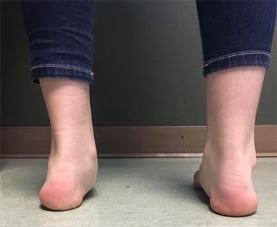
While TC may be present at birth, symptoms usually are delayed until the bone matures. This explains why most patients experience pain gradually. The typical patient is a child or teen with foot stiffness and pain with activity. Other symptoms may include discomfort when walking on uneven surfaces, frequent ankle sprains, limited side-to-side joint motion, and flat feet.

Causes

Tarsal coalition is caused by a gene mutation. The bones of the feet are divided into three parts: the hindfoot (back of the foot), midfoot (middle of the foot), and forefoot (front of the foot). Tarsal coalition involves the bones in the midfoot and hindfoot.

Of these bones, the calcaneus (heel bone), talus (lower bone of the ankle), and navicular (boat-shaped bone in the middle of the foot) are most commonly involved. Most tarsal coalitions are one of two types:

  • Talo-calcaneal coalition: In this case the talus and the calcaneus have not fully separated.
  • Calcaneo-navicular coalition: In this case the front part (beak) of the calcaneus is attached to the outside and lower part of the navicular bone.

Diagnosis

Your foot and ankle orthopedic surgeon will typically diagnose TC with X-rays of the foot and ankle. In some cases, a CT scan or MRI can help in the diagnosis. A CT scan will best determine the extent of a bony coalition while an MRI will be more useful in diagnosing small, fibrous, or cartilage coalitions.

Treatments

In asymptomatic coalitions, observation is all that is needed. In almost all symptomatic cases, non-surgical treatment is the first option.

Non-surgical Treatment

Your surgeon may suggest the following treatments to relieve symptoms:

  • Avoiding aggravating activities or walking on uneven ground
  • A supportive shoe, orthotic, or ankle brace
  • Anti-inflammatory medications
  • Immobilization with a cast or walking boot
  • Local corticosteroid injection

Surgical Treatment

When conservative management has been exhausted, surgical options should be considered. The type of surgery will depend on the location and size of the coalition, the presence of any arthritis at the joints near the coalition, and the expectations and activity level of the patient.

Resection of the coalition is performed to decrease pain and improve motion between the affected bones. Younger patients tend to do better with resection of a coalition. If the coalition is large or if arthritis is present, your surgeon may suggest a fusion of the affected joints. This improves pain but does not improve motion.

Recovery

A walking cast or boot is used to protect the surgical site. If a fusion procedure is performed, a longer period of immobilization and non-weightbearing is necessary. This is followed by physical therapy to restore range of motion and strength. Arch supports or orthotics also can be helpful in stabilizing the joint, even after surgery. Although it may take up to a year to fully recover, most patients have pain relief and improved motion after surgery.

Risks and Complications

All surgeries come with possible complications, including the risks associated with anesthesia, infection, damage to nerves and blood vessels, and bleeding or blood clots. In fusion surgery there is a small risk that the bones will not fuse together.

What is a talus fracture?

Talus Fracture

A talus fracture is a broken ankle bone. The talus is the bone in the back of the foot that connects the leg and the foot. It joins with the two leg bones (tibia and fibula) to form the ankle joint and allows for upward and downward motion of the ankle. The talus (ankle bone) sits within the ankle mortise or hinge, which is made up of the two leg bones, the tibia and fibula. There are three joints:

  • the ankle, which allows the up-and-down motion of the foot with the leg
  • the subtalar joint, which allows for side-to-side movement
  • the talonavicular joint, which has a complicated biomechanical function that controls flexibility of the foot and the arch of the foot.

The talus has no muscular attachments and is mostly covered with cartilage, which makes injuries difficult to heal.

Symptoms

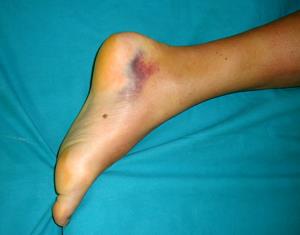
Most patients present with pain and swelling around the ankle. They also experience severe ankle pain and difficulty walking due to bruising and swelling.

Causes

Ankle fractures often are the result of high-energy injuries. Falls from ladders and automobile accidents result in the most severe injuries. However, these fractures also may occur from twisting the ankle, which can result in small chips or fragments that are broken off the edges of the ankle.

Diagnosis

In many cases the diagnosis can be made by your physician on physical examination alone. He or she will examine your foot for evidence of swelling or bruising around the ankle. X-rays help visualize the extent of joint involvement and show the location and size of bone fragments. Oftentimes a CT scan is ordered to provide the physician with more information about the fracture. Due to the high energy that is commonly associated with these injuries, your physician also may examine you for other injuries involving the back, neck, head and other extremities.

Treatments

Talus fractures may be treated in a cast or surgery may be recommended.

Non-surgical Treatment

Non-surgical treatment is recommended for fractures in which the pieces of bones remain close together and the joint surfaces are well-aligned. Patients who smoke or have diabetes or poor circulation may be treated without surgery due to the very high risk of developing complications if surgery is performed.

Surgical Treatment

For a majority of patients, surgical treatment is the correct form of treatment. The goal of surgery is to restore the size and shape of the talus. Sometimes this is a problem as the multiple fragments of bone are like putting together the pieces of a difficult puzzle. If the bone has several large pieces, your foot and ankle orthopedic surgeon may perform open reduction and internal fixation (ORIF). The procedure involves making a cut(s) on your foot and placing a metal plate and/or screws to hold the bones together until healing occurs. The procedure aids in restoring the function of your foot.

Recovery

Recovery can be prolonged. No weight or walking on the leg will be allowed for 8-12 weeks. Once the bone is healed, exercise and physical therapy is started to maximize the function of the ankle. You should expect some swelling around the foot for several months after the procedure.

This injury can be very debilitating with persistent pain, stiffness and swelling even after an excellent non-surgical or surgical treatment. However, most people, depending on the type and severity of the fracture, are able to return to most work and recreational activities.

Risks and Complications

Talus fractures are quite severe injuries and can lead to longstanding problems with the foot and ankle. There are early and late complications.

Early complications most often are related to the significant swelling that can occur after these injuries, which can cause wound problems and infection. People who smoke, diabetics, and those with poor circulation are at greatest risk for these complications.

Late complications typically are related to the severity of the initial injury. Most people experience a certain degree of stiffness within the foot and ankle. When the blood supply to the ankle is damaged it can lead to death of the bone, a condition called avascular necrosis (AVN). This condition can lead to significant deformity and arthritis and require additional surgery.

FAQs

How long will I be out of work?

This is a severe injury. Depending on the type of work performed, most people are unable to return to work for at least two weeks after the injury. Those with active jobs may not be able to return for six months to a year.

Do the plates and screws need to be removed?

Plates and screws hold the bones together so that they can heal. Once the bones are healed the hardware serves no purpose. However, most orthopedic surgeons do not recommend removal of the plate and screws unless there are problems with pain or infection.