Home > Archives for Orthopedic Specialists > Page 2
About Orthopedic Specialists
Orthopedic Specialists of Seattle provides new and advanced procedures including endoscopic carpel tunnel release surgery for carpal tunnel syrome, complex joint restoration procedures, anterior approach hip replacement surgery, and more.
The acetabular labrum is a structure attached to the outside rim of the hip socket. This labrum is made of fibrous cartilage, a flexible material present in multiple joints of the body. In the hip, the labrum is thought to act as a gasket, keeping fluid in the joint during the normal loading of the joint that occurs with movement. It also acts as a stabilizer of the joint keeping the head seated in the socket.
Causes of Labral Tears
Various conditions can lead to damage of this labrum. These include traumatic events, degenerative conditions over time, as well as situations where the shape of the hip bones is incorrect. Traumatic events leading to labral tears can occur with multiple activities including motor vehicle accidents as well as common trips and falls. Degenerative labral tears are a component of generalized hip degeneration where the cartilage throughout the hip joint becomes rough and torn.
Conditions where the shape of the hip bones is incorrect are currently falling under the term hip impingement. These conditions involve improper shape of the hip socket, junction of the thigh bone head and neck, and more commonly a combination of both. This improper shape causes the labrum to be pinched or rubbed during normal movement leading to tearing and degeneration.
Treatment of Labral Tears
Treatment of labral tears involves repairing tissue if possible and removing the tissue that is too severely torn. Attention is then directed at correcting any bony abnormalities that have caused the labral tear in the first place. This is usually possible with hip arthroscopy, but may require more invasive procedures to correctly address the underlying bony problem.
More Information
Read our answers to Frequently Asked Questions about hip impingement and labral tears.
Description
Carpal tunnel syndrome is a common source of hand numbness and pain. It is more common in women than men and affects up to 10 percent of the population. It is caused by increased pressure on a nerve entering the hand through the confined space of the carpal tunnel.
The median nerve travels from the forearm into your hand through a tunnel in your wrist. The bottom and sides of this tunnel are formed by wrist bones and the top of the tunnel is covered by a strong band of connective tissue called a ligament.
Your doctor may make the diagnosis by discussing your symptoms and examining you. If symptoms continue to bother you, electrical testing of the nerve function is often performed to help confirm the diagnosis and clarify the best treatment option in your case.
Symptoms
Symptoms usually begin gradually without a specific injury. Numbness, tingling and pain in the hand are common. You may experience an electric-like shocking feeling. The thumb side of the hand is usually most involved. Symptoms at night are common and may awaken you from sleep.
During the day symptoms frequently occur with holding a phone, reading or driving. Symptoms may occur at any time. Moving or shaking the hands often helps decrease symptoms. Sometimes strange feelings and pain will travel up the arm.
Initially symptoms come and go, but over time they may become constant. A feeling of clumsiness or weakness can make delicate motions like buttoning buttons difficult and may cause you to drop things. If the condition is very severe, muscles in the palm may become visibly wasted.
Risk Factors
The actual cause is unknown in most people. Carpal tunnel syndrome is more common in women. In women, the swelling that occurs during pregnancy may cause symptoms, but those will frequently go away after delivery. Carpal tunnel syndrome becomes more common as we grow older and seems to affect people with certain medical conditions such as diabetes, thyroid conditions and rheumatoid arthritis more frequently.
Treatment Options
Symptoms can often be relieved without surgery. Treatment often begins with a brace or splint worn at night to keep the wrist in a natural position. Splints can also be worn during activities that aggravate symptoms. Simple medications such as Tylenol® or Advil® can help decrease pain. Changing patterns of hand use to avoid aggravating positions and activities may be helpful. A corticosteroid injection will often provide temporary relief, but symptoms may come back.
If your carpal tunnel syndrome continues to bother you and you do not gain relief from non-surgical treatments, surgery can be effective in diminishing symptoms. Because carpal tunnel syndrome is not a dangerous problem, the decision whether to have surgery is based mostly on the severity of your symptoms.
If your symptoms are severe and won’t go away you may want to consider surgery.
In more severe cases, surgery is considered sooner because other treatment options are less helpful.
In very severe cases, surgery may be recommended to prevent irreversible damage.
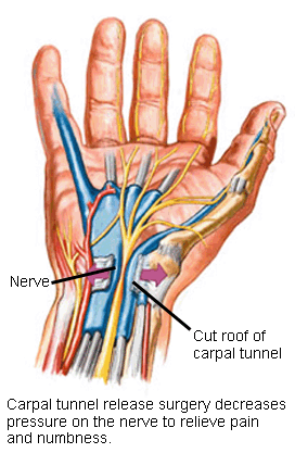
Treatment Options: Surgical
The strong roof of the carpal tunnel is cut during carpal tunnel surgery to increase the size of the tunnel and decrease pressure on the nerve. This is done through an incision in the palm or wrist. A small camera may be used to allow the surgery to be performed through a smaller incision.
Risks of the surgery include bleeding, infection and nerve injury. Some pain, swelling and stiffness are expected, but severe problems are rare.
After surgery, elevating the hand and moving the fingers helps minimize swelling and stiffness. Minor soreness in the palm is common for several months after surgery. Most patients have improvement following surgery, but recovery may be gradual.
When carpal tunnel syndrome has been present longer and the nerve is more severely affected, recovery is slower and less complete.
Carpal Tunnel Syndrome is a common condition that occurs when the median nerve is compressed at the wrist, resulting in pain, numbness, and tingling of the hand and fingers. The carpal tunnel is a tight area of the wrist that many important tendons and nerves pass through. It has a floor, walls, a ceiling, an entrance, and an exit. The floor and walls of the tunnel are made up of the bones.
The ceiling is the transverse carpal ligament. The median nerve runs from the forearm to the hand and directly through the carpal tunnel. This nerve controls some of your hand muscles and also allows sensation of your thumb, index, middle, and half of your ring finger. When this nerve is pressed and squeezed, carpal tunnel syndrome develops.
What are the symptoms of Carpal Tunnel Syndrome?
There are nine tendons that pass through the carpal tunnel and these attach muscles of this area to bones to allow your your thumb and fingers to flex. Each of these tendons has a slippery covering called the synovium. This covering allows the tendons to slide easily as the fingers are moved.
When the synovium becomes inflamed or thickened, pressure develops on the median nerve, adversely affecting its function. With a compromised median nerve, there is less blood and nutrients available, leading to numbness, weakness, and pain of the fingers and thumb. These symptoms are considerably worse at night or while driving. … read more
Big toe arthritis, also called 1st metatarsophalangeal (MTP) arthritis or hallux rigidus, is a common condition affecting the foot and ankle. It is the most common site for arthritis in the foot. Patients typically develop symptoms between age 30 and 60, and females are more commonly affected than men.
Presentation
Patients typically develop stiffness and decreased range of motion at the big toe, which affects walking, running, and other athletic activities. Some patients develop large bone spurs on the top of the foot, which can cause pain with shoe wear and discomfort when going up on the toes.
Causes
Sometimes trauma (a fracture or crush injury) can lead to this condition, but for most patients there is no specific inciting event. Some patients are more likely than others to develop big toe arthritis, either because of some anatomic abnormality or because of genetic predisposition.
Diagnosis
A clinical exam and x-rays can confirm the diagnosis. Generally, advanced imaging like MRI or CT scan is not required.
Treatment
Non-surgical options include anti-inflammatories, shoe wear modifications, and over-the-counter or custom inserts (orthotics). Physical therapy can be helpful to maintain range of motion. Occasionally cortisone injections into the joint can decrease inflammation for a period of time.
Surgery can be used to treat cases that fail non-operative treatment. Traditionally, a procedure called a cheilectomy can be used to remove bone spurs from the top of the big toe joint. This is recommended for mild to moderate cases of hallux rigidus. This is a joint-sparing procedure. Recovery involves walking in a surgical sandal for about 3-4 weeks after the surgery.
For moderate to severe arthritis, a fusion has until recently been the only proven surgical option. This is a joint-sacrificing procedure, in which the bones on either side of the joint are fused together with screws and possibly a plate.
This reliably addresses pain symptoms but eliminates all motion at the joint. Recovery involves a period of non- or heel- weight bearing followed by fully weight bearing in a surgical sandal for 8 weeks or more after the surgery.
Summary
Hallux rigidus is a common condition that involves pain, swelling, stiffness and decreased range of motion of the big toe. Diagnosis is often straightforward and involves a clinical examination and x-ray. Several non-operative treatments exist, including NSAIDs, shoe wear modifications, shoe inserts, and injections. When non-operative treatment fails, surgery is an appropriate option.
Mark Reed, MD is a fellowship-trained foot and ankle orthopedic surgeon who has undergone training on the Cartiva procedure and has incorporated it in his practice. Please contact OSS to schedule an appointment for an in-depth evaluation.
The word “arthritis” basically means “inflammation of the joint”. Inflammation is the body’s natural reaction to injury or disease. With inflammation, the area involved develops stiffness, pain, and swelling and it can last for a long time or recur, leading to tissue damage.
A joint is where two bones join together. The knee is the largest joint of the body. The bones of a joint are covered with a spongy material called cartilage to allow a cushion for the bones so the joint can move without pain. With arthritis, the area in and around the joint becomes inflamed and the cartilage cushion may be damaged, making mobility difficult.
Is There More than One Type of Arthritis?
There are more than one-hundred types of arthritis but the most common type is osteoarthritis. Two other common types include rheumatoid arthritis and gouty arthritis.
Osteoarthritis: Osteoarthritis occurs when the cartilage covering the bone ends gradually wears away, thus earning it the name “wear-and-tear arthritis.” When the cartilage is damaged, the bones begin to rub against each other leading to swelling and pain. Osteoarthritis can occur in any of the joints in the body, but it affects the knee most commonly.
Rheumatoid Arthritis: Also called RA, Rheumatoid arthritis is a long-lasting disease that leads to deformities and destruction of the joints. It most commonly involves the knees, wrists, and hands. With rheumatoid arthritis, the body’s immune system mistakenly attacks itself causing the joint lining to swell and ache. The inflammation associated with RA spreads to the surrounding tissues and will eventually damage bone and cartilage. This leads to an unstable joint, pain with movement, and profound stiffness.
Gouty Arthritis: Gout is a painful condition of the joints where the body cannot eliminate uric acid or produces too much uric acid. This natural substance builds up and forms needle-like crystals in the joint leading to severe pain and swelling. Gouty arthritis most often affects the big toe, but can involve other joints including the knee and the wrist joints.
What Are the Symptoms of Arthritis?
The various kinds of arthritis produce different symptoms and it really depends on the severity from person-to-person. The most common symptoms are swelling, pain, stiffness, tenderness, warmth of the joint, and redness.
How Is Arthritis Diagnosed?
Most forms of arthritis are diagnosed with a complete medical history and various imaging techniques. Your orthopedic specialist will take X-rays or MRIs to evaluate the condition of your joints. Sometimes it is necessary for your doctor to do tests on your blood, urine, and joint fluid to determine the type of arthritis you have.
How is Knee Arthritis Treated?
Your orthopedic specialist cares about your health so the goal of treatment is to provide pain relief for you and to increase your mobility and strength in the knee joint. Treatment options include exercises, medications, heat compresses, cold therapy, or knee surgery.
What is Involved in Surgical Treatment?
If your arthritis does not respond to the nonsurgical therapies your orthopedic specialist tries, you may benefit from surgery. There are many surgical options available. The first is knee arthroscopy where the orthopedic surgeon uses fiber optic technology to view inside the joint, repair what is damaged, and perform necessary surgical techniques.
Another procedure is an osteotomy that cuts the shinbone or the thighbone to improve the alignment of the joint. Sometimes it is necessary for the doctor to do a total or partial knee arthroplasty to replace the severely damaged knee joint cartilage with plastic and metal prostheses. Finally, there is cartilage grafting that is done when the knee has limited cartilage or loss of cartilage.

 Treatment Options: Surgical
 The ceiling is the transverse carpal ligament. The median nerve runs from the forearm to the hand and directly through the carpal tunnel. This nerve controls some of your hand muscles and also allows sensation of your thumb, index, middle, and half of your ring finger. When this nerve is pressed and squeezed, carpal tunnel syndrome develops.