mm

About Orthopedic Specialists

Orthopedic Specialists of Seattle provides new and advanced procedures including endoscopic carpel tunnel release surgery for carpal tunnel syrome, complex joint restoration procedures, anterior approach hip replacement surgery, and more.

What is lesser metatarsal shortening osteotomy?

Metatarsals

Each foot has five metatarsals. These are the long bones of the foot. They connect the toes to the rest of the foot. They also make the ball of the foot. The lesser metatarsals are the bones that connect to the second through fifth toes (not the first, or big toe).

An osteotomy is a cut made in the bone. It is similar to breaking the bone but in a very controlled manner. A lesser metatarsal shortening osteotomy changes the pressure distribution under the ball of the foot, relieving pain. It also can be used to put a chronically dislocated toe back in position.

Diagnosis

Surgery is appropriate if non-surgical methods have not adequately relieved pain. This procedure is used to alter pressure across the ball of the foot as well as correct toe angulation or dislocation. This is most commonly seen when the toes are abnormally bent (hammertoe or claw toe deformities). Other less involved options may be more appropriate depending on the deformity and symptoms of the patient. However, when the toe is dislocated, the deformity usually is addressed with a metatarsal shortening osteotomy, as just straightening the toe (without shortening) can cause loss of adequate blood flow to the toe which could cause gangrene. Another reason to perform the procedure is to relieve pressure under a metatarsal head. In this case, there is usually a callus and pain in the ball of the foot.

Reasons to avoid this surgery include lack of attempted non-surgical treatment, active infection around the surgical site, lack of adequate blood supply to allow healing, or arthritis at the joint next to the osteotomy. The shortening osteotomy would correct the toe’s position, but the patient would likely have continued pain due to the arthritis.

Treatment

This procedure typically is performed with the patient going home the same day. General and/or regional anesthesia (a block) can be utilized. The surgeon uses a saw to cut the bone. The two bone ends are shifted so that the bone shortens. This shortening gives “slack” to the toe and allows the toe to be placed in the correct position. Other soft tissue procedures often are performed at the same time to correct toe deformities.

Specific Technique

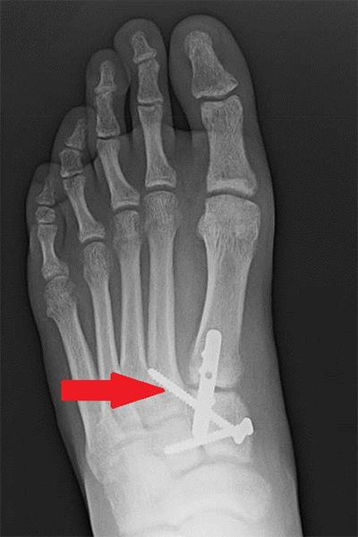
A one to two inch incision is made over the top of the foot. There are many types of metatarsal shortening osteotomies. Where the incision is made depends on the location of the osteotomy. A bone cut is made using a saw at the end of the metatarsal. The cut is made completely through the bone. After the appropriate amount of sliding has occurred, the osteotomy is held in that position with either pins or screws. The surgeon may also place a temporary wire through the toe itself. The skin is then closed.

Recovery

Immediately after surgery, a padded soft dressing is placed around the foot. The patient is given a hard-soled shoe or boot and allowed to place weight only on the heel.

Instructions are given to keep the dressing clean. Showers/bathing can be done with either a plastic bag or commercially available cover to keep the dressings dry. The patient should keep the foot elevated as much as possible to avoid swelling. Excess swelling can lead to problems with wound healing.

The patient usually is seen in the office one to two weeks after surgery. The dressing is changed and when skin has adequately healed, the stitches are removed. Range-of-motion exercises with the toe may be started. If a wire was placed in the toe, it typically is removed in the office four to six weeks after surgery. At 4-6 weeks, weight bearing usually is allowed as tolerated.

Risks and Complications

All surgeries come with possible complications, including the risks associated with anesthesia, infection, damage to nerves and blood vessels, and bleeding or blood clots. There also are complications specific to metatarsal osteotomies. Failure of the bone to heal after it has been cut may result in continued pain. This complication typically requires another surgery to get the bone to heal in order to alleviate the pain. Excessive scaring can occur as can recurrence of the deformity.

FAQs

Will I have normal function of my toes after this surgery?

You should expect some degree of stiffness of the operative toes after this procedure. The amount of motion regained depends on the severity of the deformity before surgery and any other procedures done. Range-of-motion exercises after surgery can help to decrease stiffness.

How long will my toe be swollen after surgery?

Even with appropriate healing, toes may remain swollen for months after surgery.

What is lateral ankle ligament reconstruction?

Lateral Ankle Ligament Reconstruction

Ankle sprains are one of the most common sports-related injuries. They affect 10,000 people per day. When the ligaments on the outside of the ankle are stretched or torn, patients can have pain and feelings of instability. If symptoms persist after non-surgical treatment, surgery may be required. The goal of this surgery is to restore normal stability to the ankle. This should also fix your feeling that the ankle “gives way” and any pain that is associated with an unstable ankle.

Diagnosis

Surgery is considered when you have an unstable ankle that does not respond to non-surgical treatment. Six months of non-surgical treatment is often recommended before surgery. A physical examination will show that the ankle is unstable, and X-rays are sometimes used to help with the diagnosis. Patients who have failed physical therapy and bracing and have instability of the ankle may consider surgery.

Your general health plays a role in any decision to have surgery. Patients with nerve or collagen diseases may not be helped by this type of surgery. Patients should not have this surgery if they have certain medical issues or poor circulation or are unable to follow the recommended post-surgery rehab. You should discuss any medical concerns with your foot and ankle orthopedic surgeon.

Other diagnoses, including ankle arthritis, may require different surgeries that treat the bones and joints. Patients with chronic pain may benefit most from non-surgical management with a pain specialist. Surgery is not recommended for treatment of a single ankle sprain.

Treatment

Ankle ligament reconstruction (ALR) typically is an outpatient surgery, meaning the patient can go home the same day as the procedure. It is most often done under general or regional anesthesia. Other surgeries may be performed at the same time, most often arthroscopic surgery of the ankle joint. At least one larger incision is required for the ligament reconstruction.

Several different techniques can be performed depending on the individual patient. One option is to repair the patient’s own existing ligaments with stitches. This repair is called a modified Bröstrom procedure and can result in a stronger repair because of support from other tissues. Another option is to use a tendon to replace the torn ligaments. This technique is similar to what is done in knee ligament reconstructions.

Specific Techniques

The modified Bröstrom procedure is the most commonly performed surgery for this problem. The surgeon begins by making a C- or J-shaped incision over the outside of the ankle. The ankle ligaments are identified if possible. They are then tightened using either stitches or anchors that are placed into one the bones of the ankle (the fibula bone). Stitching other tissue over the repaired ligaments further strengthens the repair.

Tendons may also be used to replace the ligaments. The surgeon weaves a tendon into the bones around the ankle. The tendon is held in place with stitches and possibly a screw in the bone. One option is to use the patient’s own hamstring tendon, which is taken through a separate incision on the inside part of the knee. Another option is to use a cadaver tendon. A different method is to take a portion of one of the tendons from the side of the ankle and weave it into the fibula bone.

Recovery

You can expect to be in a splint or cast for a minimum of two weeks. It may be up to six weeks before weight can be placed on the ankle. Weight bearing is gradually advanced in a removable walking boot. An athletic ankle brace typically is used after the boot.

Ankle strengthening begins after six weeks as pain and swelling allow. This may involve formal physical therapy. Straight-line running is allowed when the ankle is strong enough for it. Sport-specific exercises can then start gradually. The total expected recovery time is 6-12 months. It is recommended that patients wear a brace for sports activities for up to a year.

Risks and Complications

All surgeries come with possible complications, including the risks associated with anesthesia, infection, damage to nerves and blood vessels, and bleeding or blood clots.

Patients having ALR commonly have decreased feeling around their incision. It also is common to have decreased feeling that extends to the top of the foot. This occurs up to 20 percent of the time and ranges from increased sensitivity to complete loss of sensation. Other less common problems include delayed wound healing and infection. Blood clots in the leg veins also can occur. Recurrent ankle instability or stiffness also may occur.

FAQs

What are the alternatives to surgery?

Treatment for ankle instability typically involves bracing and physical therapy. Many patients will respond to this treatment.

What can happen if ankle instability goes untreated?

Repeat ankle sprains can occur. This can lead to ankle joint damage, bone and tendon injuries, and arthritis.

What is the risk of retearing my ligament after it has been repaired?

Tearing of the ligament can have many definitions. Complete tearing can occur but usually only after repeat injury. However, repaired ligaments can stretch out over time. Long-term studies that look at these surgeries and patient satisfaction have shown that more than 90 percent of patients have a good or excellent outcome.

What if my ankle instability does not improve after surgery?

The results of surgery vary based on the severity of the initial injury. Outcomes will vary as well. Patients who have persistent instability after surgery may improve with physical therapy or by wearing a brace. Additional surgeries to reconstruct the ligaments may be an option. Ankle fusion could also be considered.

What is insertional Achilles tendinitis?

Insertional Achilles Tendinitis

The Achilles tendon is the largest tendon in the body. Insertional Achilles tendinitis is a degeneration of the Achilles tendon fibers where the tendon inserts into the heel bone. It may be associated with inflammation of a bursa or tendon sheath in the same area.

Symptoms

Most patients report a gradual onset of pain and swelling at the back of the heel bone without specific injury. At first, the pain may only be noticeable after activity, but it becomes more constant over time. The pain increases with jumping or running and especially with sports requiring short bursts of these activities. Patients experience tenderness over the back of the heel bone and the bone often becomes more prominent. It is painful to position the ankle above a 90 degree position.

Causes

Insertional Achilles tendinitis primarily is caused by degeneration of the tendon over time. The average patient is in their 40s. Conditions associated with increased risk include psoriasis and Reiter’s syndrome, spondyloarthropathy (generalized inflammation of joints), gout, familial hyperlipidemia, sarcoidosis, and diffuse idiopathic skeletal hyperostosis as well as the use of medications such as steroids and fluoroquinolone antibiotics.

Diagnosis

Your foot and ankle orthopedic surgeon will perform a clinical exam to determine if you have insertional Achilles tendinosis. They may order X-rays to look for calcification (bone) deposits within the tendon at its insertion into the heel. These deposits are present approximately 60 percent of the time and are associated with a more guarded success rate for non-surgical treatment and a much longer recovery time for surgical treatment. X-rays also may reveal a Haglund’s deformity.

MRIs may be used to determine the extent of tendon degeneration as well as other factors such as bursitis, which may contribute to heel pain.

Treatments

Non-surgical Treatment

Non-surgical treatments, including liberal use of nonsteroidal anti-inflammatory drugs, heel lifts, stretching, and switching to shoes that do not put pressure over this area, are effective for the majority of patients. If symptoms persist, your surgeon may recommend night splints, arch supports, physical therapy, or the use of a cast or brace. Nitroglycerin patches also may be of benefit to increase the blood supply to this area.

What is heel pain?

Every mile you walk puts tons of stress on each foot. Your feet can handle a heavy load, but too much stress pushes them over their limits. When you pound your feet on hard surfaces playing sports or wear shoes that irritate sensitive tissues, you may develop heel pain, the most common problem affecting the foot and ankle.

Heel Pain

A sore heel will usually get better on its own without surgery if you give it enough rest. However, many people ignore the early signs of heel pain and keep on doing the activities that caused it. When you continue to walk on a sore heel, it will only get worse and could become a chronic condition leading to more problems.

Diagnosis

Heel pain can have many causes. If your heel hurts, see your primary care doctor or foot and ankle orthopedic surgeon right away to determine the cause and get treatment. Tell him or her exactly where you have pain and how long you’ve had it. Your doctor will examine your heel, looking and feeling for signs of tenderness and swelling. You may be asked to walk, stand on one foot, or do other physical tests that help your doctor pinpoint the cause of your sore heel. Conditions that cause heel pain generally fall into two main categories: pain beneath the heel and pain behind the heel.

Pain Beneath the Heel

If it hurts under your heel, you may have one or more conditions that inflame the tissues on the bottom of your foot:

  • Stone bruise: When you step on a hard object such as a rock or stone, you can bruise the fat pad on the underside of your heel. It may or may not look discolored. The pain goes away gradually with rest.
  • Plantar fasciitis (subcalcaneal pain): Doing too much running or jumping can inflame the tissue band (fascia) connecting the heel bone to the base of the toes. The pain is centered under your heel and may be mild at first but flares up when you take your first steps after resting overnight. You may need to do special exercises, take medication to reduce swelling, and wear a heel pad in your shoe.
  • Heel spur: When plantar fasciitis continues for a long time, a heel spur (calcium deposit) may form where the fascia tissue band connects to your heel bone. Your doctor may take an X-ray to see the bony protrusion. Treatment usually is the same as for plantar fasciitis: rest until the pain subsides, do special stretching exercises and wear heel pad shoe inserts. Having a heel spur may not cause pain and should not be operated on unless symptoms become chronic.

What is hammertoe surgery?

Hammertoe

A hammertoe is a deformity that causes a toe to become bent upward in the middle so it resembles a hammer. This can cause the toe to rub against the top of the shoe or irritate the end of the toe by jamming it into the ground. A hammertoe also can be associated with a contracture of the joint at the base of the toe and can often occur in conjunction with a bunion or other foot problems.

Non-surgical treatment for a hammertoe include padding the toe and changing or stretching shoes. If you still have pain, cannot participate in your normal activities, and/or cannot wear shoes comfortably, surgery may be an option. The main goal of hammertoe surgery is to correct the deformity, which will relieve the pain.

The hammertoe can be flexible or stiff. Depending on the flexibility of the toe and the preference of your foot and ankle orthopedic surgeon, several different surgical procedures can be used to treat the hammertoe.

Treatment

If you have active infections, poor circulation, or a serious illness (for example, heart disease), surgery may not be recommended. You should discuss your health history with your foot and ankle orthopedic surgeon prior to considering hammertoe surgery.

Most often, hammertoe correction is done as an outpatient procedure, meaning you can go home the same day. You may be fully asleep during surgery or given an injection (nerve block) that makes the foot go numb temporarily. This is similar to what a dentist does to your tooth when filling a cavity.

If you have other toe problems in addition to the hammertoe, you should consider addressing the other deformities at the same time as one problem may contribute to the other.

Specific Technique

The type of surgery depends to a large degree on whether the toe can be easily straightened during your examination.

Flexible Hammertoe: If your toe can be straightened, it is called a flexible hammertoe. A flexible hammertoe can be corrected with ligament and tendon lengthening or detachment procedures. One of the most common procedures involves transferring a tendon from the bottom of the toe to the top of the toe. This procedure stops the tendon from pulling the joint into a bent position and places the tendon in a position where it will help hold the toe down.

Fixed (Stiff) Hammertoe: If your toe cannot be straightened by the foot and ankle orthopedic surgeon during the examination, it is called a fixed hammertoe. This means that the joint has become so contracted that releasing and lengthening the tendons and ligaments alone cannot restore the toe to a straight position. Some bone needs to be removed as well to get the toe to be straight. Usually, the end of the bone at the fixed (stiff) joint is removed to allow the toe to straighten completely.

Once the bone is removed, there are two options to stabilize the area:

  • Pins can temporarily hold the toe in a straight position. This allows the area to fill in with scar tissue after the pins are removed.
  • Fusing the joint is done by using pins, screws, or other implants to keep the toe straight. The goal is for the bone ends to heal together to become a solid bone rather than scar tissue.

For either option, as well as the flexible hammertoe treatment, your surgeon may need to release the joint and lengthen the tendon at the base of the toe to allow the toe to lay completely flat.

Recovery

Usually, you are able to place your full weight on your foot after surgery without the need for crutches or a walker. You may also have a special shoe or boot to wear after surgery. You will need to lie down with your foot elevated at the level of your heart for the first few days after surgery.

Stitches usually are removed 10-14 days after surgery. If pins were placed, these will be taken out in the office 4 weeks after the surgery. This usually is not painful because the pins loosen over time after the surgery and come out easily. You may not put your foot under water until the stitches and pins are removed.

Recovery usually takes several weeks depending on the type of surgery that was done. If the hammertoe is on your right foot, you may not be able to drive a car for several weeks.

Your foot and ankle orthopedic surgeon may ask you to do exercises to stretch and move the toe at home after surgery. This can help with flexibility and motion.

It is normal to have swelling after surgery. It may take many months for the swelling and symptoms to fully resolve.

Risks and Complications

All surgeries come with possible complications, including the risks associated with anesthesia, infection, damage to nerves and blood vessels, and bleeding or blood clots.

Complications specific to hammertoe surgery include the chance that the hammertoe may come back after your surgery. There is a risk that after the surgery you may feel like the toe is unstable. This is due to the cutting of bone, ligaments, and tendons. If you have a fusion there is a risk of the bones not healing. These complications are not common. Discuss the potential complications with your foot and ankle orthopedic surgeon prior to undergoing hammertoe correction.

FAQs

If I am treated with surgery will the hammertoe ever come back?

A hammertoe usually develops as a result of progressive muscle imbalance that deforms the toe. This progression does not necessarily stop at the time of surgery. It is therefore possible but unlikely that your hammertoe may come back after surgery. If this happens and you have discomfort in the toe, reevaluation and possible surgery may be an option.

Can I bend my toe after surgery?

Most hammertoe surgical procedures will result in some stiffening of your toe. Depending on the specific surgery performed, you may or may not be able to bend your toe once it has healed. The goals are to have a toe that is not painful and that you can place into a shoe easily. Your foot and ankle orthopedic surgeon can discuss your surgery and possible outcomes in more detail.