mm

About Orthopedic Specialists

Orthopedic Specialists of Seattle provides new and advanced procedures including endoscopic carpel tunnel release surgery for carpal tunnel syrome, complex joint restoration procedures, anterior approach hip replacement surgery, and more.

Forearm Fractures in Children

Description

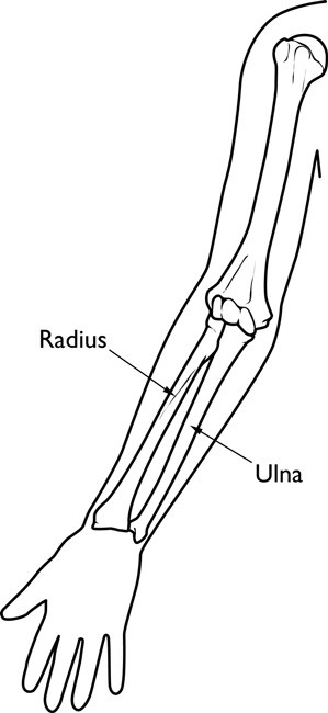
The bones of the forearm are the radius and the ulna. If you hold your arm naturally by your side, the ulna is the bone closer to you and the radius is farther away.

Fractures of the forearm can occur near the wrist at the farthest (distal) end of the bone, in the middle of the forearm, or near the elbow at the top (proximal) end of the bone.

A child’s bones are also subject to a unique injury called a growth plate fracture. Growth plates are made of cartilage near the ends of children’s bones. They help determine the length and shape of the mature bone.

a00039f02

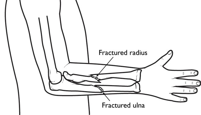
Fractures of both bones in the forearm.
Fractures in a child’s bones begin to heal much more quickly than an adult’s bones. If you suspect a fracture, you should obtain prompt medical attention for the child so that the bones can be set for proper healing.

Statistics
Forearm fractures account for 40% to 50% of all childhood fractures. About three out of four forearm fractures in children involve the wrist-end of the radius.

Cause
Children love to run, hop, skip, jump and tumble. But if a child falls onto an outstretched arm, he or she might break one or both of the bones in the forearm.

Classifications
Torus fracture. This is also called a “buckle” fracture. The topmost layer of bone on one side of the bone is compressed, causing the other side to bend away from the growth plate. This is a stable fracture and the broken pieces of bone have not separated apart (displaced).

Metaphyseal fracture. The fracture is across the upper, or lower, portion of the shaft of the bone and does not affect the growth plate.

Greenstick fracture. The fracture extends through a portion of the bone, causing it to bend on the other side.

Galeazzi fracture. The injury affects both bones of the forearm. There is usually a displaced fracture in the radius and a dislocation of the ulna at the wrist, where the radius and ulna come together.

Monteggia fracture. The injury affects both bones of the forearm. There is usually a fracture in the ulna and the top (head) of the radius is dislocated. This is a very severe injury and requires urgent care.

Growth plate fracture. Also called a physeal fracture, this fracture occurs at or across the growth plate. Usually these fractures affect the growth plate of the radius near the wrist.

a00039f01

Symptoms
In most cases, a broken forearm causes severe pain. Your child’s forearm and hand may also feel numb.

Examination, Signs

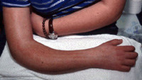
This child’s forearm fracture has resulted in a bent appearance of the forearm.
(Courtesy of Texas Scottish Rite Hospital for Children)

  • Any type of deformity about the elbow, forearm, or wrist
  • Tenderness
  • Swelling
  • An inability to rotate or turn the forearm

Your doctor will also test to make sure that the nerves and circulation in your child’s hand and fingers have not been affected.

Investigation, Tests
The hand, wrist, arm, and elbow can all be injured during a fall on an outstretched arm. To determine exactly what injuries have occurred, your doctor will probably want to see x-rays of the elbow and wrist, as well as the forearm.

Nonsurgical Treatment
Treatment depends on the type of fracture and the degree of displacement.

a00039f03

Casts support and protect broken bones while they heal.
Some mild fractures, such as buckle fractures, may simply need the support of a splint or cast until they heal. For more severe fractures that have become angled, your doctor may be able to push (manipulate) the bones into proper alignment without surgery, as long as the bones have not broken through the skin.

A stable fracture, such as a buckle fracture, may require 3 to 4 weeks in a cast. A more serious injury, such as a Monteggia fracture-dislocation, may need to be immobilized for 6 to 10 weeks.

Surgical Treatment
Surgery to align the bones and secure them in place may be required if:

  • The skin is broken
  • The fracture is unstable — the ends of the broken bones will not stay lined up
  • Bone segments have been displaced
  • The bones cannot be aligned properly through manipulation alone
  • The bones have already begun to heal at an angle or in an improper position

After the bones are aligned, the physician may use pins, metal implants, or a cast to hold them in place until they have healed.

Long-Term Outcome
When the cast is removed, the wrist joint and elbow joint may be stiff for 2 to 3 weeks. This stiffness will go away without the need for physical therapy.

The forearm bones may temporarily be weaker due to the immobilization in the cast. Children should avoid playground structures, such as monkey bars, for 3 to 4 weeks after the cast is removed, in order to allow the bone to safely regain its normal strength.

If the fracture disrupts the growth plate at the end of the bone, your doctor will probably want to watch it carefully for several years to ensure that growth proceeds normally.

Elbow Fractures in Children

Description
Whether your child is an active athlete or just a toddler jumping on the bed, there’s a good possibility that he or she will take a spill at home or on the field or court at some time. These falls are usually harmless; but when a child falls on an outstretched arm, the velocity of the fall combined with the pressure of hitting the ground could be enough to break a bone.

That’s how most fractures around the elbow joint occur. These fractures account for about 10 percent of all fractures in children.

If your child complains of elbow pain after a fall and refuses to straighten his or her arm, see a doctor immediately. The doctor will first check to see whether there is any damage to the nerves or blood vessels. X-rays will help determine what kind of fracture occurred and whether the bones moved out of place.

Because a child’s bones are still forming, the doctor may request X-rays of both arms for comparison.

Types of Fractures

A child can experience a fracture in several places about the elbow, including:

Above the elbow (supracondylar): The upper arm bone (humerus) breaks, slightly above the elbow. These fractures usually occur in children younger than 8 years of age. This is the most common elbow fracture, and one of the more serious because it can result in nerve damage and impaired circulation.

At the elbow knob (condylar): This type of fracture occurs through one of the bony knobs (condyles) at the end of the upper arm bone. Most occur through the outer (lateral) knob. These fractures require careful treatment, because they can disrupt both the growth plate (physeal) and the joint surface.

At the inside of the elbow tip (epicondylar): At the top of each bony knob is a projection called the epicondyle. Fractures at this point usually occur on the inside (medial) epicondyle in children between 9 and 14 years of age.

Growth plate: The upper arm bone and both lower arm bones have growth plates located near the end of the bone. A fracture that disrupts the growth plate can result in arrested growth and/or deformity if not treated promptly.

Forearm: An elbow dislocation can break off the head of the thumb-side lower arm bone (radius), and excessive force can cause a compression fracture to the bone as well. Fractures of the tip (olecranon) of the other lower arm bone (ulna) are rare.

Fracture dislocation: A fracture of the inside bone (ulna) can be combined with the top of the thumb-side bone (radius) coming out of the socket at the elbow. This is called a Monteggia fracture. If the dislocation is not seen, and only the fracture is treated, this can lead to permanent impairment of elbow joint function.

cons1_276_250

Risk Factors / Prevention

If your child is an active athlete, make sure that he or she wears the proper protective equipment. Elbow guards and pads can help reduce the risk of a fracture about the elbow.

Symptoms

Regardless of where the break is, the symptoms of a broken elbow are similar:

  • Acute pain
  • Tenderness
  • Swelling (may be severe or mild)
  • Limited movement

Treatment Options
Treatment depends on the type of fracture and the degree of displacement. If there is little or no displacement, the doctor may immobilize the arm in a cast or splint for 3 to 5 weeks.

During this time, another set of X-rays may be needed to determine whether the bones are staying properly aligned.

If the fracture forced the bones out of alignment, the doctor will have to manipulate them back into place. Sometimes, this can be done without surgery, but more often, surgery will be needed. Pins, screws or wires are used to hold the bones in place. The child will have to wear a cast for several weeks before the pins are removed.

Range of motion exercises can usually begin about a month after surgery.

Elbow (Olecranon) Bursitis

Description
The bursa is a slippery sac between the loose skin and the bones of your elbow. The bursa allows the skin to move freely over the underlying bone. It is located at the tip of the elbow. Normally, the bursa is flat and it’s hard to tell it is even there. If the bursa becomes irritated or inflamed, a condition known as elbow bursitis develops.

Risk Factors/Prevention
Common causes of elbow bursitis include:

  • Trauma: A hard blow to the tip of the elbow could cause the bursa to produce excess fluid and swell.
  • Prolonged pressure: Leaning on the tip of the elbow for long periods of time on hard surfaces such as a tabletop may cause the bursa to swell. Typically, this type of bursitis would develop over several months.
  • Infection: If the tip of the elbow has an injury that breaks the skin, such as an insect bite or a scrape, bacteria may get inside the bursa and cause an infection. The infected bursa produces fluid, redness and swelling. If the infection goes untreated, the fluid may turn to pus.
  • Medical conditions: Certain conditions such as rheumatoid arthritis and gout are associated with development of elbow bursitis.

Symptoms
Swelling is often the first symptom. The skin on the back of the elbow is loose, so you may not notice small amounts of swelling right away. As the swelling continues, the bursa gets larger. This causes pain as the bursa is stretched, since the bursa contains nerve endings. The swelling may get large enough to restrict motion in the elbow.

If the bursitis is infected, the skin becomes red and warm. If the infection is not treated right away, it may spread to other parts of the arm or move into the bloodstream. This can cause serious illness.

cons1_414_166

See your doctor to diagnose elbow bursitis. You may need an X-ray so the doctor can look for a foreign body or a bone spur. Bone spurs are often found on the tip of the bone in the elbow in patients who have recurrent problems with elbow bursitis.

Treatment Options: Nonsurgical
First, the doctor must determine whether the bursitis is due to an infection. If the doctor suspects this, fluid removal (aspiration) of the swollen area may be recommended. This is commonly performed as an office procedure. Fluid removal helps relieve symptoms and gives the doctor a sample that can be looked at in a laboratory to identify if any bacteria are growing.

This also lets the doctor know if a specific antibiotic is needed to fight the infection.

Often, the doctor may start you on antibiotics before the exact bacteria can be identified. This is done to prevent the infection from progressing. The antibiotic that the doctor recommends in this case will cover a number of possible infections.

If the bursitis is not from an infection, it is treated with elevation, ice and other nonoperative treatments such as an elbow pad and avoidance of direct pressure on the swollen elbow. Oral medications such as ibuprofen or other anti-inflammatories may also be used.

If the swelling and pain do not respond to these measures, your doctor may recommend removing fluid from the bursa and injecting a corticosteroid medication into the bursa. The steroid medication is an anti-inflammatory that is stronger than the medication that can be taken by mouth.

Treatment Options: Surgical
Infected bursa that do not improve with antibiotics and/or removing fluid from the elbow may require surgery. Patients who have surgery for elbow bursitis may need to stay in the hospital for a period of time.

If elbow bursitis is not a result of infection, surgery may be needed if nonoperative treatments don’t work. Surgery to remove the bursa is usually performed as an outpatient procedure. The surgery does not disturb any muscle, ligament or joint structure. Physical therapy after surgery is not always needed. Postoperative casting or prolonged immobilization is not typically required.

Broken Arm

A broken arm is a common injury. Counting all fractures, about one in every 20 involve the upper arm bone (humerus). Children are more likely to break the lower arm bones (radius and ulna). Falling on an outstretched hand or being in a car crash or some other type of accident is usually the cause of a broken arm.

Most people know right away if their arm broke, because there may be a snap or a loud cracking sound. The broken arm may appear deformed and be swollen, bruised and bleeding. A person with a broken arm usually has:

  • Extreme pain at the site of the injury.
  • Pain increased by any movement.
  • Loss of normal use of the arm.

First aid
First make sure the injured person is out of the way of further harm. Is he or she breathing normally? Is there a good pulse? Call 911 if there is serious bleeding, reason to suspect multiple broken bones or other injuries. To slow bleeding and reduce swelling, elevate the injured arm above the level of the person’s heart.

If a broken bone sticks out from the skin (open fracture), do not try to push it back in. Use a clean, dry cloth or bandage to cover it until medical help arrives.

It is important that the injured person not try to use the broken arm. Moving a broken arm would also cause more damage to blood vessels, nerves and other tissues. To immobilize a broken arm:

  • Make a temporary splint. Immobilize the joints above and below the site of the injury. You can use wood or rolled up magazines, making sure both ends of the splint extend far beyond the injured region. You can use cloth, belts or tape to fasten the splint. Avoid any constriction of the arm with the supporting strap.
  • Make a sling. This stabilizes the injury and supports the splint. A broken arm sling can be as simple as a loop of cloth supported from the neck. Take the injured person to a doctor right away.

Doctor’s treatment

Exam: Tell the doctor exactly what happened. He or she will physically examine the broken arm and check for other injuries, such as nerve damage. The doctor may want to see if the patient can flex and extend the wrist and fingers. Sometimes the doctor may use X-rays or other diagnostic imaging tools to see the bones of both the injured and uninjured arms.

If the patient is a child, the long bones of the arm are probably still growing. So the doctor will look carefully for any damage to growth plates.

Reduction: The doctor may need to move pieces of bone back into their correct positions (a process called reduction). Depending upon the severity of injury, the patient may or may not need anesthesia. Those with more serious fractures may require surgery.

Immobilization: With the broken bone back in place, the doctor immobilizes the arm. Most patients get a cast or splint. The doctor tells the patient how long to wear the cast or splint, and removes it at the right time.

Rehabilitation
It may take from several weeks to several months for the broken arm to heal completely. Rehabilitation involves gradually increasing activities to restore muscle strength, joint motion and flexibility. The patient’s cooperation is essential to the rehabilitation process. The patient must complete range of motion, strengthening and other exercises prescribed by the doctor.

Rehabilitation lasts until tissues perform their functions normally. After rehabilitation, the doctor may want to see the arm again to make sure healing is complete.

Biceps Tendinitis

The biceps muscle, in the front of the upper arm, helps stabilize the upper arm bone (humerus) in the shoulder socket. It also helps accelerate and decelerate the arm during overhead movement in activities like tennis or pitching. Strong, cord-like structures called tendons connect one end of the biceps muscle to the shoulder in two places.

At the other end of the muscle, tendons connect the biceps muscle to the smaller bone (radius) in the lower arm. If the tendons become inflamed or irritated, the condition is called tendinitis.

Signs and symptoms
Injuries to the biceps tendons are commonly caused by repetitive overhead activity. Symptoms include:

  • Pain when the arm is overhead or bent.
  • Localized tenderness as the tendon passes over the groove in the upper arm bone.
  • Occasionally, a snapping sound or sensation in the shoulder area.

cons1_139_209Diagnosis and treatment
During the physical examination, the doctor will assess the shoulder area for range of motion, tenderness and signs of shoulder instability. He or she may ask you to raise or rotate the arm.

X-rays may be requested to uncover associated conditions that might cause irritation. The doctor may also request an MRI that can show any damage to the tendons. Overuse, aging and stress can cause the tendon to deteriorate, even if there is no inflammation present.

Initial treatment is conservative. The first step is to rest the arm and shoulder. Switch to another sport or activity for awhile. Ice applications and nonsteroidal anti-inflammatory medications such as ibuprofen can help reduce inflammation.

Your physician can also recommend stretching and progressive strengthening exercises to build muscle endurance and restore range of motion. Then you can gradually return to overhead activity.

Surgical options
If the pain results from shoulder instability or from pressure on the tendon from the shoulder bones, your orthopaedist may recommend arthroscopic surgery. Using fiber optic technology and miniature instruments inserted through a small incision, the surgeon can examine the shoulder joint and anchor the tendon properly.

After surgery, your orthopaedist will prescribe a rehabilitation program that includes stretching and strengthening exercises. Early movement is important, but you should wait for your physician’s approval before doing any heavy lifting or returning to sports.