mm

About Orthopedic Specialists

Orthopedic Specialists of Seattle provides new and advanced procedures including endoscopic carpel tunnel release surgery for carpal tunnel syrome, complex joint restoration procedures, anterior approach hip replacement surgery, and more.

Nerve Injuries

What are nerves?

cons1_71_49
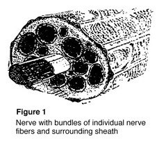
Nerves are the “electrical wiring” system in all people that carry messages from the brain to the rest of the body. A nerve is like an electrical cable wrapped in insulation. A ring of tissue forms a cover to protect the nerve, just like the insulation surrounding an electrical cable (Figure 1).

Nerves serve as the “wires” of the body that carry information to and from the brain. Motor nerves carry messages from the brain to muscles to make the body move. Sensory nerves carry messages to the brain from different parts of the body to signal pain, pressure, and temperature. While the axon (nerve fiber) carries only one type of message, either motor or sensory, most nerves in the body are made up of both.

What happens when a nerve is injured?
Nerves are fragile and can be damaged by pressure, stretching, or cutting. Injury to a nerve can stop signals to and from the brain causing muscles not to work properly, and you may lose feeling in the injured area. When a nerve is cut, both the nerve and the insulation are broken. Pressure or stretching injuries can cause the fibers carrying the information to break and stop the nerve from working, without damaging the cover.

When nerve fibers are cut, the end of the fiber farthest from the brain dies, while the insulation stays healthy. The end that is closest to the brain does not die, and after some time may begin to heal. If the insulation was not cut, new fibers may grow down the empty cover of the tissue until reaching a muscle or sensory receptor.

If both the nerve and insulation have been cut and the nerve is not fixed, the growing nerve fibers may grow into a ball at the end of the cut, forming a nerve scar or neuroma. A neuroma can be painful and cause an electrical feeling when touched.

How is it treated?
cons1_72_49
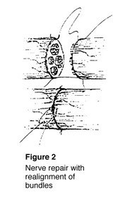
To fix a cut nerve, the insulation around both ends of the nerve are sewn together. The goal in fixing the nerve is to save the cover so that new fibers may heal and work again (Figure 2). If a wound is dirty or crushed, your physician may wait to fix the nerve until the skin has healed.

If there is a space between the ends of the nerve, the doctor may need to take a piece of nerve (nerve graft) from another part of the body to fix the injured nerve. This may cause permanent loss of feeling in the area where the nerve graft was taken.

Once the nerve cover is fixed, the nerve generally begins to heal three or four weeks after the injury. Nerves usually grow one inch every month depending on the patient’s age and other factors. This means that with an injury to a nerve in the arm above the fingertips, it may take up to a year before feeling returns to the fingertips. The feeling of pins and needles in the fingertips is common during the recovery process. While this can be uncomfortable, it usually passes and is a sign of recovery.

What is my role in recovery and what kind of results can I expect? The patient must do several things to keep up muscle activity and feeling while waiting for the nerve to heal. Your doctor may recommend therapy to keep joints flexible.

If the joints become stiff, they will not work even after muscles begin to work again. When a sensory nerve has been injured, the patient must be extra careful not to burn or cut fingers since there is no feeling in the affected area.

After the nerve has recovered, the brain gets “lazy,” and a procedure called sensory re-education may be needed to improve feeling to the hand or finger. Your doctor will recommend the appropriate therapy based on the nature of your injury.

Factors that may affect results after nerve repair include age, the type of wound and nerve, and location of the injury. While nerve injuries may create lasting problems for the patient, care by a physician and proper therapy help two out of three patients return to more normal use.

Mallet Finger (Baseball Finger)

Description
Mallet finger injuries occur when the tip of a finger or the thumb is forcefully bent. This condition is also known as baseball finger. It happens when a ball or other object strikes the tip of the digit. The force tears the thin tendon that allows you to straighten the finger. The force of the blow may even pull away a piece of bone along with the tendon.

Symptoms
A person with this injury has pain, swelling and bruising of the fingertip. The fingertip may droop noticeably. Occasionally, blood collects beneath the nail and the nail can even become detached from beneath the skin fold at the base of the nail.

cons1_397_151

Treatment Options
If you experience a mallet finger injury, immediately apply ice and elevate your hand above the level of your heart. Seek medical attention within the first week after this injury. It is very important to seek urgent attention if you have blood beneath the nail or if the nail is detached because you could also have a nail bed laceration or open (compound) fracture.

In most cases, the doctor will order X-rays to look for a major fracture or malalignment of the joint.

The majority of mallet finger injuries can be treated without surgery. The doctor may apply a splint to hold the fingertip straight (in extension) until it heals. Most of the time, you will have to wear the splint full-time for six weeks.

After six weeks, most patients gradually wean from the splint by wearing it less frequently over a three to four week period The finger usually regains acceptable function and appearance with this treatment plan; however, many patients may not regain full fingertip extension.

In children, mallet finger injuries may involve the cartilage that controls bone growth. A doctor must carefully evaluate and treat this type of injury, so the finger does not become stunted or deformed.

Treatment Options: Surgical

In certain cases, surgical repair may be considered. These cases include mallet finger injuries with:

  • Large fracture fragments
  • Joint malalignment

Surgical treatment in these cases often involves repair of the fracture using pins, pins and wire, or even small screws.

Surgery may also be considered if non-operative treatment fails. Repairing soft tissue mallet fingers is uncommon and usually reserved for patients with very severe deformities and functional problems. Surgical treatment can include tightening the stretched tendon tissue, using tendon grafts, or even fusing the joint straight. Consultation with an orthopaedic surgeon should be sought in these cases.

Kienböck’s Disease

Bone is living tissue that requires a regular supply of blood for nourishment. If the blood supply to a bone stops, the bone can die, a condition known as osteonecrosis. That’s what happens in Kienböck’s disease, which affects the lunate, one of the small bones of the hand near the wrist.

Kienböck’s Disease: Signs and Symptoms

The cause of Kienböck’s disease is unknown. Many people with Kienböck’s disease think they have a sprained wrist at first. They may have experienced some form of trauma to the wrist, such as a fall. This type of trauma can disrupt the blood flow to the lunate. In most people, two vessels supply blood to the lunate, but in some people there is only one source. This puts them at greater risk for developing the disease.

As the disease progresses, other signs and symptoms are noted, including:

  • A painful and sometimes swollen wrist
  • Limited range of motion in the affected wrist (stiffness)
  • Decreased grip strength in the hand
  • Tenderness directly over the bone (on the top of the hand at about the middle of the wrist)
  • Pain or difficulty in turning the hand upward

Progression

Kienböck’s disease follows a specific progressive pattern through four stages.

  • Stage 1: Symptoms are similar to those of a wrist sprain. X-rays may be normal or show a line indicating a possible fracture. Magnetic resonance imaging (MRI) may also be helpful in making the diagnosis in this early stage.
  • Stage 2: The lunate bone begins to harden. On an X-ray, it may appear brighter or whiter than the surrounding bones. These changes indicate that the bone is dying. Either an MRI or a computed tomography (CT) scan may be used to assess the condition of the bone. Recurrent pain, swelling and wrist tenderness are common.
  • Stage 3: The dead bone begins to collapse and break into pieces. The surrounding bones may begin to shift position. Patients experience increasing pain, weakness in gripping, and limited motion.
  • Stage 4: The surfaces of adjoining bones are affected, resulting in arthritis of the wrist.

Diagnosis and treatment
In its early stages, Kienböck’s disease may be difficult to diagnose because the symptoms are so similar to those of a sprained wrist. Even X-rays of the wrist may appear normal. At this point, the goal of treatment is to relieve the pressure on the lunate and help restore blood flow within the bone. Your physician may splint or cast your wrist for two to three weeks.

Anti-inflammatory medications such as aspirin or ibuprofen will help relieve any pain and reduce swelling. If the pain continues, your physician may refer you to an orthopaedic or hand surgeon for further evaluation.

Surgical options
Although there is no cure, there are several surgical options for treating the more advanced stages of Kienböck’s disease. The right procedure for you will depend on several factors, including disease progression, your personal activity levels and goals and your surgeon’s experience with various procedures.

Do not hesitate to discuss these options with your orthopaedic or hand surgeon and to ask why he or she is recommending a particular procedure.

In some cases, returning the blood supply to the bone (revascularization) may be possible. This procedure uses a bone graft from the inner bone of the lower arm. It may be combined with an external fixator, a metal device that helps relieve pressure on the lunate and preserve the spacing between bones.

If the bones of the lower arm are uneven in length, a joint leveling procedure may be recommended. Bones can be made longer with bone grafts, or shortened by removing a section of the bone.

This reduces the compressive forces on the lunate and seems to halt progression of the disease. If the lunate is severely collapsed or fragmented, it can be removed. The two bones on either side of it are also removed. This procedure is called a proximal row carpectomy and will relieve pain while maintaining partial wrist motion.

Another way to ease pressure on the bone is to fuse several of the small bones of the hand together. However, this will not improve range of motion. If the disease has progressed to severe arthritis of the wrist, fusing the bones will reduce pain and help maintain function, although motion is limited.

Human Bites

Human bite wounds may not sound very dangerous, but they are. Human bite wounds contain very high concentrations of bacteria, so the risk of infection is high. Even though the wound may appear insignificant, an infection can lead to a severe joint infection. About one third of all hand infections result from human bite wounds. These infections can progress quickly and result in significant complications, so early treatment is necessary.

Bites can transmit the human immunodeficiency virus (HIV), as well as the hepatitis B virus and syphillis. Joint infections can lead to septic arthritis. Someone who has a human bite wound should get prompt first aid and see a doctor within 24 hours of the injury if the skin is broken.

Sometimes the wound is incurred directly (an actual bite). Other times, it is indirect (a clenched fist strikes a tooth, which breaks the skin on the hand). In a fight, a punch to the mouth can result in an indirect bite injury at the knuckle joint (MP or metacarpophalangeal joint), which can lead to a severe infection and possible destruction of the joint if it is not promptly treated.

Signs and Symptoms
In some cases, the bite will not break the skin but may cause damage to underlying tendons and joints. If the skin is broken, there is the additional possibility of infection as well as injury to joints, tendons and nerves. An injury to the top of the hand can result in significant swelling within hours.

Signs of an infection include:

  • Warmth around the bite wound
  • Swelling
  • Pain
  • A pus discharge

Signs of damage to tendons or nerves include:

  • An inability to bend or straighten the finger
  • A loss of sensation over the tip of the finger

First aid
Don’t put the bitten area in your mouth!

If there is no bleeding, wash the wound thoroughly. Use soap and water or an antiseptic such as hydrogen peroxide or alcohol. Apply an antibiotic ointment and cover with a non-stick bandage. Watch the area carefully to see if there are signs of damaged nerves or tendons. If you see any, seek medical help immediately.

If there is bleeding, apply direct pressure with a clean dry cloth. Elevate the area. Do not clean a wound that is actively bleeding. Cover the wound with a clean dressing and seek medical help. In these situations, early intervention (within 24 hours) is needed to prevent complications.

Medical assistance
Be sure to tell your doctor how you got the bite. Your physician will measure the wound, note its location, and check for signs of nerve or tendon damage. The doctor may examine your arm to see if there are signs of a spreading infection. You may need to get X-rays and a blood test.

You may also need to get a tetanus shot and a prescription for antibiotics. You may also have to return in a day or two so that the physician can confirm that an infection has not developed. If the tendons or nerves have been injured, you may need to see a specialist for additional treatment.

Hand Surgery

Our hands serve many purposes. Hands help us eat, dress, write, earn a living, create art and do many other activities. To accomplish these tasks and activities, our hands require sensation and movement, such as joint motion, tendon gliding and muscle contraction.

When a problem takes place in the hand, care must be given to all the different types of tissues that make function of the hand possible.

Hand surgeons are specifically trained to give that care:

  • Hand surgery is the field of medicine that deals with problems of the hand, wrist and forearm.
  • Hand surgeons care for these problems without surgery, and they are specially trained to operate when necessary.
  • Many hand surgeons are also experts in diagnosing and caring for shoulder and elbow problems.
  • Hand surgeons are orthopedic, plastic or general surgeons who have additional training in surgery of the hand.
  • To become a member of the American Society for Surgery of the Hand, a hand surgeon must complete a full year of additional training and must pass a rigorous certifying examination.