What is a Morton’s neuroma?

Morton’s Neuroma

Morton’s neuroma is a thickening of the tissue that surrounds the small nerve leading to the toes. It occurs as the nerve passes under the ligament connecting metatarsal bones in the forefoot (front part of the foot).

Morton’s neuroma most frequently develops between the third and fourth toes. It often occurs in response to irritation, trauma, or excessive pressure, and is more common in women.

Symptoms

Morton’s neuroma may feel like walking on a stone or marble. You may have burning pain in the ball of your foot that radiates into the toes. The pain can worsen with activity or wearing shoes. You may also experience numbness or a “clicking” feeling in the toes.

Runners may feel pain as they push off. High heeled and narrow toe box shoes also can aggravate the condition.

Diagnosis

During the examination, your foot and ankle orthopedic surgeon will feel for a mass or a “click” between the metatarsal bones. They will squeeze the spaces between the toes to try to recreate the pain. Range of motion tests are used to rule out arthritis or joint inflammation. X-rays can help rule out a stress fracture or arthritis.

Treatment

Initial treatment can involve several non-surgical options:

Changing shoes: Avoid high heels or tight shoes. Wear wider shoes with lower heels and a soft sole. This helps to decrease compression of the nerve.

Orthotics: Custom shoe inserts and pads may help relieve irritation by lifting and separating the bones, reducing the pressure on the nerve.

Injection: An injection of a corticosteroid will reduce the swelling and inflammation of the nerve, which should provide relief.

Several studies have shown that a combination of shoe changes, oral anti-inflammatory medications, orthotics, and/or cortisone injections will provide relief in more than 80% of people with Morton’s neuroma. If conservative treatment does not relieve your symptoms, or if symptoms return, you may require surgery.

Surgery involves either removing a small portion of the nerve including the neuroma or releasing the tissue around the nerve to decompress it. It is an outpatient procedure, meaning the patient can go home the same day as surgery. Patients may be immobilized for 1-2 weeks to allow for healing of the incision, and then transitioned to regular shoes as tolerated.

What is a Lisfranc injury?

Lisfranc Injury

A Lisfranc injury involves the joints and/or the ligaments of the midfoot (middle of the foot). The Lisfranc is a ligament of the foot that runs between two bones called the medial cuneiform and the second metatarsal. The name comes from French surgeon Jacques Lisfranc de St. Martin (1790-1847), who was the first physician to describe injuries to this ligament.

There are a variety of causes for a Lisfranc injury such as a car accident, sports injury, or a simple slip and fall. Sometimes the injury can be mistaken for a foot sprain when X-rays do not show any broken bones. Delaying treatment can sometimes lead to more significant problems. Proper diagnosis from a foot and ankle orthopedic surgeon is key.

Symptoms

The common symptoms of a Lisfranc injury are swelling and pain on the top of the foot near the instep. Bruising is common, and a bruise on the bottom of the foot can be a clue that this injury has occurred. With a severe injury, the foot may be distorted and putting any weight on it may be very painful. With a mild injury, the foot may appear normal and you may be able walk on it with only mild pain.

Causes

Low-energy injuries can happen when the back of the foot twists or presses down with the ball of the foot planted on the ground. This can happen during athletic activities such as football but also can occur from a misstep or even missing a stair and stumbling over the top of the foot. High-energy injuries occur from direct trauma such as a car accident or a fall from a height.

Specialized ligaments in the midfoot hold the bones in this area together like puzzle pieces to maintain the arch of the foot. When the ligaments, joints, or bones in this area are injured, they may shift out of place, making the arch unstable.

Diagnosis

A Lisfranc injury diagnosis is made based on what happened at the time of injury, your symptoms, and an examination of the foot and ankle that compares the injured foot to your uninjured foot. Your foot and ankle orthopedic surgeon will examine the middle part of your foot to identify the location of your pain and perform tests to check the stability of this area. X-rays may show broken or shifted bones in the middle of the foot. Sometimes X-rays will be taken while you are standing in order to better identify the shifting of bones in the foot. An MRI scan may be helpful to see if the ligaments in the foot are damaged. A CT scan can help determine the extent of the bone injury and is useful when planning surgery if needed.

Treatments

Non-surgical Treatment

If the ligaments and the bones in the middle of the foot are not severely injured, and bones are not shifted out of their normal positions, non-surgical treatment can be successful. A cast or CAM boot may be needed for at least 6 weeks in order for the ligament and/or bone to heal. Your foot and ankle orthopedic surgeon will follow up regularly with X-rays to make sure the bones maintain good position during the recovery.

Surgical Treatment

If the bones or ligaments are injured in a way that causes them to shift out of their normal positions, Lisfranc surgery may be necessary to restore the anatomy of the foot. Surgery may involve the placement of plates and screws that may need to be removed later, once the bones and ligaments have healed.

Recovery

Recovery from Lisfranc surgery depends on the severity of the injury. Most patients will be in a non-weightbearing cast for 6 weeks, followed by 6 weeks in a walking boot. Physical therapy may be needed to strengthen the foot and ankle and help regain walking ability. Return to maximal function, running, and sports can take up to one year.

Risks and Complications

Lisfranc injuries may cause arthritis and chronic pain in the middle of the foot. This may require additional treatment. With surgery, injury to the nerves and tendons may occur. Because of the swelling that often occurs with this injury, complications such as wound opening, infection, and/or further swelling of the foot may occur after surgery.

The outcome for Lisfranc injuries depends on their severity. Some patients will not be able to return to their pre-injury level of functioning or athletic activities even with well-performed treatment. The cartilage joint surfaces commonly are injured and some patients may develop midfoot arthritis (arthritis of the middle of the foot). It is also common for pain to continue in the joints after this injury. For some patients, surgery such a fusion of the joints may be necessary to relieve arthritis pain.

FAQs

How can I tell if it’s a sprain or a Lisfranc injury?

Unrecognized and untreated Lisfranc injuries can have serious complications, including joint degeneration and a buildup of pressure within muscles that can damage nerves and blood vessels. If the standard treatment for a sprain (rest, ice, and elevation) doesn’t reduce the pain and swelling within a day or two, or there is extensive bruising on the bottom of the foot, see your foot and ankle orthopedic surgeon immediately.

How soon can I get back to normal activity?

It is important to follow your doctor’s orders and refrain from activities until you are given the go-ahead. If you return to activities too soon after a Lisfranc injury or surgery, you may suffer another injury that results in damage to blood vessels, arthritis, or an even longer healing time.

What is a Jones fracture?

Jones Fracture

A Jones fracture is the name often used for a fracture at the base of the fifth metatarsal, the bone on the outer side of your foot. Jones fractures are one of the most common foot injuries. This broken bone may heal slowly because of poor blood supply to the area and the amount of force placed on this part of the foot. Those with high-arched feet especially are at risk for a Jones fracture because they put more pressure on the outside of their feet.

Symptoms

Jones fractures produce pain, swelling, bruising, and difficulty walking. Some people may experience pain before the fracture occurs.

Causes

A Jones fracture can occur when the foot twists. Stress fractures can occur in this area when there is a sudden increase in high-impact activity (for example, marathon training).

Diagnosis

A Jones fracture typically is visible on X-rays of the injured foot. An MRI may be helpful if the fracture is not seen on X-ray. A CT scan can be useful to gauge fracture healing.

Treatments

Your foot and ankle orthopedic surgeon may recommend treating the Jones fracture without surgery. This involves a period of immobilization in a non-weightbearing cast or boot. Studies have shown that some of these injuries fail to heal. For this reason, repeat X-rays are necessary to ensure appropriate healing. If X-rays do not show healing in six weeks, then surgical treatment should be considered.

Surgery is most common for Jones fractures in athletes or when non-surgical treatment isn’t successful. During surgery, your foot and ankle orthopedic surgeon will insert a screw to stabilize the fracture while it heals.

Recovery

Recovery is about eight weeks with or without surgery. After the bone has healed, some patients will need physical therapy to regain motion and strength.

Risks and Complications

All surgeries come with possible complications, including the risks associated with anesthesia, infection, damage to nerves and blood vessels, and bleeding or blood clots.

Sometimes the bone will take longer than eight weeks to heal. If this happens, either surgery or a bone stimulator is recommended to help healing. Rarely, the fracture may not heal and the screw can break. If this happens, a second surgery may be performed. Jones fractures also have a high rate of refracture, which occurs more frequently with non-surgical treatment.

FAQs

When can I return to playing sports after a Jones fracture?

Athletes typically return to playing sports at around 8 weeks. Athletic trainers and physical therapists help with this process. Some may wear a clamshell orthosis or turf toe plate when returning to sports, particularly athletes who play on hard surfaces such as artificial turf.

Can I wait to have surgery until I see if the fracture doesn’t heal?

Yes, you can wait. However, postponing surgery may make it more difficult.

Does the hardware need to come out?

No, the hardware does not need to come out for you to return to activities. However, if the screw head or hardware is prominent and irritated by shoes, it may need to be removed.

What is a joint injection?

Joint Injection

A joint injection is a procedure your foot and ankle orthopedic surgeon uses to introduce medication into a joint. The injection is done under sterile conditions using a syringe and needle.

The goals of a joint injection are to relieve pain and improve joint function. Your doctor also may confirm your diagnosis when giving a joint injection.

Diagnosis

An injection may be needed if you have redness, pain, swelling, loss of smooth motion, and trouble with walking normal distances.

Joint injection should be avoided in certain situations. Some of these include the presence of skin or blood infections and a history of allergic response to the injectable medication or its components. In addition, your surgeon may rule out a joint injection if there was little improvement after a previous injection, you have a bleeding disorder or are on blood thinning medication, you have poorly controlled diabetes, or your body has problems fighting infections.

Treatment

Your surgeon will position your foot for the injection procedure. The skin over the joint will be cleaned and sterilized with topical iodine, chlorhexidine or alcohol. The medication will be injected using a syringe and needle. The injection site may need to be covered with a bandage or have pressure applied for a few minutes.

Specific Techniques

The ankle, subtalar, and metatarsophalangeal joints are the common joints for injections. An ultrasound or X-ray may be used to help guide the injection. In addition, fluid can be drawn from the joint before an injection and sent for testing. The symptoms of infection, gout, and autoimmune disease can be similar, and lab testing can help determine a diagnosis.

Recovery

You may be asked to remain in the office for 30 minutes or so to be observed for side effects of the injection. You may be instructed to avoid or limit activity for a day or so after the injection. Your foot and ankle orthopedic surgeon may prescribe other medication, splinting, or physical therapy as part of your treatment plan. Your specialist will tell you when to schedule your next appointment.

Risks and Complications

Potential complications include infection at the injection site, infection of the joint, tenderness, swelling, and warmth. There can sometimes be nerve or blood vessel injury, or damage to the joint surfaces.

When corticosteroids are used, they may cause loss of skin pigment or thinning of the skin. Corticosteroids also can cause weakening of a nearby ligament or tendon with the possibility of complete tears. This medication also may temporarily increase blood sugar and disrupt the body’s own steroid hormone balance, particularly in patients with hormone disorders.

Local anesthetic may cause flushing, hives, chest or abdominal discomfort, and nausea. Viscosupplements, substances that act like naturally occurring joint fluid, may cause joint pain, swelling, and inflammation. Contrast agent may cause allergic reaction.

FAQs

Should I apply ice or heat after a joint injection?

It is generally recommended that you apply ice once or twice per hour for 10-15 minutes for the first few hours after a joint injection. You should avoid applying heat to the affected joint.

How should I clean the injection site at home?

No specific cleaning of the injection site is typically needed. You generally are able to resume normal showering or bathing after joint injections.

Will a joint injection interfere with my other medications?

You doctor will be able to answer this question for you. Be sure to bring a current list of your medications to your appointment.

Will a steroid injection raise my blood sugar?

If you are diabetic, a joint injection with a corticosteroid may cause your blood sugar to increase for a short time. It is a good idea to talk to the doctor who manages your diabetes medication and your surgeon before you receive a joint injection.

What is a high ankle sprain?

High Ankle Sprain

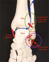
The “high ankle sprain” is named in distinction to the “low ankle sprain.” You may have heard the term while watching American football or other sports broadcasts. The high ankle ligaments (also called the syndesmosis) are located above the ankle, as opposed to the more commonly injured ligaments on the outside of the ankle. These high ankle ligaments connect the tibia to the fibula. It is important to have stability between the tibia and fibula at this level because walking and running place a tremendous amount of force at this junction.

A high ankle sprain, also called a syndesmotic injury, occurs when there is tearing and damage to the high ankle ligaments. These injuries are much less common than a traditional ankle sprain.

Symptoms

If there is an associated fracture around the ankle, patients typically won’t be able to bear weight on the foot/ankle and often will need surgery. If there is not an associated ankle fracture, patients may notice increased pain with activities that cause the ankle to be flexed up placing more stretch on the injured ligaments. Activities like climbing stairs are the most common as the ankle joint is loaded and the talus bone is driven upwards placing stress on the high ankle ligaments.

Causes

A high ankle sprain occurs from a twisting or rotational injury. They are common in sports, especially impact sports. An external rotation mechanism most commonly causes these tears, when the foot is turned towards the outside with respect to the leg. A high ankle sprain also can occur if the ankle is broken.

In some cases, the ligament on the inside of the ankle (the deltoid) will be torn. In this event, the energy of the injury (indicated on the diagram with blue arrows) passes from the deltoid, through the high ankle ligaments (syndesmosis), and up the leg through the fibula. This causes the fibula to be broken at a very high level. This type of fracture is called a Maisonneuve fracture. Patients with a high ankle sprain without fracture may be able to bear weight, but will have pain over the junction between the tibia and fibula just above the level of the ankle (green circle). This is higher than the more traditional sprains (purple circle).

Anatomy

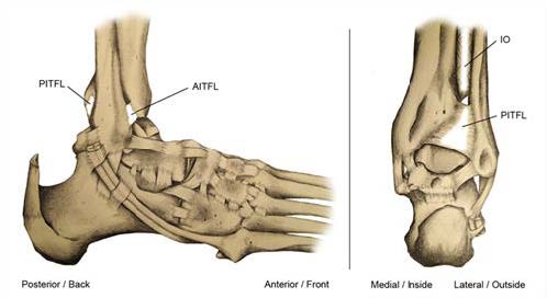
As noted above, the syndesmosis or high ankle ligaments connect the tibia and fibula and allow some rotation. There are three major components of this ligament complex. Ligaments connect bone to bone which prevents bones from moving away from each other and maintains normal movement between the bones, whereas tendons connect muscle to bone, allowing them to move parts of the body.

  1. The first ligament is called the anterior inferior tibiofibular ligament, or AITFL, which runs in front of the two bones.
  2. The second is called the posterior inferior tibiofibular ligament, or PITFL, which runs in the back.
  3. The interosseous (IO) membrane runs down the middle of these and provides a major support between the two bones.

Diagnosis

Patients who have a high ankle ligament tear usually will have pain just above the level of the ankle, thus a “high” ankle sprain. They may also have tenderness over the deltoid ligament if they have a Maisonneuve injury, as noted above. It is important to touch the area to assess whether pain is just around the lateral ankle ligaments or higher.

Your foot and ankle orthopedic surgeon may also perform two tests called the squeeze test and the external rotation test. The squeeze test is performed by squeezing the leg just below the knee to see if pain radiates to the ankle area, which would suggest a high ankle sprain. With the external rotation test, your surgeon will bend your knee and place your ankle in neutral or 90 degrees with the foot in relation to the leg, and the foot is turned to the outside. If there is pain at the ankle area, this suggests a high ankle sprain.

X-rays are very important. A broken bone must be looked for. Three views of the ankle including the whole leg are needed. A fracture on the back portion of the tibia may indicate an injury to the high ankle ligaments given that this is where the PITFL attaches. It also is important to look for increased space between the tibia and the fibula as the high ankle ligaments keep these bones in place. Special imaging may be used, such as an MRI or CT scan, to assess the relationship of the tibia with the fibula.

Treatments

The goals of treatment are to move the tibia and fibula to the correct positions with respect to each other and to heal in those positions. This allows the ankle joint to function as intended. It is very important to note that these injuries can take a lot longer to heal than “low” ankle sprains. If you have a sprain but do not have a broken bone, the treatment immediately following the injury is to rest the leg, ice for 20 minutes every two to three hours, gently compress the leg with an ACE wrap, and elevate the leg with the toes higher than the nose. You may have enough tenderness to require a removable walking boot. Physical therapy may include strengthening the tendons on the outside of the ankle called the peroneals, as well as getting back the movement in the ankle and returning to activities.

It can take up to 6-8 weeks to return to normal activity, but can sometimes take even longer. A general rule of thumb is that bones take roughly six weeks to heal while soft tissues (e.g., ligaments) take around three months to heal. One good indication that you are ready to go back to sports is if you can hop on the foot 15 times. This hopping test is acceptable if there is no obvious widening between the tibia and the fibula on X-rays. If there is widening, which is called diastasis, or if there is a broken bone, surgery may be needed. There is debate as to how to properly fix these injuries, but the idea is to put the fibula and tibia back together and hold them with either screws or new devices that contain a suture, which is the same type of material used to close wounds.

Recovery

As mentioned above, the recovery for high ankle sprains can take considerably longer than typical ankle sprains. In those cases in which a separation of the tibia and fibula or fracture has occurred and surgery is necessary, patients will likely need to be non-weightbearing in a cast followed by a walking boot for about 12 weeks. It is important to do early range of motion passively, meaning with the help of a physical therapist who moves the ankle, to help avoid stiffness. The screws commonly are removed in a second, small surgery before full weightbearing is allowed so they will not break.

Outcomes generally are good if the injury is recognized and treated appropriately. It is more likely, however, to have some stiffness of the ankle after a high ankle sprain as compared to a standard ankle sprain. This is especially true if a fracture has occurred.

Risks and Complications

Stiffness can occur in the ankle. If surgery is performed, one can have an infection or damage to one of the nerves that provides sensation to the top of the foot called the superficial peroneal nerve. This is because that nerve runs very close to the outside of the leg where the incision is commonly made. Arthritis also can develop from a very severe sprain if the cartilage of the ankle is damaged at the time of the original injury.

FAQs

Should I be concerned if I have sprained my ankle and it is not better after six weeks?

Yes. It is probably worth seeing a foot and ankle orthopedic surgeon to assess if any other injuries have occurred. Sometimes high ankle sprains can take that long or longer to heal, but it is wise to make sure that another injury has not occurred.