Treatment of Cubital Tunnel Syndrome (Cell Phone Elbow)

Ulnar nerve entrapment at the elbow, also known as cubital tunnel syndrome is a condition where the ulnar nerve in your arm becomes irritated or compressed. This nerve is one of the three important arm nerves that travel from your neck all the way down into your hand. Constriction can occur in a number of places along this path, and depending on the site of irritation or compression, this pressure causes numbness, elbow pain, hand and wrist discomfort, or finger pain. When the ulnar nerve is compressed at the elbow, it is called, Cubital Tunnel Syndrome.   This condition is now also commonly called “cell phone elbow”.

Causes of Cubital Tunnel Syndrome

The ulnar nerve gives you feeling in your little finger and half of your ring finger. Additionally, it controls the muscles of the hand that allow you to pick stuff up and do other fine movements. It also controls bigger muscles of the forearm that allow you to grip objects.  The exact cause of cubital tunnel syndrome is not completely understood, but it is believed that the ulnar nerve is susceptible to compression at the elbow because it passes through a narrow space where there is not much tissue for protection.

Keeping your elbow bent for long periods of time (like when you hold a cell phone to your ear) may cause ulnar nerve irritation and symptoms.   Other common reasons for this condition include:

  • -A direct blow to the inside of the elbow or “hitting the funny bone”
  • -Fluid buildup in the elbow that leads to swelling and nerve compression
  • -Irritation when the nerve slides in and out of place with bending
  • -Pressure on the nerve from prolonged leaning on your elbow
  • -Sleeping with your elbow bent

Home Remedies for Cubital Tunnel Syndrome

The simplest thing you can do is to lay down your cell phone and avoid other activities that require you to bend your arm for long periods of time. Also, make sure your computer chair is not too low, and do not rest your elbow on the armrest a lot. Keep your elbow straight when sleeping, if possible, by wrapping a towel around your elbow region or wear an elbow pad backwards.

What the Doctor May Do at Your Visit

If the orthopedic specialist suspects you have cubital tunnel syndrome, he may order special X-rays to see if bony deformities are the cause of the problem.  Additionally, he may order electrical nerve conduction studies to determine how well your ulnar nerve is working and to identify exactly where the compression site is located.

Nonsurgical Treatment

Sometimes, non-steroidal anti-inflammatory medicines can alleviate your symptoms. The orthopedic specialist will want to decrease the swelling around the nerve with these medications. Also, he may inject a “steroid”, like cortisone around the ulnar nerve area of compression. It is not uncommon for the doctor to recommend a brace or splint for you to wear at night to keep your elbow straight. Finally, there are certain nerve gliding exercises that may help your nerve slide through the cubital tunnel so that symptoms can improve or resolve completely. These special exercises help keep the wrist and forearm from getting stiff and sore.

Surgical Treatment

For some people, nonsurgical measures are not enough to relieve the symptoms of cubital tunnel syndrome. In these cases, the orthopedic specialist recommends surgery to take the pressure off the ulnar nerve. Also, surgery is indicated for those who have severe nerve compression or muscle wasting due to the condition. The surgical procedures available include:

Endoscopic or Open Cubital Tunnel Release:  In this surgery, the ligament “roof” of the cubital tunnel is divided. This allows for an increased tunnel space and a decreased nerve pressure.   This procedure minimizes the dissection around the nerve and allows for the quickest recovery.  Dr. Weil is one of the only surgeons in the northwest performing Endoscopic Cubital Tunnel Ulnar Nerve Decompression surgery.  This method is the least invasive and allows for the fastest recovery of all ulnar nerve decompression surgeries.  Dr. Weil was highlighted on King 5 news Health Link for his treatment of cubital tunnel syndrome.

Ulnar Nerve Anterior Transposition:  With this procedure, the nerve is moved from the cubital tunnel and placed in front of that region. Ulnar nerve anterior transposition allows the nerve to lie under the skin and fat but on the muscle, within the muscle, or under the muscle. Placement will depend on your particular problem and the surgeon’s choice.

Medical Epicondylectomy:  One great option to release the ulnar nerve is to remove part of a bony section called the medial epicondyle. This technique prevents the nerve from becoming caught on one of the bony ridges so that it can adequately stretch with bending motions.

Surgical Recovery

If you must undergo a surgical procedure, the orthopedic specialist may put you in a splint following the surgery. For the endoscopic technique no splint is required, for the transposition technique, you may have to wear it as long as 6 weeks. Also, your doctor may recommend that go to physical therapy to learn exercises that will help you regain strength and motion in your arm.

Common Forearm Fractures in Children

Fractures of the forearm in children can occur near the wrist (at the distal bone end), in the middle of the bone, or near the elbow (at the proximal bone end). The two forearm bones are the radius and the ulna. A child’s bones are at risk for growth plate fractures because these sections are made of cartilage. Fortunately, children’s bones heal faster than adults’.

Forearm fractures make up around 50 percent of all childhood fractures, and the majority of these involve the wrist-end of the radius bone. Most forearm fractures in children occur from a fall onto an outstretched arm.

Types of Childhood Forearm Fractures

The main categories of fractures are non-displaced or displaced fractures and closed or open fractures. Displaced fractures involve the bone snapping into two or more parts and these parts do not line up. A non-displaced fracture is one in which the bones break but are still in anatomic position.

These fractures require a procedure to re-align the bones. A closed fracture is where the bone breaks but does not protrude through the skin. An open fracture, however, involves the bone breaking through the skin.

Torus Fracture

A torus fracture, is also called a “buckle” fracture, results in the top layer of the bone on one side compressing and buckling away from the growth plate. This is a stable fracture, with the broken segments not separated or displaced. These breaks hurt but do not cuase a deformity.

Metaphyseal Fracture

With a metaphyseal fracture, the break is across the upper or lower portion of the bone shaft. With this type of injury, the growth plate is not affected.

Greenstick Fracture

A greenstick fracture runs through a portion of the bone, causing it to bend on the other side. This term came about because doctors described this fracture like a green stick breaks. These types of injuries occur only in children, as adult bones won’t break in this manner.

Galeazzi Fracture

The Galeazzi fracture affects the radius and the ulna bones. These types of injuries are usually displaced with dislocation of the ulna at the wrist area.

Monteggia Fracture

With a Monteggia fracture, both forearm bones are affected. The ulna breaks and the top and the radius dislocates. This is a severe injury that requires immediate urgent care.

Growth Plate Fracture

Growth plate fractures, also called physeal fractures, occur at or across the growth plate, typically in the area where the radius attaches with the wrist. This area heals well, with less than 5 percent having complications.

Symptoms

Symptoms really depend on the type of fracture, but most fractures cause severe pain and numbness of the hand and forearm. Sometimes, the fracture causes a bent appearance of the forearm. Other symptoms include swelling, bruising, and inability to rotate or turn the arm.

Treatment

Treatment depends on the degree of displacement and the specific type of fracture. The severity of a fracture depends on the location and the amount of damage done to the tissue and bone. A minor fracture could heal within a few weeks, whereas a serious fracture could take months to heal.

Treatment also depends on the age of the child, the degree of deformity, and the stability of the break. The goal of treatment to a fracture involving the growth plate is restoration of normal alignment with minimal trauma.

Nonsurgical Treatment

Many fractures of the forearm in children can be treated without surgery. Casts and immobilizing devices protect the bones while they heal. Sometimes it is necessary for the orthopedic specialist to manipulate the bones into proper alignment, a process called reduction.

Surgical Treatment

Surgery to realign the bones and secure them in place is necessary if the skin is broken, if the fracture is unstable, if bone segments have been displaced, and if the bones cannot be manipulated to realign through reduction. Once the surgeon aligns the bone segments, he may use metal implants, pins, or a cast to hold these in place.

Long-Term Outcome

Once your child’s cast comes off, the wrist joint or elbow joint may be stiff for a few weeks. This will resolve without the need for physical therapy in most cases. Your child’s healing bones will be temporarily weak following immobilization, so you should not allow rough play, climbing, or contact sports for 3 to 4 weeks after the cast comes off.

OSS Offering Xiaflex Treatment for Dupuytren’s

The Orthopedic Specialists of Seattle offer Xiaflex injections for the treatment of Dupuytren’s contracture. This innovative medication can completely straighten your fingers without invasive surgery and anesthesia.

Dupuytren’s Contracture Caused by Excessive Thick Collagen
Dupuytren’s contracture is a progressive hand disorder that limits movement of the fingers. This condition results from an abnormal buildup of a substance called collagen. For people with Dupuytren’s, the collagen accumulates over time in the palm and thickens into a rope-like cord. The fingers bend toward the palm and you cannot straighten them back into normal position. Because arthritis and other disorders have similar symptoms to Dupuytren’s contracture, this condition is only diagnosed by an orthopedic specialist. … read more

Dr. Ruhlman Traveled to Kenya to Treat Orthopedic Injuries

Written by Dr. Scott Ruhlman

I had the privilege in March of serving at Tenwick Hospital, Bomet Kenya. I was nervous about going as this was quite a new experience for me and I was traveling during the midst of their national elections, which 4 years earlier was quite violent with thousands killed and hundreds of thousands displaced. I was relieved to have a very smooth trip with no major problems. The country was quite beautiful and the people are very generous and accommodating.

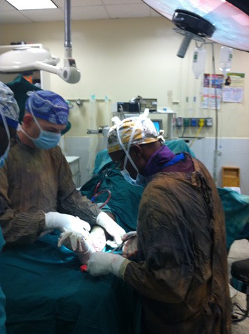
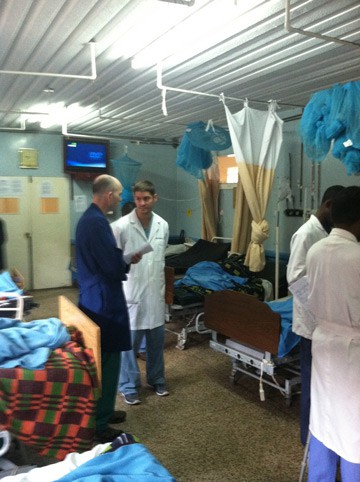
Just about a 4-hour drive out of Nairobi, Tenwick Hospital is a rural 300- bed hospital serving a very large undeserved area.

The hospital employs over 500 local Kenyan workers and has significant international support. World Medical Mission supports the hospital with logistics and helps volunteers travel to Tenwick and that is how I got involved to help with their Orthopedic Surgical load.

There were many fractures and infections when I arrived and a team of Kenyan residents and surgeons who were certainly grateful for the additional help. I was able to operate every day, performing between 3-5 surgeries, working with the resident and attending surgeons to help teach them techniques I often use. Not surprisingly, they taught me so much about themselves, their culture, techniques, and how to perform fairly sophisticated surgery with often less than ideal equipment.

Overall, it was a very positive experience and I plan on going back, perhaps even with my family. The Kenyan people were very enjoyable and genuine and the country has many beautiful areas and safaris that will be fun to explore in the future.

kenya-img-1


kenya-img-3

kenya-img-4


kenya-img-2


Find OSS on Facebook, Google+, and follow on Twitter to keep up to date on new articles and news.

Dr. Mark Reed to Host Foot and Ankle Seminar – April 15th

Top 5 Foot & Ankle Conditions

Hosted by Dr. Mark Reed

Monday, April 15th 6:00pm—8:00pm
Swedish Ballard Conference Room A
5300 Tallman Ave. N.W. Seattle, WA 98107

Our foot and ankle orthopedic specialists and newest addition, Dr. Mark Reed, is presenting a foot and ankle seminar this April 15th. Join this seminar to hear Dr. Reed discuss the top five foot and ankle conditions including:

  • Toe Deformities
  • Foot and Ankle Arthritis
  • Sports Injuries
  • Arch Deformities
  • Trauma

Orthopedic Specialists of Seattle is excited for this upcoming seminar and invites all who are interested to attend. Join us and listen to the top orthopedic conditions of the foot and ankle from our highly trained orthopedic surgeon. … read more