
Wrist Sprains

When you fall forward, your natural response is to put your hands out in front to catch yourself. As you land on your palm, your wrist bends backwards, which can stretch and perhaps tear the ligaments that connect bone to bone within the joint. The resulting injury is a sprain.

Signs and symptoms
Signs and symptoms of a wrist sprain may vary in intensity and location, depending on the degree of injury. Indications of a wrist sprain include:

  • Swelling in the wrist
  • Pain at the time of the injury
  • Persistent pain when you move the wrist
  • Bruising or discoloration of the skin
  • Tenderness at the injury site
  • A feeling of popping or tearing inside the wrist
  • A warm or feverish feeling to the skin

Degrees of injury
Wrist sprains are graded, depending on the degree of injury to the tissues.

  • Grade 1 or mild sprains occur when the ligaments are stretched, but not torn.
  • Grade 2 or moderate sprains occur when some of the ligaments are torn. Grade 2 sprains may involve some loss of function.
  • Grade 3 or severe sprains occur when there is a complete tear all the way through the ligament. These are significant injuries that require medical or surgical care to ensure recovery. As the ligament tears away from the bone, it may also take a small chip of bone with it, resulting in an avulsion fracture.

If you have any doubts about the severity of your injury, arrange to have a doctor examine your wrist. Sometimes, a sprain can have only mild swelling but still be serious enough to require surgery. To avoid problems later, proper diagnosis and treatment is important.

Diagnosing a wrist sprain
Your doctor will review your symptoms and ask how the injury happened. He or she will also ask about any numbness in your hand as well as about your medical and orthopaedic history, especially any previous injuries to your hand or wrist. The physical exam will cover your entire arm and hand to ensure that there are no other injuries. Tenderness in certain areas may suggest a broken bone.

If a severe sprain is suspected, your doctor will likely order X-rays. In some cases, a magnetic resonance image (MRI) or computed tomography (CT) scan may also be ordered.

Treating a wrist sprain
Mild wrist sprains can be usually be treated at home with the RICE protocol.

  • Rest the joint for at least 48 hours.
  • Ice the injury to reduce swelling. Do not apply ice directly to the skin. Use an ice pack or wrap a towel around the ice or a package of frozen vegetables. Apply ice for about 20 minutes at a time.
  • Compress the swelling with an elastic bandage.
  • Elevate the injury above the level of your heart.

You may also wish to take a pain reliever such as aspirin or ibuprofen. If pain and swelling persist for more than 48 hours, however, see a doctor. Moderate sprains may need to be immobilized with a wrist splint for 7 to 10 days. Severe sprains may require surgery to repair the fully torn ligament. Afterwards, you will need a period of rehabilitation and exercises to strengthen the wrist and restore motion. A wrist sprain can take anywhere from 2 to 10 weeks to heal completely, depending on the severity of the sprain.

Prevention
Because wrist sprains usually result from a fall, be careful if you must walk in wet or slippery conditions. Wrist sprains also occur during sports such as skating, skateboarding, and skiing. You can help prevent sports-related wrist sprains by wearing wrist guard splints or protective tape to support the wrist and prevent it from bending too far backwards. If you ski, try to remember to drop your poles during a fall to prevent wrist sprains.
Thumb Fractures

A broken thumb is a serious problem because it affects your ability to grasp items in your hand and can increase the risk of arthritis later in life.

Anatomy
The thumb has two finger bones connected to a hand bone. The first finger bone (distal phalange) extends from the tip of the thumb to the knuckle. The second finger bone (proximal phalange) extends from the knuckle to the webbing between the thumb and the first finger. There, it connects with the hand bone (first metacarpal), which extends from the webbing down to the wrist.
Although a break can occur in any of these bones, the most serious breaks happen near the joints, particularly at the base of the thumb near the wrist.

These fractures have specific names, depending on the type of break. The Bennett and Rolando fractures are breaks at the base of the thumb, involving the joint between the thumb metacarpal and a specific wrist bone. Fractures that involve the joints are always more difficult to treat and are at increased risk of an unfavorable outcome.

How it happens
Thumb fractures are usually caused by direct stress, such as when you fall or when a ball catches and pulls the thumb back. Some fractures may be caused indirectly, from twisting or muscle contractions. People who play contact sports such as wrestling, hockey or football; skiers; and people with a history of bone disease or calcium deficiency are especially at risk.
The risk of a thumb fracture can be lessened by using protective taping, padding or other equipment and by developing strength in your hands through exercise and proper nutrition.

Symptoms of a fracture

  • Severe pain at the fracture site
  • Swelling
  • Limited or no ability to move the thumb
  • Extreme tenderness
  • A misshapen or deformed look to the thumb
  • Numbness or coldness in the thumb

Treatment options
See a doctor as soon as possible. Without immediate treatment, the joint will be unstable and you will be unable to grip or pinch properly. Continued swelling may make it more difficult to align the bones properly. Delayed treatment will make the fracture much more difficult to treat and can lead to a poor outcome. Use a padded splint to prevent the bone from moving further out of alignment is encouraged prior to definitive treatment.

The physician will examine the injury, take your medical history and have X-rays taken of the injury. You may need surgery, depending on the location of the fracture and the amount of movement between the broken pieces of bone. If there is little movement (displacement) or if the break is located in the middle (shaft) of the bone, an orthopaedist may be able to use a specially designed cast (spica cast) to hold the bone fragments in place. You will have to wear the cast for at least four to six weeks, and your doctor may request regular X-rays to ensure that the bone hasn’t slipped out of alignment.

Often, Bennett and other more severe fractures of the thumb require surgical treatment. The hand surgeon may use one of several operative fixation techniques to restore boney anatomy and hold the bones in place while they heal. These techniques include the use of wire, pins, plates and screws as internal fixation. Another technique uses pins in bone that exit the skin and are attached to an external fixation device You will likely be required to wear a cast or splint for two to six weeks after surgery. When the cast is removed, your physician may recommend hand therapy to restore movement. It can take three months or more to regain full use of the hand, depending on the severity of the injury.

Complications and outcomes
If the bones remain in a stable position, thumb fractures generally heal well. There is a possibility of infection and tenderness around the surgical site and around the pins, if external fixation is used. As with all joint injuries, there is an increased tendency to develop arthritis, which can limit motion and reduce strength in the thumb.

To help restore motion, soak your hand in warm water and move your thumb in a circular direction. Try to touch your little finger with your thumb. To help restore strength, get a spring-type hand squeezer and use it regularly.

Sprained Thumb

When people start to fall, they usually extend their arm to reduce the force of the impact when they hit the ground. If you try to break your fall on the palm of your hand or take a spill on the slopes with your hand strapped to a ski pole, your thumb may be injured. The main ligament (ulnar collateral), which supports pinch and grasp activities, may be torn (sprained). The ligament helps your hand to function properly, acting like a hinge to keep your thumb joint (metacarpophalangeal) stable.

When you have a sprained thumb, you lose some or all of your ability to grasp items between your thumb and index finger. It may or may not hurt right away. Other signs include bruising, tenderness and swelling. To make sure your injury won’t cause long-term weakness, pain and instability, see your doctor for evaluation and treatment.

Partial and complete tears
Your thumb ligament may have a partial or complete tear. Your doctor will probably move your thumb joint to test its stability and take X-rays to make sure you don’t also have a broken bone. You may also get a stress X-ray showing what the joint looks like when your ligament is tested. If it hurts to do this, a shot of local anesthetic may help. Your doctor will probably also X-ray your uninjured thumb to compare it.

If you have a partial tear, your doctor will probably immobilize your thumb joint with a splint or other bandage until it heals. You wear the splint for about three weeks straight, then start taking it off to do flexion and extension exercises with your thumb. Put the splint back on for protection when you are not doing the exercises. Keep doing this for another two or three weeks until your thumb’s swelling and tenderness are gone. You may also put ice on your injury twice a day for 2-3 days after the fall.

If your thumb ligament is completely torn, you may need surgery. Fragments of bone that sometimes get pulled away when your ligament tears may be removed or put back in their correct positions. After surgery, you’ll probably need to wear a short-arm cast or a splint to protect your thumb ligament for six to eight weeks while it heals.

Restoring Hand Function after Spinal Cord Injury

An injury to the upper part of the spinal cord can leave an individual with little or no sensation or movement in both the arms and the legs, a condition called tetraplegia (tet-rah-PLEE-gee-ah). A surgical technique called a tendon transfer can help restore function to arms and hands by giving working muscles different jobs.

This can greatly enhance the quality of life for people with tetraplegia by enabling them to do many more tasks for themselves. The types of tendon transfer surgeries that can be performed depend on where the spinal cord injury occurred and which muscles are affected.

How it works
Tendons are the strong cords that connect muscle to bone. When a tendon crosses a joint, it helps transmit muscle action into joint movement. A tendon transfer repositions the tendons of a working muscle so that they take over the functions of a paralyzed muscle. This enables the working muscle to do what the paralyzed muscle can no longer do.

For example, in the upper arm, the triceps muscle is used to straighten the elbow. The larger deltoid muscle pulls the arm backwards and forwards away from the body. If the triceps muscle is paralyzed but the deltoid is still functional, surgeons can split the deltoid muscle and graft a portion of it to the triceps. This restores elbow function without greatly diminishing shoulder function.

Tendon transfers can help restore three critical capabilities necessary for self-care and increased independence:

  • the ability to straighten (extend) and bend (flex) the elbow
  • the ability to bend and straighten the wrist
  • the ability to grip with the fingers and hand

Planning for a tendon transfer
Usually, a tendon transfer is not scheduled until about a year after injury. During the first months after the injury, rehabilitation focuses on retaining passive range of motion. These exercises help prevent shoulder stiffness and pain. As time progresses, strength and range of motion (both active and passive) must be evaluated frequently. Severe muscle contractures or muscle spasms may necessitate another type of surgery rather than a tendon transfer. Usually tendon transfer surgery is scheduled only after there is no more progress in function.

Before surgery is scheduled, several assessments must be made, including:

  • identifying which muscles still work and measuring how well they work to determine whether they can be used in the transfer
  • assessing the individual’s abilities to see which functions need to be restored
  • matching available muscles with functional requirements
  • determining if an additional procedure such as a joint fusion or electrical stimulation implant is needed to restore function
  • verifying that the individual has a strong support system that can provide the care needed during rehabilitation after surgery
  • assessing the individual’s motivation and commitment to the process
  • determining which surgeries should be performed, when, and in what order

Elbow Extension
The ability to bend and straighten the elbow adds greatly to a person’s independence, so this is often the first surgery to be performed. In most cases, a portion of the deltoid muscle in the shoulder is used to provide elbow extension. The back (posterior) portion of the deltoid is brought down toward the elbow. Because the deltoid portion is not long enough to reach the attachment point in the lower arm, a graft is taken from an upper leg muscle (fascia lata) to provide the necessary length.

In some cases, the biceps muscle in the upper arm is used instead of the deltoid muscle. After the surgery, the arm is immobilized in a slightly bent position for up to four weeks. When the cast is removed, a hinged brace is used to allow a gradual stretching and strengthening of the muscles. Initially, the brace is worn night and day, but as the individual gains the ability to fully extend the arm, the brace is worn only at night.

Tendon transfer to achieve elbow extension is done on one arm at a time because the arm is totally immobilized during rehabilitation. This means that the person becomes even more dependent on others for the simple activities of daily living. However, the results are impressive. It can eliminate the need for many adaptive devices and enable the person with tetraplegia to propel a wheelchair, to move independently from bed to chair, to shift weight within a chair or bed and to reach up and outwards.

Key Pinch
Restoring key pinch enables the individual to grip items between the thumb and the hand. This greatly enhances the ability of the patient with tetraplegia to accomplish activities of daily living, such as writing or feeding themselves. In this surgery, one of the forearm muscles (brachioradialis) is grafted to the tendons that move the wrist and thumb. The surgeon may also stiffen the joint so that when the wrist is extended, the grip forms automatically.

These two surgeries significantly improve hand and arm function in many patients, providing them with much greater independence.

Electronic Implants
A relatively recent advance uses an electronic implant, similar to a pacemaker, to stimulate muscles. Tiny electrodes are attached to the functioning muscles of the arm and hand. The electrodes are connected to a control device implanted in the front of the chest. An external unit delivers the signals to initiate grasp and key pinch. This is an option when tendon transfers cannot be used.

Outcomes
Because tendon transfers use the patient’s own tissues, the risk of infection is lessened. However, the risk of developing a latex allergy is increased so precautions should be taken. The length of the surgery (approximately six hours) also increases the risk of postoperative respiratory problems.

In general, the results using tendon transfer surgery to restore arm and hand function after spinal cord injury are good. People with tetraplegia can often benefit from the increased self-confidence and independence they gain after tendon transfer surgery.

Nerve Injuries

What are nerves?

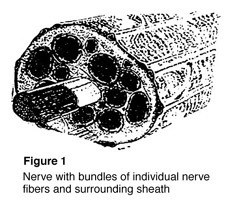
cons1_71_49Nerves are the “electrical wiring” system in all people that carry messages from the brain to the rest of the body. A nerve is like an electrical cable wrapped in insulation. A ring of tissue forms a cover to protect the nerve, just like the insulation surrounding an electrical cable (Figure 1).
Nerves serve as the “wires” of the body that carry information to and from the brain. Motor nerves carry messages from the brain to muscles to make the body move. Sensory nerves carry messages to the brain from different parts of the body to signal pain, pressure, and temperature. While the axon (nerve fiber) carries only one type of message, either motor or sensory, most nerves in the body are made up of both.

What happens when a nerve is injured?
Nerves are fragile and can be damaged by pressure, stretching, or cutting. Injury to a nerve can stop signals to and from the brain causing muscles not to work properly, and you may lose feeling in the injured area. When a nerve is cut, both the nerve and the insulation are broken. Pressure or stretching injuries can cause the fibers carrying the information to break and stop the nerve from working, without damaging the cover.

When nerve fibers are cut, the end of the fiber farthest from the brain dies, while the insulation stays healthy. The end that is closest to the brain does not die, and after some time may begin to heal. If the insulation was not cut, new fibers may grow down the empty cover of the tissue until reaching a muscle or sensory receptor.

If both the nerve and insulation have been cut and the nerve is not fixed, the growing nerve fibers may grow into a ball at the end of the cut, forming a nerve scar or neuroma. A neuroma can be painful and cause an electrical feeling when touched.

How is it treated?
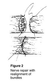
cons1_72_49To fix a cut nerve, the insulation around both ends of the nerve are sewn together. The goal in fixing the nerve is to save the cover so that new fibers may heal and work again (Figure 2). If a wound is dirty or crushed, your physician may wait to fix the nerve until the skin has healed.

If there is a space between the ends of the nerve, the doctor may need to take a piece of nerve (nerve graft) from another part of the body to fix the injured nerve. This may cause permanent loss of feeling in the area where the nerve graft was taken.

Once the nerve cover is fixed, the nerve generally begins to heal three or four weeks after the injury. Nerves usually grow one inch every month depending on the patient’s age and other factors. This means that with an injury to a nerve in the arm above the fingertips, it may take up to a year before feeling returns to the fingertips. The feeling of pins and needles in the fingertips is common during the recovery process. While this can be uncomfortable, it usually passes and is a sign of recovery.

What is my role in recovery and what kind of results can I expect? The patient must do several things to keep up muscle activity and feeling while waiting for the nerve to heal. Your doctor may recommend therapy to keep joints flexible.

If the joints become stiff, they will not work even after muscles begin to work again. When a sensory nerve has been injured, the patient must be extra careful not to burn or cut fingers since there is no feeling in the affected area.

After the nerve has recovered, the brain gets “lazy,” and a procedure called sensory re-education may be needed to improve feeling to the hand or finger. Your doctor will recommend the appropriate therapy based on the nature of your injury.

Factors that may affect results after nerve repair include age, the type of wound and nerve, and location of the injury. While nerve injuries may create lasting problems for the patient, care by a physician and proper therapy help two out of three patients return to more normal use.