Hand Fractures

Have you ever been so frustrated that you wanted to slam your fist into a wall? If you do, you could break one of the bones in your hand (metacarpals). Fractures of the hand bones account for about one-third of all hand fractures. In fact, fractures of the fifth bone (the one that leads to your little finger) are commonly known as “boxer’s fractures.”

The hand bones can break near the knuckle, mid-bone, or near the wrist. Signs and symptoms of a broken bone include:

  • Swelling
  • Tenderness
  • Deformity
  • Inability to move the finger
  • Shortened finger
  • Depressed knuckle
  • Finger crosses over its neighbor when you make a partial fist

Diagnosis and Treatment
Your physician will request X-rays to identify the fracture location and type. The physical examination may include some range of motion tests and an assessment of sensation in the fingers to ensure that there is no damage to the nerves.

Most of the time, the physician can realign the bones by manipulating them without surgery. Then, a cast, splint or fracture-brace is applied to immobilize the bones and hold them in place. The cast will probably extend from the fingertips down past the wrist almost to the elbow to ensure that the hand bones remain fixed in place.

Your physician will probably request a second set of X-rays about a week later to ensure that the bones remain in the proper position.

You will usually have to wear the cast for three to four weeks, but you can probably begin gentle hand exercises after three weeks. Afterwards, the finger may be slightly shorter, but this should not affect your ability to use your hand and fingers.

Surgical options
Some hand fractures, such as those that break through the skin or result from a crushing accident, require surgery to stabilize and align the bones. The orthopaedic surgeon implants wires, screws or plates in the hand to hold the fracture in place. If the bone rotates while healing, loss of function could result.

After the bone has healed, the surgeon may remove the implants, or they may be left in the hand. Research to develop implants that are resorbed into the body is ongoing. Your physician may ask you to return frequently for check-ups to ensure that the joint doesn’t tighten (contract) during healing. You may experience some joint stiffness in your hand because of the long immobilization period.

Your physician may prescribe exercises to help restore strength and range of motion or recommend that you see a physical therapist.

Fracture of the Finger

You might think a broken finger is a minor injury, but without proper treatment it can cause major problems. The bones in a normal hand line up precisely, letting you perform many specialized functions like grasping a pen or manipulating small objects in your palm. When you fracture a finger bone, it can put your whole hand out of alignment. Without treatment, your broken finger might stay stiff and painful.

Signs of a fractured finger

  • Swelling
  • Tenderness
  • Inability to move it completely
  • Deformity

Diagnosis

If you think you broke your finger, tell your doctor right away exactly what happened and when. You have three bones (phalanges) in each finger and two in each thumb. Your doctor must determine not only which bone fractured, but also how it broke: straight across, in a spiral, into several pieces, or shattered completely.

Your doctor may want to see how your fingers line up when you extend your hand or make a fist. Does any finger overlap its neighbor? Angle in the wrong direction? Look too short? Your doctor may X-ray both of your hands for comparison.

Treatment and rehabilitation

Your doctor will put your broken bone back into place, usually without surgery. Sometimes you need pins, screws or wire to hold it together, especially if you have a complicated injury. You’ll get a splint or cast to hold your finger straight and protect it from further injury while it heals. Sometimes your doctor may splint the fingers next to the fractured one for support.

Your doctor will tell you how long to wear the splint – usually about three weeks. Sometimes you may need more X-rays as you heal so your doctor can check your progress.

Begin using your hand again as soon as your doctor determines it is okay to move your finger. Doing simple rehabilitation exercises each day will help reduce the finger’s stiffness and swelling. You may be required to see a physical therapist to assist you in these exercises.

Flexor Tendon Injuries

Tendons are tissues that connect muscles to bone so that joints can bend or straighten. The flexor muscles that move your fingers and thumb are located in your forearm, above your wrist. Long tendons extend from the flexor muscles through the wrist and attach to the small bones of your fingers and thumb.

Anatomy
Each finger has two tendons; the thumb has one tendon. The tendons run along the palm side of the fingers and are very close to the surface of the skin, particularly where the skin folds as you bend your fingers.

The longer tendon (flexor digitorum profundus or FDP) attaches to the last bone of the finger and bends the tip; the shorter tendon (flexor digitorum sublimis or FDS) bends the middle joint of the finger.

If you tear (rupture) or cut (sever) the tendon anywhere along its route, at the wrist, in the palm of the hand, or along the finger, you may be unable to bend your finger. If you injure the FDS tendon, you may still be able to bend the finger, but not completely, and bending the finger will be painful.

Tendons are stretched tightly as they connect the muscle to the bone. If the tendon tears, the end connected to the muscle will be pulled back in toward the palm. Because the tendon can’t heal unless the ends are touching, a severed tendon must be sewn back together again (a surgical repair).

Types of injuries
Most often the flexor tendons are damaged by a cut. Because the nerves to the fingers are also very close to the tendons, a cut may damage them as well, resulting in a feeling of numbness on one or both sides of the finger.

Athletic injuries are also common, usually in football, wrestling or rugby. One player grabs another’s jersey, and a finger(usually the ring finger)gets caught and pulled. This type of injury is so common, it even has a name: “jersey finger.” You can also strain or rupture the tendon while rock climbing.

People with rheumatoid arthritis may experience a spontaneous rupture of the flexor tendons. You may notice that the finger no longer bends, but cannot recall when you lost the ability to bend it.

Signs and symptoms

  • An inability to bend one or more joints of the finger
  • Pain when you bend your finger
  • An open injury, such as a cut, on the palm side of the hand, particularly in the joint area where the skin folds as the finger bends
  • Mild swelling over the joint closest to your fingertip
  • Tenderness along the finger on the palm side of the hand

Diagnosing your injury
See your physician whenever you injure your fingers, especially if you “jam” the finger and notice that you cannot bend or straighten the tip. For immediate first aid, apply ice and compression to slow the flow of blood to the damaged site.

Your doctor will ask you to bend and straighten the fingers and may apply resistance to test the strength of the fingers. Your doctor may also test the sensation and blood flow to your fingers to see if any nerves or blood vessels were also injured.

You may need to get an X-ray to see if there is any damage to the bone; if you have an open wound, you may need a tetanus shot or antibiotics.

Treatment
Your doctor may first clean and treat any superficial wounds and put your hand in a splint. Flexor tendon injuries require surgical repair and it’s best to have the surgery as soon as possible after the injury. The hand surgeon will sew the tendon together using special stitches on both the inside and outside of the tendon.

However, it can take up to two months before the repair is healed and strong enough to use your hand without protection. It may take another month or so before you can use your hand with any force.

In the meantime, you will need to wear a splint and see a physical therapist. The therapist will give you special exercises to perform. Follow your doctor’s and your therapist’s instructions carefully to ensure the best possible result.

Even after surgery, you may experience some stiffness in your finger. However, it will be considerably less than if you did not have the surgery.

Fingertip Injuries/Amputations

Your child accidentally slams a fingertip in a car door. Or you cut off the end of your finger while chopping vegetables. Or you lose the tops of several fingers trying to clear debris from a lawnmower or snowblower.

Fingertip injuries are very common. Accidents can happen at work, play and in the home, crushing, tearing (lacerating) or cutting off (amputating) the tips of fingers and thumbs. The tips of longer fingers get injured more often because they are last to escape when you pull your hand out of harm’s way. You may have damage to skin and soft tissue, bone (distal phalanx) and/or nail and nailbed.

Fingertip injuries

What to do

Always see a doctor right away if you injure the tip of a finger or thumb. Fingertips are rich with nerves and extremely sensitive. Without prompt and proper care, a fingertip injury can disrupt the complex function of your hand, resulting in permanent deformity and disability.

First aid:

  • Elevate the injury and apply ice to reduce bleeding and swelling.
  • Cover the fingertip wound with a dry, sterile dressing.
  • Immobilize the affected hand and wrist with a short splint.

If a fingertip is completely cut-off:

  • Gently cleanse the amputated part with water (preferably saline).
  • Cover it in gauze wrap.
  • Put it in a watertight bag.
  • Place the bag on ice.
  • Do not put the amputated part directly on ice. You could further damage it.
  • Take the amputated part with you to the emergency room.

Medical treatment

Doctors provide individualized treatment for a fingertip injury/amputation based on the angle and extent of the injury, as well as factors related to your health and lifestyle. Tell your doctor how and when the injury happened and:

  • If the injury is on your dominant hand.
  • What you do for a living and recreational activities.
  • If you have other hand problems, osteoarthritis or systemic diseases (i.e., diabetes, rheumatoid arthritis).
  • Whether your tetanus immunization is current.

The doctor will probably give you an injection (digital block anesthesia) to stop pain in the affected finger. Then he or she may irrigate the wound with a saline solution; inspect it for exposed bone, soft tissue loss and nail/nailbed injury; and clean (debride) it, removing dead (devitalized) tissue and foreign contaminants to reduce risk of infection.

You may need X-rays to check for fractures. If blood has accumulated beneath the nail (subungual hematoma), your doctor may drain it by piercing through the fingernail. You may also need an antibiotic and/or tetanus shot.

Your doctor formulates a plan for treatment after completely assessing your injury. The goal is a painless fingertip that has durable and sensate skin. Your hand should be able to pinch, grip and perform other normal functions. If possible, your doctor may also try to maintain the finger’s length and appearance and preserve its fingernail.

Soft tissue injury with no exposed bone

Small wound: A small wound to a fingertip’s skin and fleshy tissue (pulp) may close on its own (healing by secondary intention). Your doctor may put a protective dressing over the wound, splint your hand and instruct you to change the bandage at regular intervals. After 48 hours, you may begin range-of-motion finger exercises.

After about a week, you may start daily finger soaks in a warm water-peroxide solution. Complete healing usually takes 3-5 weeks. Then you may need a program of fingertip desensitization.

Large wound: If you let a larger fingertip wound heal itself, you may not get a durable fingertip. Therefore, your doctor may need to transplant a piece of skin from the palm of your hand or other donor sites (skin graft) to cover the injury. The donor site is surgically closed.

Exposed bone

If your injury exposes bone, there is probably not enough tissue available on the fingertip to surgically close it. Your doctor may need to shorten the bone, which generally does not hurt hand function. He or she may also need to transfer a piece of skin and underlying fat and blood vessels from a healthy part of your body to the injury site (reconstructive flap surgery). Depending upon the angle of injury or amputation, the flap may come from:

  • The injured finger (triangular volar advancement flap).
  • An uninjured finger (cross-finger flap).
  • The palm of the hand (thenar flap).

Your doctor sews (sutures) the flap to the defect and surgically closes the donor site. A bulky dressing and splint supports your hand after surgery, with uninjured fingers left free to exercise. A second operation may be necessary in a few weeks to detach the flap from its origin.

If the amputated part is large (includes the entire nail and dorsal skin), your doctor may discuss the pros and cons if replantation is right for you. This involves a long, complicated surgical procedure which may keep you hospitalized for several days.

Young children

Doctors treat fingertip amputations somewhat differently in children under age 6. After thoroughly cleaning and removing fat from an amputated fingertip, your doctor may suture it back on the finger (composite flap). Especially in children under age 2, a relatively normal looking fingertip may form, even if bone was exposed.

Outcome

In many cases, fingertip repair surgery can give you back a large degree of feeling and function. Infection, poor healing, loss of feeling or motion, blood clots and adverse reactions to anesthesia are all possible complications of surgery. You may have mild to severe pain and sensitivity to cold following treatment for a fingertip injury/amputation.

Recovery may take months, and you will probably need hand therapy. This may include hand exercises to improve movement and strength, heat and massage therapy, electrical nerve stimulation, splinting, traction and special wrappings to control swelling.

Boutonnière Deformity

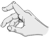
a00004f01Boutonnière deformity is an injury to the tendons in your fingers that usually prevents the finger from fully straightening. The result is that the middle joint of the injured finger bends down, while the fingertip bends back. This is the characteristic shape of a boutonnière deformity. Unless this injury is treated promptly, the deformity may progress, resulting in permanent deformity and impaired functioning.

There are several tendons in your fingers that work together to bend and straighten the finger. These tendons run along the side and top of the finger. The tendon on the top of the finger attaches to the middle bone of the finger (the central slip of tendon). When this tendon is injured, the finger is not able to be fully straightened.

Boutonnière deformity is generally caused by a forceful blow to the bent finger.

It also can be caused by a cut on the top of the finger, which can sever the central slip from its attachment to the bone. The tear looks like a buttonhole (“boutonnière” in French). In some cases, the bone actually can pop through the opening.

Boutonnière deformities may also be caused by arthritis. About one third of all people with rheumatoid arthritis also have fingers with boutonnière deformities.