Rotator Cuff Tears – Dr. Franklin

From orthoinfo.aaos.org

From orthoinfo.aaos.org

Anatomy
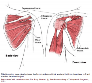
The rotator cuff is made up of 4 muscles surrounding the shoulder.

Three muscles on the back of the shoulder (supraspinatus, infraspinatus and teres minor) converge as a tendon to insert on the outer edge of the humeral head.

They act to elevate and externally rotate the shoulder. The fourth muscle (subscapularis) is on the front of the shoulder and helps internally rotate the shoulder.

Injury Mechanism
Rotator cuff tears can occur after an acute injury such as a fall or catching a heavy falling object, or they can occur over time as a gradual wearing of a hole in the rotator cuff from ongoing rubbing on the acromion such as overuse with overhead or throwing sports.

Symptoms
With acute complete tears of the rotator cuff, patients often describe a burning or tearing sensation at the time of injury. Early along, it is difficult to raise the arm overhead. Patients describe pain and weakness. The pain is usually located over the outer (lateral) aspect of the upper arm. Rotator cuff tears are often painful at night. Older patients may not recall an injury, but may just describe a gradual aching in the shoulder that has progressed over several months or years.

Diagnosis
The physician’s work-up will start with a careful history and exam.

X-rays are often obtained to see that no fractures have occurred with the injury and to help assess the overall condition of the shoulder joint. An MRI scan is often obtained to confirm the diagnosis and to evaluate any associated injuries to the labrum, rotator cuff or damage to the joint surfaces. Often the radiologist will inject contrast into the shoulder joint with a small needle to coat the undersurface of the rotator cuff and to see if the contrast leaks through the rotator cuff suggesting a complete tear. An MRI with contrast is called an arthro/MRI.

Plain x-rays show the bones of the shoulder, while MRI scans reveal the soft tissues around the shoulder including the labrum (lip of cartilage around the socket) and the rotator cuff tendons.

Treatment
Your physician will discuss treatment options with you. Treatment decisions are based on age, activity level and the severity of symptoms. In older patients with less activity demands and less severe symptoms, one will usually start with non-operative treatment including rest, ice, anti-inflammatories, stretching and occasional injections to see if the symptoms become tolerable.

In younger, more active patients, surgery is almost always recommended when a full thickness rotator cuff tear has occurred, since the rotator cuff has poor blood supply, therefore poor healing potential. Rotator cuff surgery is usually done on an outpatient basis. In most cases, the orthopedist will start with an exam under anesthesia to see that full motion of the shoulder is present and to see that the joint is stable. Next, one usually looks into the joint with a small arthroscope (a small lens and camera) so the surgeon can see and probe all of the structures in the joint. Once the complete rotator cuff tear is confirmed, the torn tendon is repaired back to the bony attachment site.

Because there is poor blood supply at the attachment site to the bone, one usually creates a small groove in the bone and pulls sutures through drill holes or uses anchors in the bone for fixation.

What to expect after surgery
Rotator cuff surgery is performed as an outpatient procedure. The surgery takes about 60 minutes. Patients go home in a sling that they use for 3 or 4 weeks. Most patients are uncomfortable for the first 2 or 3 days, but prescription medication is used to help alleviate the pain. Patients are seen back in the office one week after surgery to check their incisions and to start their exercise routine.

For the first 4 to 6 weeks, patients avoid any active elevation of their arm or lifting with that arm. At that point, formal physical therapy is started to improve range of motion and strength. It takes about 3 months from surgery before most of the strength and use of the shoulder returns. Full recovery may take 4 to 6 months.

Shoulder Impingement

Shoulder Impingement (Rotator Cuff Tendinitis)

The rotator cuff is made up of tendons and muscles that allow for a great range of motion of your arm. This is a frequent source of pain for athletes and an area that is at risk for injury during sporting activities. Shoulder impingement is often referred to as rotator cuff tendinitis and is one of the most common causes of shoulder pain.

What causes rotator cuff tendinitis?
When you raise your arm to shoulder height, the space between the bone and rotator cuff narrows. The bone can rub against (or impinge on the tendon and the bursa, causing irritation and pain when the arm is used repeatedly. Young athletes who use their arms for overhead action are particularly vulnerable. This includes those who play tennis, softball and baseball, and swimmers.

What are the symptoms of shoulder impingement?
When the rotator cuff is irritated this can lead to local swelling and tenderness in the front aspect of the shoulder. You may also have pain and stiffness when you lift your arm. There is also a sensation of tenderness when the arm is lowered from an elevated position. Other symptoms include sudden pain when reaching or lifting, pain radiating from the front of the shoulder to the side of the arm, minor pain at rest, and pain when throwing or using the arm.

How is rotator cuff tendinitis treated without surgery?
Your orthopedic specialist wants to reduce your pain and restore function of your shoulder. He will consider your activity level, your age, and your general state of health. Many times shoulder impingement can be treated with medications and rest. It is not uncommon for athletes to be ordered physical therapy to help restore normal motion of the shoulder. Your therapist will teach you specific stretching and strengthening exercises to relieve your shoulder pain and help you get back to normal activities.

What is involved with surgical treatment?
The goal of surgery is to create more space for the rotator cuff and this involves removing a portion of the inflamed bursa. Your orthopedic specialist will perform an anterior acromioplasty, where part of the bone is removed to allow for movement of the rotator cuff. Many times, the surgeon opts to perform this procedure by way of arthroscope.

The arthroscopic technique allows for use of small thin surgical instruments to be inserted around puncture wounds around the shoulder. The doctor can see inside the shoulder through a small camera inserted into the joint that displays images onto a computer TV monitor.

Biceps Tendon Tears

From orthoinfo.aaos.org

From orthoinfo.aaos.org

Anatomy: The biceps is a large muscle in the front of the shoulder. The upper end of the biceps has to heads (attachment sites). The long head of the biceps or its attachment to the labrum (cartilage lip at the top of the shoulder socket) is commonly injured.

When it is torn at the upper end or at the labral attachment, there is often pain in the front of the shoulder. When the long head of the biceps is completely torn, the biceps muscle has a typical bulging appearance, but oftentimes does not cause much discomfort of weakness.

Injury Mechanism: Acute injuries to the long head of the biceps occur with heavy lifting or by grabbing the railing to support one’s body weight during a fall. Tears of the biceps can also occur with repetitive use of the shoulder in sports or work.

Symptoms: Acute complete tears of the long head of the biceps usually present as a sharp pain in the front of the shoulder, followed by bruising and the “Popeye muscle” deformity of the biceps (bulging in the middle of the upper arm where the muscle has retracted). Chronic tears or fraying of the biceps usually presents as aching in the front of the shoulder with use of the biceps.

From orthoinfo.aaos.orgDiagnosis: The physician’s work-up will start with a careful history and exam. X-rays will be normal as they show the bones and joints, not the muscles and tendons. An MRI scan can be obtained to confirm the diagnosis, especially if the diagnosis is not obvious by history and exam.

Treatment: Your physician will discuss treatment options with you. Most complete tears of the rotator cuff can be treated without surgery. Rarely patients notice weakness or cramping in the biceps. Stretching, strengthening, anti-inflammatories, and time will usually settle down symptoms to a tolerable level.

The partial tears are usually more painful. Injections can sometimes manage the discomfort, but occasionally surgery is necessary to help alleviate symptoms.

From orthoinfo.aaos.orgSurgery is usually accomplished through the arthroscope to remove the frayed tissue.

If the partial tear is nearly complete, the tendon is released from its attachment to the socket (labrum) and attached further down on the upper arm bone (humerus). This procedure is called a biceps tenodesis. Sometimes a small incision is necessary to re-attach the tendon to the bone.

What to expect after surgery: If the frayed tendon is just removed through the arthroscope, the recovery is rapid. Usually, after a few days of rest, gentle exercise is started. A full recovery is expected after 4 to 6 weeks. If the tendon is re-attached to bone, a sling is used with the elbow bent for 3 or 4 weeks to protect the repair. No resisted use of the biceps is started until 6 to 8 weeks after the repair.

Collateral Ligament Injuries

Anatomy
collateral-300x208
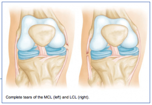
The medial collateral ligament (MCL) runs from the inner side (medial side) of the femur (thigh bone) to the inner (medial side) of the tibia (lower leg bone). It prevents the knee from opening on the inside when struck from the outside of the knee joint. The MCL lies on the outside of the joint capsule and has a good blood supply that contributes to its good healing potential.
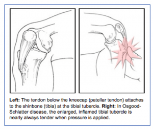
The lateral collateral ligament (LCL) runs from the outer side (lateral side) of the femur (thigh bone) to the top of the fibula (the smaller of the two lower leg bones). It prevents the knee from opening on the outer side when struck from the inner side of the knee joint. The LCL is thinner and when completely disrupted often requires surgical repair.

Injury Mechanism
The MCL is often injured in sports when one is struck from the outer or lateral side of the knee, such as having an opponent fall against the outside of one’s knee in football. Another common mechanism of injury to the MCL is when the foot is forced out to the side away from the body, such as with a simultaneous kick of a soccer ball with the inside of the foot. LCL injuries are much more rare and usually occur when the knee is struck from the inside while the foot is planted, forcing a distraction force to the outside of the knee.

Symptoms
When patients sustain an injury to the collateral ligaments they often experience pain, localized swelling and bruising on the involved side of the knee. With partial tears, there is stiffness and pain when fully bending the knee, but no sense of instability. With a complete tear, the knee will feel unstable and will give way to the side with any lateral movements.

Diagnosis
The physician’s work-up will start with a careful history and exam. Often the description of a direct blow to either side of the knee can lead the physician to the suspected injury to the MCL or ACL. On examination, the physician can feel the instability when pulling the foot to one side or the other while stabilizing the knee. X-rays are often obtained to see that no fractures have occurred. Occasionally, a small avulsion fracture might hint that a collateral ligament injury has occurred. An MRI scan is often obtained to confirm the diagnosis and to evaluate any associated injuries to the menisci, other ligaments, and damage to the joint surfaces.

Treatment
Your physician will discuss treatment options with you. Treatment decisions are based on degree of instability. Minor tears (sprains) can be treated with rest, ice, elevation and compression. More significant tears in which many of the fibers of the ligament have been torn may require bracing for 6 weeks to keep the fibers from healing in a stretched out position. Occasionally physical therapy is needed to help regain full range of motion and strengthen the surrounding muscles after the period of bracing. Rarely is surgery recommended for an isolated MCL tear, but occasionally LCL injuries can benefit from surgical repair or reconstruction.

Osgood-Schlatter Disease

What is Osgood-Schlatter Disease?
Osgood-Schlatter Disease is a condition that occurs in adolescents that involves pain and inflammation that occurs where the patellar tendon attaches to the lower let bone (tibia). It is commonly seen during a sports season that involves running and jumping (basketball, soccer, track).

osgood-300x252Diagnosis
The diagnosis is usually made by identifying swelling and tenderness at the attachment of the patellar tendon to the front of the tibia. Often a bump is present at that location. Occasionally x-rays will reveal a bony prominence or sometimes an extra piece of bone where the patellar tendon attaches to the tibia.

MRI scans are not usually necessary to help make the diagnosis in these patients.

Treatment
Surgery is almost never needed, as most cases of Osgood-Schlatter Disease will resolve as one reaches the end of adolescence. Often, activity restrictions are necessary to help resolve the symptoms. Warming up and stretching before activities and icing after can be helpful. Non-steroidal anti-inflammatories are sometimes necessary to help with the discomfort.