
Football Related Knee Injuries

The knee is a complex joint that is vulnerable to a variety of injuries. The knee is made up of the femur (the thigh bone), the tibia (the shin bone), and the patella (the knee cap). The femur rotates on the upper end of the tibia and the patella fits on the end of the femur.

There are also many large ligaments that connect the bones of the knee and help control knee motion. The meniscus is a wedge of cartilage that serves as a cushion between the femur and tibia and also absorbs shock. Many football athletes experience injuries to these knee structures.

Ligament Injuries

Football players frequently injure one or more of the knee ligaments. These ligaments include the anterior cruciate ligament (ACL), the posterior cruciate ligament (PCL), the medial collateral ligament (MCL), and the lateral collateral ligament (LCL).

  • ACL Injury – When an athlete changes direction rapidly, lands wrong from a jump, or simply slows down when running, the ACL could tear. With this injury, knee swelling immediately occurs and walking is painful. The knee may have loss of range of motion and tenderness with an ACL injury. Treatment for this type of injury depends on the degree of tear to the ligament, whether or not there are other associated injuries, and how much physical demand the patient puts on their knee. Sometimes, the orthopedic specialist needs to operate to repair a complete tear of the ACL. Recovery is measured in months, rather than weeks for this type of injury.
  • MCL Injury – The MCL is generally injured from a direct blow to the outside portion of the knee. The ligament is torn or stretched when the foot is planted firmly on the ground and a sideways force hits the knee. An injured MCL causes pain, difficulty walking, and tenderness. Therapy involves the use of a knee immobilizer, rest, ice applications, compression with a support bandage, and frequent knee elevation. Surgery is only necessary for severe tears of the MCL.
  • PCL Injury – The PCL is injured when a football player receives a blow to the front aspect of the knee or makes a simple misstep on the turf. Most PCL tears and injuries will heal with conservative treatment. An injured PCL leads to pain with walking, instability, and swelling of the knee. Surgery may be necessary with complete tearing and extensive damage to the PCL.
    LCL Injury – The LCL is the least likely ligament to be injured during football activities. When severe force is applied to the inside of the knee, a LCL injury could occur. Symptoms include pain, swelling, weakness, tenderness, and discomfort to the outside of the knee. Treatment involves the RICE method, anti-inflammatory medications, and immobilization. Surgery to reattach the ligament to the bone is sometimes required.

Cartilage Injuries
Torn Cartilage – Most of the time, the meniscus is the cartilage that is torn during a football game. This rubbery, tough structure serves as a shock absorber during athletic activities. The meniscus tears with cutting, decelerating, pivoting, twisting, or from being tackled. Most torn meniscus injuries cause gradual pain and swelling, worse with climbing steps or uphill.

Not all meniscus tears require surgery, but frequently the damage can only be repaired through an operation.

Fractures
The patella can break if the football player falls directly onto it or receives a direct blow in that area. If the bone is fragmented, surgery will be required for repair. If the bone is in appropriate position, the orthopedic specialist may prescribe an immobilizer and rest for the injury.

The head of the fibula on the outside area of the knee joint is easily fractured from direct blows or as part of an injury to the lower leg. If the bone is not out of alignment, immobilization and conventional therapy will treat the injury.

Sometimes, however, the fibula fracture is complex and requires surgical repair. With jumping types of injuries, the tibia bone can be damaged. If the fracture occurs in the tibial plateau, surgery is often necessary.

Bursa Inflammation
Bursa inflammation is also called ‘housemaid’s knee’ or prepatellar bursitis and is the result of repetitive kneeling or crawling on the knees. The space between the kneecap and skin is called the bursa and it becomes irritated and fills with fluid. Bursa inflammation is a common type of knee injury of football players. Treatment includes using anti-inflammatory medications and rest. Occasionally the bursa needs to be drained for resolution of the problem.

Patellar Injuries
The patella can dislocate if it receives a direct blow. The blow can force this bone toward the outside area of the knee. Most dislocations of the patella easily return to normal alignment by simply straightening out the knee. However, some patella dislocations are serious and require surgery.

Also, patella-femoral syndrome is inflammation to the underside of the patella. This condition causes localized pain, which is worse with running and walking down stairs.

Treatment involves strengthening exercises, the use of ice therapy, and anti-inflammatory medications. Severe cases of this disorder require arthroscopic surgery to remove the damaged cartilage and realign parts of the quadriceps muscle.

Muscle and Tendon Strain
Most strains of the knee are treated with rest, ice therapy, elevation, and compression. Crutches help with walking, and the doctor may order an anti-inflammatory medication. These injuries are often the result of hyper-extension involving the hamstring muscles or hyperflexion causing the quadriceps to be injured.

If the patellar or quadriceps tendon is ruptured, there is inability to extend the knee. Surgery is necessary to repair this type of injury.
Common Football Injuries

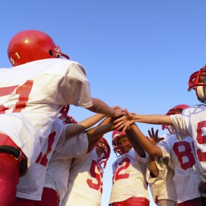
Football is a rough sport, regardless of the protective equipment worn. Injuries go with the territory when it comes to this sport. Because the players are big, the game is fast, and the nature is physical, football injuries are quite common. In the United States alone, there are an estimated 1.2 million football injuries each year, and around 50 percent of these injuries occur during training. … read more
Knee Dislocations

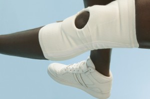
Knee Dislocations | Treatment of Knee Dislocation Knee dislocations are uncommon orthopedic injuries and occur when the bones that form the knee are out of place. A knee dislocation involves damage to multiple ligaments, resulting in severe instability. Knee dislocations also often occur with injuries to the meniscus and the nerves and vessels that surround the knee. A knee dislocation occurs when the femur (thigh bone) and tibia (shin bone) lose contact with each other. In some injuries, the knee cap (patella) also is disrupted. Most knee dislocations are the result of high-energy traumatic injury, such as a motor vehicle accident or severe fall or impact. … read more

Common Sports Shoulder Injuries

Most injuries to the shoulder occur during athletic activities that involve repetitive, excessive overhead motion. These include pitching, weightlifting, tennis, and swimming.  Some sports related shoulder injuries include shoulder instability, shoulder impingement, shoulder separation, shoulder dislocation, rotator cuff tears, acromioclavicular joint sprains, and SLAP lesions.

Shoulder Instability

When the shoulder joint is forced out of normal position, the condition is known as instability. Shoulder instability can result in a dislocation, which is quite painful. Most people who suffer with shoulder instability have pain when raising the arm and the shoulder feels as if it is slipping out of place. If this instability becomes a chronic, recurring problem, the surgeon may find it necessary to perform an arthroscopy. This procedure allows for the orthopedic specialist to look inside the shoulder with a tiny camera to assess the extent of the injury and perform surgery on the area to repair the soft tissues.

Shoulder Impingement

Impingement of the shoulder is caused by excessive rubbing of the tendons against the upper portion of the shoulder blade (the acromion). When there is repeated use of the arm overhead, shoulder impingement is likely. Injections and physiotherapy can improve this syndrome, but surgery is often necessary to remove bony spurs that trap the rotator cuff tendons and worsen the condition.

Shoulder Separation

With a separated shoulder, the acromioclavicular (AC) joint is injured. The AC joint is located where the collarbone (clavicle) meets the upper area of the shoulder blade (acromion). Most of these injuries are the result of a fall where the ligaments attaching to the underside of the clavicle become torn. A separated shoulder causes pain and deformity of the shoulder region. A mild separation involves AC ligament sprain and will appear normal on X-rays. With a more serious injury, the AC ligament could tear, putting the collarbone out of alignment.  Most minor shoulder separations can be treated conservatively with the use of slings, cold packs, and medications for pain.

For more severe injuries, the orthopedic specialist may need to surgically trim back part of the end of the collarbone to prevent rubbing against the acromion. Also, the torn ligaments may need to be addressed by attaching them back to the underside of the collarbone to restore stability of the AC joint therefore allowing motion, flexibility, and strength to return.

Shoulder Dislocation

The shoulder joint is the most mobile joint of the body, making it potentially unstable and at risk for dislocation. Repeated dislocations result in instability and stretching of the shoulder joint, which can lead to poor sports performance and long periods out of the game. In order to reduce a shoulder dislocation, the surgeon will position the ball of the upper arm bone back into the joint socket by means of a closed reduction. For severely dislocated shoulders, however, surgery is often necessary to repair the torn or stretched tissues around the shoulder that normally support the joint.

Rotator Cuff Tears

The rotator cuff is a group of tendons and muscles that allow for movement and stability of the shoulder. The rotator cuff allows an individual to lift the arm and reach overhead. When this structure is injured, pain and weakness occurs. If tearing is significant, the surgeon may need to perform a rotator cuff repair through small incisions (arthroscopy) or by an open method.

Acromioclavicular Joint Sprain

The AC joint is important for athletes who throw and put their arms overhead. It is often sprained from repeated falls and can dislocate easily. When this joint is sprained, there will be pain and loss of normal movement of the shoulder. The orthopedic specialist can provide injections and physiotherapy to improve an AC sprain. Occasionally, with more significant AC sprains, an operation may be necessary to help alleviate persistent, long-term pain.

SLAP Lesions

Tears of the Superior Labral Antero-Posterior (SLAP) region of the shoulder occur with overhead throwing, tackling sports, and heavy lifting. Because the biceps anchors the shoulder, it is easily pulled off the bone by force. The symptoms of this type of injury include pain within the shoulder with lifting and sports. Many complain of a clicking sensation that extends down the upper arm. If the SLAP tears are not serious, the orthopedic specialist will prescribe non-steroidal anti-inflammatory medications and physical therapy. Some tears, however, will require surgical repair via arthroscopy or open techniques. This way, the surgeon can determine the extent of your injury and repair it at the same time.

Common Sports Related Joint Dislocations

Elbow Dislocations

The elbow becomes dislocated when the joint surfaces are separated. With a complete dislocation, the joint surfaces are separated completely. The joint surfaces are only partly separated with a partial dislocation, also called a subluxation. The elbow is a hinge joint as well as a ball-and-socket joint.

Injuries and dislocations to this structure can affect the normal range of motion. Elbow dislocations are either simple or complex. With simple dislocations, there is no major bone injury. With complex dislocations bones and ligaments are injured.

What causes an elbow dislocation?

The most common reason an elbow is dislocated is when a person falls onto an outstretched hand. There is considerable force sent to the elbow when the hand hits the ground. Most often there is a turning motion in this force and this can rotate and put the elbow out of its socket. Elbow dislocations often occur during car accidents, when the passenger reaches forward in an attempt to cushion the impact.

What are the symptoms of an elbow dislocation?

With a complete elbow dislocation, there is considerable pain and obvious deformity. With a partial dislocation, the joint appears fairly normal. However, there usually is pain and bruising on the inside or outside area where ligaments have been torn or stretched. If the ligaments never heal, partial dislocations can recur.

How is an elbow dislocation treated?

Elbow dislocations require immediate treatment and are emergency injuries. The goal of treatment is to return the joint to its normal alignment and restore function of the arm. The orthopedic specialist can realign the elbow joint using a reduction maneuver. Once the joint is reduced, an immobilizer is required for two to three weeks.

Physical therapy is often required to restore full range of motion. For complex dislocations, surgery is often required to restore bone alignment and repair ligaments. After the operation, the elbow will be protected with an external hinge device to prevent further dislocation.

Shoulder Dislocations

The shoulder is susceptible to dislocation because it can turn in many directions. A partial dislocation of the shoulder is also called a subluxation. This means the head of the upper arm bone is partially out of the socket. With a complete dislocation, the structure is all the way out of the socket.

What are the symptoms of a dislocated shoulder?

Most shoulder dislocations lead to swelling, bruising, numbness, and weakness. Many times, a dislocation results in torn ligaments or tendons or damaged nerves. It is possible for the shoulder to dislocate downward, forward, or backward.

What is the treatment for a dislocated shoulder?

Many times the shoulder can be reduced. This is where the surgeon places the ball of the upper arm bone back into the socket. After the reduction, the orthopedic specialist will immobilize the shoulder in a sling or device for a few weeks. Ice is recommended 3 to 4 times a day to reduce pain and swelling. Also, the doctor will prescribe rehabilitation exercises for you to help restore the shoulder’s range of motion and strengthen the muscles. If recurrent dislocations occur, surgery is sometimes necessary.

Hip Dislocations

The hip becomes dislocated when the head of the thighbone slips out of the socket in the hip bone. For around 90% of people, the thighbone is pushed out of position in a backwards direction. This is known as posterior dislocation. Rarely, the thighbone slips out of socket in a forward direction. This is called an anterior dislocation.

What causes hip dislocations?

The hip joint is formed of a ball-and-socket construction. The ball is the head of the thighbone (femur) and it fits into a cup-shaped socket in the pelvis. This joint has a great deal of stability and moves freely. The most common reason for a dislocated hip is motor vehicle accidents. Falls can also result in dislocated hips.

How is a dislocated hip treated?

With a dislocated hip, the orthopedic specialist must administer an anesthetic or sedative and then position the bones back into proper position. This is called a reduction and surgery is often required. The surgeon will make an incision over the hip region in order to reduce the hip.

It takes up to three months for a hip to heal once it has been dislocated. Many times it is necessary for the patient to be in traction for a short period of time. Special controlled exercises are prescribed to help the hip regain strength and function.