Broken Arm

A broken arm is a common injury. Counting all fractures, about one in every 20 involve the upper arm bone (humerus). Children are more likely to break the lower arm bones (radius and ulna). Falling on an outstretched hand or being in a car crash or some other type of accident is usually the cause of a broken arm.

Most people know right away if their arm broke, because there may be a snap or a loud cracking sound. The broken arm may appear deformed and be swollen, bruised and bleeding. A person with a broken arm usually has:

  • Extreme pain at the site of the injury.
  • Pain increased by any movement.
  • Loss of normal use of the arm.

First aid
First make sure the injured person is out of the way of further harm. Is he or she breathing normally? Is there a good pulse? Call 911 if there is serious bleeding, reason to suspect multiple broken bones or other injuries. To slow bleeding and reduce swelling, elevate the injured arm above the level of the person’s heart.

If a broken bone sticks out from the skin (open fracture), do not try to push it back in. Use a clean, dry cloth or bandage to cover it until medical help arrives.

It is important that the injured person not try to use the broken arm. Moving a broken arm would also cause more damage to blood vessels, nerves and other tissues. To immobilize a broken arm:

  • Make a temporary splint. Immobilize the joints above and below the site of the injury. You can use wood or rolled up magazines, making sure both ends of the splint extend far beyond the injured region. You can use cloth, belts or tape to fasten the splint. Avoid any constriction of the arm with the supporting strap.
  • Make a sling. This stabilizes the injury and supports the splint. A broken arm sling can be as simple as a loop of cloth supported from the neck. Take the injured person to a doctor right away.

Doctor’s treatment

Exam: Tell the doctor exactly what happened. He or she will physically examine the broken arm and check for other injuries, such as nerve damage. The doctor may want to see if the patient can flex and extend the wrist and fingers. Sometimes the doctor may use X-rays or other diagnostic imaging tools to see the bones of both the injured and uninjured arms.

If the patient is a child, the long bones of the arm are probably still growing. So the doctor will look carefully for any damage to growth plates.

Reduction: The doctor may need to move pieces of bone back into their correct positions (a process called reduction). Depending upon the severity of injury, the patient may or may not need anesthesia. Those with more serious fractures may require surgery.

Immobilization: With the broken bone back in place, the doctor immobilizes the arm. Most patients get a cast or splint. The doctor tells the patient how long to wear the cast or splint, and removes it at the right time.

Rehabilitation
It may take from several weeks to several months for the broken arm to heal completely. Rehabilitation involves gradually increasing activities to restore muscle strength, joint motion and flexibility. The patient’s cooperation is essential to the rehabilitation process. The patient must complete range of motion, strengthening and other exercises prescribed by the doctor.

Rehabilitation lasts until tissues perform their functions normally. After rehabilitation, the doctor may want to see the arm again to make sure healing is complete.

Biceps Tendinitis

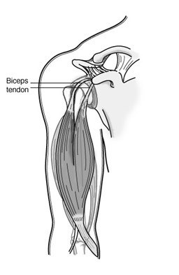
The biceps muscle, in the front of the upper arm, helps stabilize the upper arm bone (humerus) in the shoulder socket. It also helps accelerate and decelerate the arm during overhead movement in activities like tennis or pitching. Strong, cord-like structures called tendons connect one end of the biceps muscle to the shoulder in two places.

At the other end of the muscle, tendons connect the biceps muscle to the smaller bone (radius) in the lower arm. If the tendons become inflamed or irritated, the condition is called tendinitis.

Signs and symptoms
Injuries to the biceps tendons are commonly caused by repetitive overhead activity. Symptoms include:

  • Pain when the arm is overhead or bent.
  • Localized tenderness as the tendon passes over the groove in the upper arm bone.
  • Occasionally, a snapping sound or sensation in the shoulder area.

cons1_139_209Diagnosis and treatment
During the physical examination, the doctor will assess the shoulder area for range of motion, tenderness and signs of shoulder instability. He or she may ask you to raise or rotate the arm.

X-rays may be requested to uncover associated conditions that might cause irritation. The doctor may also request an MRI that can show any damage to the tendons. Overuse, aging and stress can cause the tendon to deteriorate, even if there is no inflammation present.

Initial treatment is conservative. The first step is to rest the arm and shoulder. Switch to another sport or activity for awhile. Ice applications and nonsteroidal anti-inflammatory medications such as ibuprofen can help reduce inflammation.

Your physician can also recommend stretching and progressive strengthening exercises to build muscle endurance and restore range of motion. Then you can gradually return to overhead activity.

Surgical options
If the pain results from shoulder instability or from pressure on the tendon from the shoulder bones, your orthopaedist may recommend arthroscopic surgery. Using fiber optic technology and miniature instruments inserted through a small incision, the surgeon can examine the shoulder joint and anchor the tendon properly.

After surgery, your orthopaedist will prescribe a rehabilitation program that includes stretching and strengthening exercises. Early movement is important, but you should wait for your physician’s approval before doing any heavy lifting or returning to sports.

Wrist Sprains

When you fall forward, your natural response is to put your hands out in front to catch yourself. As you land on your palm, your wrist bends backwards, which can stretch and perhaps tear the ligaments that connect bone to bone within the joint. The resulting injury is a sprain.

Signs and symptoms
Signs and symptoms of a wrist sprain may vary in intensity and location, depending on the degree of injury. Indications of a wrist sprain include:

  • Swelling in the wrist
  • Pain at the time of the injury
  • Persistent pain when you move the wrist
  • Bruising or discoloration of the skin
  • Tenderness at the injury site
  • A feeling of popping or tearing inside the wrist
  • A warm or feverish feeling to the skin

Degrees of injury
Wrist sprains are graded, depending on the degree of injury to the tissues.

  • Grade 1 or mild sprains occur when the ligaments are stretched, but not torn.
  • Grade 2 or moderate sprains occur when some of the ligaments are torn. Grade 2 sprains may involve some loss of function.
  • Grade 3 or severe sprains occur when there is a complete tear all the way through the ligament. These are significant injuries that require medical or surgical care to ensure recovery. As the ligament tears away from the bone, it may also take a small chip of bone with it, resulting in an avulsion fracture.

If you have any doubts about the severity of your injury, arrange to have a doctor examine your wrist. Sometimes, a sprain can have only mild swelling but still be serious enough to require surgery. To avoid problems later, proper diagnosis and treatment is important.

Diagnosing a wrist sprain
Your doctor will review your symptoms and ask how the injury happened. He or she will also ask about any numbness in your hand as well as about your medical and orthopaedic history, especially any previous injuries to your hand or wrist. The physical exam will cover your entire arm and hand to ensure that there are no other injuries. Tenderness in certain areas may suggest a broken bone.

If a severe sprain is suspected, your doctor will likely order X-rays. In some cases, a magnetic resonance image (MRI) or computed tomography (CT) scan may also be ordered.

Treating a wrist sprain
Mild wrist sprains can be usually be treated at home with the RICE protocol.

  • Rest the joint for at least 48 hours.
  • Ice the injury to reduce swelling. Do not apply ice directly to the skin. Use an ice pack or wrap a towel around the ice or a package of frozen vegetables. Apply ice for about 20 minutes at a time.
  • Compress the swelling with an elastic bandage.
  • Elevate the injury above the level of your heart.

You may also wish to take a pain reliever such as aspirin or ibuprofen. If pain and swelling persist for more than 48 hours, however, see a doctor. Moderate sprains may need to be immobilized with a wrist splint for 7 to 10 days. Severe sprains may require surgery to repair the fully torn ligament. Afterwards, you will need a period of rehabilitation and exercises to strengthen the wrist and restore motion. A wrist sprain can take anywhere from 2 to 10 weeks to heal completely, depending on the severity of the sprain.

Prevention
Because wrist sprains usually result from a fall, be careful if you must walk in wet or slippery conditions. Wrist sprains also occur during sports such as skating, skateboarding, and skiing. You can help prevent sports-related wrist sprains by wearing wrist guard splints or protective tape to support the wrist and prevent it from bending too far backwards. If you ski, try to remember to drop your poles during a fall to prevent wrist sprains.

Wrist Joint Replacement (Arthroplasty)

Most people are familiar with joint replacement surgery for the hip, knee, and shoulder joints. Joint replacement surgery in the wrist is less common but can be an option if you have painful arthritis that does not respond to other treatments.

Anatomy of the wrist
The wrist is a more complicated joint than the hip or the knee. At the base of the hand are two rows of bones, with four bones in each row. These are called the carpals. The long thin bones of the hand radiate out from one row of carpals toward the fingers and thumb. The two bones of the lower arm (radius and ulna) form a joint with the other row of carpals. All the bone ends are covered with a slick, elastic tissue called cartilage, which enables the bones to move smoothly against each other. However, if the cartilage is worn away or damaged by injury, infection or disease, the bones will rub against each other causing pain. During any total joint replacement, the worn-out bone ends are removed and replaced by an artificial joint (prosthesis).

Reasons for wrist replacement surgery
The typical candidate for wrist replacement surgery has severe arthritis but does not need to use the wrist to meet heavy demands in daily use. The primary reasons for wrist replacement surgery are to relieve pain and to maintain function in the wrist and hand.

  • Osteoarthritis, the most common form of arthritis, results from a gradual wearing away of the cartilage covering on bones.
  • Rheumatoid arthritis is a chronic inflammatory disease of the joints that results in pain, stiffness and swelling. Rheumatoid arthritis usually affects several joints on both the right and left sides of the body.

Both forms of arthritis may affect the strength of your fingers and hand, making it difficult for you to grip or pinch. In some cases, fusing the wrist bones together will reduce or eliminate pain and improve grip strength. However, if the bones are fused together, you will not be able to bend the wrist. Wrist replacement surgery may enable you to retain or recover wrist movements and improve your ability to perform daily living activities, especially if you also have arthritis in the elbow and shoulder.

Implant design
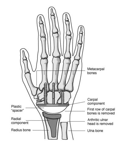
Wrist implants are made of the same kind of materials used for hip and knee joint replacements. There are several different designs. Most have two components and are made of metal; a high quality plastic called polyethylene is used as a spacer between the two components. Newer implant designs try to replicate the anatomy of the wrist.

The piece that attaches to the lower arm (radius bone component) fits into the bone of the lower arm. The top of this component has a curve that matches to the wrist part. The piece that attaches to the hand (carpal component) may have one long stem and one or two shorter stems that insert into the hand bones, or use small screws. The surface of this component is flat. The plastic spacer comes in different sizes so it can be matched to your hand. It is normally flat on one side and rounded on the other. This design enables it to fit into the carpal component while it rocks on the radial component, creating a more natural wrist motion.

Implant insertion
A wrist joint replacement can be done as an outpatient procedure, unlike a hip or knee replacement. Wrist replacement surgery is often combined with other procedures to correct deformities or disorders in the tendons, nerves, and small joints of the fingers and thumb.

The incision is made on the back of the wrist. The damaged ends of the lower arm bones are removed and the first row of carpal bones may also be removed. The radial component of the prosthesis is inserted into the center of the radius bone on the outside of the lower arm. It is held in place with bone cement. Depending on the component design, the carpal component is then inserted into the center hand bone (third metacarpal) or screwed into the remaining row of carpal bones. Bone cement may be used to hold the component in place. The carpal bones may be linked or fused together to better secure this component.

An appropriately sized spacer is used between the metal components.

After your surgery
You will have to wear a cast for the first several weeks. When the cast is removed, you will have to wear a protective splint for the next six to eight weeks. Although pain relief is immediate, you will have to do gradual exercises for several weeks to restore movement and, eventually, to increase power and endurance. Wrist arthroplasty can improve motion to about 50 percent of normal.

The physical demands that you place on the wrist prosthesis will have an effect on how long the implant lasts. You will not be able to use a hammer often or pneumatic tools. You may only be able to lift a limited amount of weight. A fall on the outstretched hand may break the prosthesis, just as it might fracture a normal wrist. So you will want to avoid activities such as roller sports that could result in a fall.

Although there have been significant advances in wrist prostheses, the implant may loosen or fail due to wear or deformation. In these cases, additional surgery may be necessary. On average, a wrist replacement can be expected to last 10 to 15 years with careful use. As with all implants, long-term follow-up is advised. Generally, you should see your hand surgeon every year or two years so that x-rays can be taken and used to identify any developing conditions or problems.

cons1_161_347

Wrist Arthroscopy

Arthroscopy (ar-THROS-ka-pea) is an outpatient surgical procedure used by orthopaedic surgeons to diagnose and treat problems inside a joint. The surgeon makes small incisions, less than half an inch long, and inserts a pencil-sized instrument called an arthroscope. The arthroscope contains a small lens, a miniature camera and a lighting system.

This projects three-dimensional images of the joint on a television monitor, enabling the surgeon to look directly inside the joint and identify the trouble. Tiny probes, forceps, knives and shavers can then be used to correct many problems at the same time.

Arthroscopy has been used for several years to treat conditions in large joints such as the knee and the shoulder. As cameras and instruments became smaller and more refined, arthroscopy was applied to other joints, including the wrist. The wrist is a complex joint, with eight small bones and many connecting ligaments. Arthroscopy enables the surgeon to see the anatomic parts and their movements and to make a more accurate diagnosis.

Diagnostic arthroscopy
Diagnostic arthroscopy may be used if the cause of your wrist pain cannot be identified or if wrist pain continues for several months despite nonsurgical treatment.

Before surgery, your doctor will do:

  • A physical examination that focuses on your hand and wrist. Your doctor will also ask about your medical history.
  • Provocative tests that involve moving your hand in ways that reproduce the pain.
  • Imaging studies, such as X-rays of your hand and wrist. In some cases, additional imaging studies may be needed. These can include an MRI (magnetic resonance image) or an arthrogram, in which a contrast agent is injected into the joint before the image is taken.

Usually, regional anesthesia is used during arthroscopic surgery. This numbs your arm and hand. You may also be given a sedative to further relax you and enable you to doze through the surgery. Two or more small incisions (portals) are made on the back of the wrist, through which the arthroscope and instruments are inserted. After the surgery, the incisions are closed with a small stitch and a dressing is applied. Sometimes a splint may also be used.

Arthroscopic surgical treatment

Several conditions can be treated using arthroscopic surgery, including chronic wrist pain, wrist fractures, ganglion cysts and tears in the ligaments or the triangular fibrocartilage complex (TFCC). Wrist arthroscopy may also be used to smooth the bone surfaces and remove inflamed tissue. Arthroscopy can also be used to surgically treat carpal tunnel syndrome, but in that diagnosis, the arthroscope is not inserted into the wrist joint itself.

  • Chronic wrist pain: Arthroscopic exploratory surgery may be used to diagnose the cause of chronic wrist pain when other tests are inconclusive. Often, there may be areas of inflammation, cartilage damage, or other findings after a wrist injury. In some cases, after the diagnosis is made, the condition can be treated arthroscopically as well.
  • Wrist fractures: Doctors can remove small fragments and fracture debris, align the broken pieces of bone, and stabilize them by using pins, wires, or screws.
  • Ganglion cysts: These cysts commonly grow from a stalk between two of the wrist bones. During an arthroscopic procedure, the surgeon can remove the stalk, which may also reduce recurrence of the cysts.
  • Ligament/TFCC tears: Ligaments are fibrous bands of connective tissue that link or hinge bones. They provide stability and support to the joints. The TFCC is a cushioning structure within the wrist. A fall on an outstretched hand can tear ligaments, the TFCC or both, resulting in pain with movement or a clicking sensation. During arthroscopic surgery, the surgeon can trim or repair the tears.
  • Carpal tunnel release: Carpal tunnel syndrome is characterized by numbness or tingling in the hand, and sometimes with pain up the arm. It is caused by pressure on a nerve that passes through the carpal tunnel, which is formed by the wrist bones and a thick tissue roof. Pressure can build up within the tunnel for many reasons, including irritation and swelling of the tissue (synovium) that covers the tendons. If the syndrome does not respond to conservative treatment, your doctor may recommend surgery to cut the ligament roof and enlarge the tunnel, thus reducing pressure on the nerve and relieving symptoms. This can sometimes be done using an arthroscope.

After surgery, you will need to keep your wrist elevated for the first two or three days and keep your bandage clean and dry. You can ice your wrist to help keep swelling down. Your doctor and/or your physical therapist will teach you exercises to help maintain motion and rebuild your strength. Analgesic medications will help relieve any postoperative pain, which is usually mild.

Complications
Complications during or after arthroscopic wrist surgery are unusual, but may include infection, nerve injuries, excessive swelling or bleeding, scarring or tendon tearing. An experienced surgeon, particularly one who specializes in treating the hand, can reduce the likelihood of complications.

Summary
Arthroscopic surgery is a valuable diagnostic and treatment tool. It is minimally invasive, and patients generally experience fewer problems and a more rapid recovery than with open surgery. Because it is an outpatient procedure, most patients are home several hours after surgery.