Trigger Finger

Description
A trigger finger is a common problem that causes pain and catching. Tendons that help bend the fingers and thumb slide through a snug tunnel. Irritation as the tendons slip into the tunnel can cause the opening of the tunnel to become smaller, or the tendon to thicken so that it can’t easily pass through the tunnel.

As you try to straighten the finger, the tendon becomes momentarily stuck at the mouth of the tunnel then pops as the tendon slips past the tight area. This can cause pain and catching as you try to move the finger. Your doctor can diagnose the problem by talking with you and examining your hand. No X-rays or other testing are usually needed.

Risk Factors/Prevention
It isn’t usually known what causes the problem. Trigger fingers are more common in women than men. They occur most frequently in people who are between the ages of 40 to 60 years. Trigger fingers are more common in people with certain medical problems such as diabetes and rheumatoid arthritis.

Symptoms
Symptoms usually start without any injury. You may feel pain in the palm, sometimes along with swelling or a small lump. You may feel the catching or popping sensation in the finger or thumb joints. Stiffness and catching tend to be worse after inactivity, such as when you wake in the morning, but will often loosen up with movement. When it breaks free, it may feel like the finger joint is dislocating. In severe cases the finger may become stuck in a bent position so that it can’t be straightened even with the help of other fingers. One or more fingers can be involved.

Treatment Options
If your symptoms are mild, simply resting the finger may be enough to relieve the problem. Over-the-counter medications such as Tylenol® or Advil® can be used for pain. Splints are sometimes used to rest the finger. Your doctor may recommend an injection that helps in most cases, but sometimes the improvement is temporary. More than one injection may be needed. Injections are less likely to bring about permanent relief when the triggering has been present for a long time, or if you have associated medical problems like diabetes. If the problem remains troubling, you may want to consider surgery.

cons1_385_168

Treatment Options: Surgical
A trigger finger isn’t a dangerous condition, so the decision whether to proceed with surgery is a personal one based on the severity of your symptoms. If the finger is stuck in a bent position, surgery may be recommended to prevent permanent stiffness. Surgery widens the opening of the tunnel so the tendon can slide through more easily. This is usually done through a small incision in the palm as an outpatient surgery.

Sometimes the tunnel can be safely opened with the tip of a needle in your doctor’s office. You can move the fingers immediately after surgery. Elevating the hand after surgery can help minimize swelling and pain. Some soreness in the palm is common but recovery is usually complete in a few weeks. If the finger was quite stiff before surgery, a therapist may help you to loosen up the finger.

Thumb Fractures

A broken thumb is a serious problem because it affects your ability to grasp items in your hand and can increase the risk of arthritis later in life.

Anatomy
The thumb has two finger bones connected to a hand bone. The first finger bone (distal phalange) extends from the tip of the thumb to the knuckle. The second finger bone (proximal phalange) extends from the knuckle to the webbing between the thumb and the first finger. There, it connects with the hand bone (first metacarpal), which extends from the webbing down to the wrist.
Although a break can occur in any of these bones, the most serious breaks happen near the joints, particularly at the base of the thumb near the wrist.

These fractures have specific names, depending on the type of break. The Bennett and Rolando fractures are breaks at the base of the thumb, involving the joint between the thumb metacarpal and a specific wrist bone. Fractures that involve the joints are always more difficult to treat and are at increased risk of an unfavorable outcome.

How it happens
Thumb fractures are usually caused by direct stress, such as when you fall or when a ball catches and pulls the thumb back. Some fractures may be caused indirectly, from twisting or muscle contractions. People who play contact sports such as wrestling, hockey or football; skiers; and people with a history of bone disease or calcium deficiency are especially at risk.
The risk of a thumb fracture can be lessened by using protective taping, padding or other equipment and by developing strength in your hands through exercise and proper nutrition.

Symptoms of a fracture

  • Severe pain at the fracture site
  • Swelling
  • Limited or no ability to move the thumb
  • Extreme tenderness
  • A misshapen or deformed look to the thumb
  • Numbness or coldness in the thumb

Treatment options
See a doctor as soon as possible. Without immediate treatment, the joint will be unstable and you will be unable to grip or pinch properly. Continued swelling may make it more difficult to align the bones properly. Delayed treatment will make the fracture much more difficult to treat and can lead to a poor outcome. Use a padded splint to prevent the bone from moving further out of alignment is encouraged prior to definitive treatment.

The physician will examine the injury, take your medical history and have X-rays taken of the injury. You may need surgery, depending on the location of the fracture and the amount of movement between the broken pieces of bone. If there is little movement (displacement) or if the break is located in the middle (shaft) of the bone, an orthopaedist may be able to use a specially designed cast (spica cast) to hold the bone fragments in place. You will have to wear the cast for at least four to six weeks, and your doctor may request regular X-rays to ensure that the bone hasn’t slipped out of alignment.

Often, Bennett and other more severe fractures of the thumb require surgical treatment. The hand surgeon may use one of several operative fixation techniques to restore boney anatomy and hold the bones in place while they heal. These techniques include the use of wire, pins, plates and screws as internal fixation. Another technique uses pins in bone that exit the skin and are attached to an external fixation device You will likely be required to wear a cast or splint for two to six weeks after surgery. When the cast is removed, your physician may recommend hand therapy to restore movement. It can take three months or more to regain full use of the hand, depending on the severity of the injury.

Complications and outcomes
If the bones remain in a stable position, thumb fractures generally heal well. There is a possibility of infection and tenderness around the surgical site and around the pins, if external fixation is used. As with all joint injuries, there is an increased tendency to develop arthritis, which can limit motion and reduce strength in the thumb.

To help restore motion, soak your hand in warm water and move your thumb in a circular direction. Try to touch your little finger with your thumb. To help restore strength, get a spring-type hand squeezer and use it regularly.

Sprained Thumb

When people start to fall, they usually extend their arm to reduce the force of the impact when they hit the ground. If you try to break your fall on the palm of your hand or take a spill on the slopes with your hand strapped to a ski pole, your thumb may be injured. The main ligament (ulnar collateral), which supports pinch and grasp activities, may be torn (sprained). The ligament helps your hand to function properly, acting like a hinge to keep your thumb joint (metacarpophalangeal) stable.

When you have a sprained thumb, you lose some or all of your ability to grasp items between your thumb and index finger. It may or may not hurt right away. Other signs include bruising, tenderness and swelling. To make sure your injury won’t cause long-term weakness, pain and instability, see your doctor for evaluation and treatment.

Partial and complete tears
Your thumb ligament may have a partial or complete tear. Your doctor will probably move your thumb joint to test its stability and take X-rays to make sure you don’t also have a broken bone. You may also get a stress X-ray showing what the joint looks like when your ligament is tested. If it hurts to do this, a shot of local anesthetic may help. Your doctor will probably also X-ray your uninjured thumb to compare it.

If you have a partial tear, your doctor will probably immobilize your thumb joint with a splint or other bandage until it heals. You wear the splint for about three weeks straight, then start taking it off to do flexion and extension exercises with your thumb. Put the splint back on for protection when you are not doing the exercises. Keep doing this for another two or three weeks until your thumb’s swelling and tenderness are gone. You may also put ice on your injury twice a day for 2-3 days after the fall.

If your thumb ligament is completely torn, you may need surgery. Fragments of bone that sometimes get pulled away when your ligament tears may be removed or put back in their correct positions. After surgery, you’ll probably need to wear a short-arm cast or a splint to protect your thumb ligament for six to eight weeks while it heals.

Scaphoid (Wrist Bone) Fracture

cons1_383_253Description
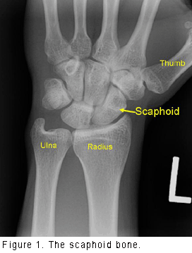
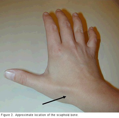
The scaphoid is one of the small bones in the wrist (carpal bones), and the one that is most likely to break. The scaphoid is located on the thumb side of the wrist, in the area where the wrist bends (see Figure 1).When you hold your thumb in a “hitch hiking” position, the scaphoid is at the base of the depression made by your thumb tendons. Pain or tenderness in this area can be a sign that the scaphoid is injured (see Figure 2).

A fracture of the scaphoid usually happens when you fall on an outstretched hand, with your weight landing on the palm. The end of the forearm bone (radius) may also break in this type of fall, depending on the position of the hand when you land. Pain and swelling in the wrist will usually cause a person with a scaphoid fracture to see a doctor. If the wrist is not deformed, however, you might not know that you have a broken bone–people sometimes think they have just sprained the wrist.

It is important to see your doctor if you have pain on the thumb side of the wrist that starts after a fall or accident and does not go away within a few days. The doctor will probably order X-rays to look at the bone.

cons2_383_253Sometimes, a broken scaphoid does not show up on an X-ray right away. When this is the case, the doctor may put your wrist in a splint and wait to take a new X-ray in a week or two, when the fracture may become visible. Keep the splint on and do not do any heavy lifting during the waiting time.

Your doctor might also recommend an MRI (magnetic resonance image) to see the bones and soft tissues. An MRI can sometimes show a fracture of the scaphoid before it can be seen on an X-ray.

Risk Factors/Prevention
Fractures of the scaphoid occur in people of all ages, including children. The injury often happens during sports activities or a motor vehicle accident. Men aged 20 to 30 years are most often affected.

There are no specific risks or diseases that increase the chance of having a scaphoid fracture. Some studies have shown that use of wrist guards during activities like inline skating and snowboarding can decrease the chance of breaking a bone around the wrist.

Symptoms
Scaphoid fractures usually cause pain in the base of the thumb, with swelling in the same area. You may have severe pain when you move your thumb or wrist or grip anything. Other times, the pain is not so bad, and you may mistakenly think that you just sprained your wrist.

Treatment Options
Treatment of scaphoid fractures depends on the location of the break in the bone. Sometimes, the scaphoid is broken at the end near the thumb. This part of the bone has a good blood supply. Scaphoid fractures in this location usually heal in a matter of weeks with proper protection. The time frame to healing is best determined by X-rays or other imaging studies such as a CT scan, which confirm that the bone has healed. Most fractures here heal well when they are placed in a cast. The cast will usually be below the elbow. It may or may not include the thumb.

More commonly, the scaphoid is broken in the middle (waist) or at the part closer to the forearm (proximal pole). This part of the scaphoid does not have a very good blood supply, so it is more difficult to heal. If your orthopaedist recommends that your scaphoid fracture be treated in a cast, it will probably include the thumb. Sometimes the cast will extend to above your elbow and may also include your index finger.

Treatment Options: Surgical
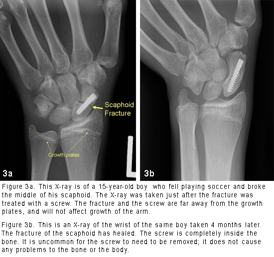
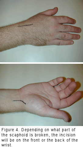
Often, when the scaphoid is broken at the waist or proximal pole, an orthopaedist will recommend surgery using a screw or wire to stabilize the scaphoid from the inside while the bone heals (see Figure 3). Depending on what part of the scaphoid is broken, the incision will be on the front or the back of the wrist (see Figure 4). Sometimes the screw or wire is placed with just a small incision.

Other times a bigger incision is needed to ensure that the pieces of the scaphoid are put back together well. Usually your wrist will be placed in a cast after the surgery, sometimes for a few months. Even with surgery, fractures in this area can take a long time to heal, and it is possible that a nonunion or avascular necrosis will occur.

cons1_386_253

If the scaphoid does not heal, either with or without initial surgery, your orthopaedist may recommend an operation to add bone graft to the scaphoid to help it heal. The surgeon may order an MRI or CT (computed tomography) scan to show more detail about position of the broken pieces of the scaphoid, to help plan the operation. Many kinds of bone graft can be used–your surgeon will discuss which one is best for you. Bone graft may be taken from a bone in the forearm or the pelvis or it may be manufactured. Sometimes a special kind of bone graft with its own blood supply (vascularized graft) may be used.

cons2_386_253

Nonunions, Avascular Necrosis and Arthritis
A bone that fails to heal is called a nonunion; this happens more often in scaphoid fractures than in fractures of many other bones. To heal properly, the bones need blood to carry oxygen and nutrients to the site of the fracture. The scaphoid bone has a poor blood supply. When the scaphoid is broken, especially when broken parts have moved apart (displaced), the blood supply may be disrupted.

Sometimes, the blood supply to one of the broken pieces is so poor that the piece does not get enough nutrients and the cells in the piece die. This is called avascular necrosis. Both nonunion and avascular necrosis of the scaphoid can lead to arthritis of the wrist.

Symptoms of arthritis in the wrist that results from scaphoid nonunion or avascular necrosis include:

  • Aching in the wrist
  • Decreased range of motion of the wrist
  • Pain with activities such as lifting or gripping

If X-rays show arthritis in the wrist because of an old break in the scaphoid, treatment focuses on improving the symptoms of arthritis. At first, this may include taking anti-inflammatory medicine (such as ibuprofen) and wearing a splint when the wrist is painful. Sometimes the doctor may give you a steroid injection into the wrist to help to lessen wrist pain. If this does not work, the doctor may recommend surgery. Many types of operations can be performed for wrist arthritis. Your surgeon will discuss the risks and benefits of the surgery that will be most helpful to you.

Rehabilitation
You will have to wear a cast or splint while the scaphoid fracture is healing, perhaps for as long as six months. Your doctor will give you specific limitations.

Most commonly, you should:

  • Avoid heavy lifting, carrying, pushing, pulling or throwing with the injured arm
  • Not participate in contact sports
  • Not climb ladders or trees
  • Avoid activities with a risk of falling onto your hand (for example, inline skating, jumping on a trampoline.

Some people have stiffness in the wrist after scaphoid fractures; this is more common when a cast was needed for a long time and when surgery was done through a long incision. Your orthopaedist may recommend hand therapy to help regain the motion and strength in your wrist. Even with therapy, some people do not recover the same motion and strength in their wrist that they had before the injury.

Research on the Horizon/What’s New?
Some orthopaedists are placing a screw in the scaphoid through a very small incision–about one-fourth to one-half inch. This may decrease the chances of long-term stiffness in the wrist by decreasing the time that a full-time cast needs to be worn. This is done using a special X-ray machine during surgery to help guide the placement of the screw. Some surgeons use this method even when the broken pieces of the scaphoid are out of place. By looking directly at the scaphoid with a tiny camera (an arthroscope), the surgeon can tell when the broken pieces are lined up.

There is a lot of research being done about the best way to treat fractures of the scaphoid that are not healing well. Some of this research looks at different types of bone graft, including vascularized grafts. New types of manufactured bone graft with special bone cell components to increase healing are being tried to help bones that are not healing.

Restoring Hand Function after Spinal Cord Injury

An injury to the upper part of the spinal cord can leave an individual with little or no sensation or movement in both the arms and the legs, a condition called tetraplegia (tet-rah-PLEE-gee-ah). A surgical technique called a tendon transfer can help restore function to arms and hands by giving working muscles different jobs.

This can greatly enhance the quality of life for people with tetraplegia by enabling them to do many more tasks for themselves. The types of tendon transfer surgeries that can be performed depend on where the spinal cord injury occurred and which muscles are affected.

How it works
Tendons are the strong cords that connect muscle to bone. When a tendon crosses a joint, it helps transmit muscle action into joint movement. A tendon transfer repositions the tendons of a working muscle so that they take over the functions of a paralyzed muscle. This enables the working muscle to do what the paralyzed muscle can no longer do.

For example, in the upper arm, the triceps muscle is used to straighten the elbow. The larger deltoid muscle pulls the arm backwards and forwards away from the body. If the triceps muscle is paralyzed but the deltoid is still functional, surgeons can split the deltoid muscle and graft a portion of it to the triceps. This restores elbow function without greatly diminishing shoulder function.

Tendon transfers can help restore three critical capabilities necessary for self-care and increased independence:

  • the ability to straighten (extend) and bend (flex) the elbow
  • the ability to bend and straighten the wrist
  • the ability to grip with the fingers and hand

Planning for a tendon transfer
Usually, a tendon transfer is not scheduled until about a year after injury. During the first months after the injury, rehabilitation focuses on retaining passive range of motion. These exercises help prevent shoulder stiffness and pain. As time progresses, strength and range of motion (both active and passive) must be evaluated frequently. Severe muscle contractures or muscle spasms may necessitate another type of surgery rather than a tendon transfer. Usually tendon transfer surgery is scheduled only after there is no more progress in function.

Before surgery is scheduled, several assessments must be made, including:

  • identifying which muscles still work and measuring how well they work to determine whether they can be used in the transfer
  • assessing the individual’s abilities to see which functions need to be restored
  • matching available muscles with functional requirements
  • determining if an additional procedure such as a joint fusion or electrical stimulation implant is needed to restore function
  • verifying that the individual has a strong support system that can provide the care needed during rehabilitation after surgery
  • assessing the individual’s motivation and commitment to the process
  • determining which surgeries should be performed, when, and in what order

Elbow Extension
The ability to bend and straighten the elbow adds greatly to a person’s independence, so this is often the first surgery to be performed. In most cases, a portion of the deltoid muscle in the shoulder is used to provide elbow extension. The back (posterior) portion of the deltoid is brought down toward the elbow. Because the deltoid portion is not long enough to reach the attachment point in the lower arm, a graft is taken from an upper leg muscle (fascia lata) to provide the necessary length.

In some cases, the biceps muscle in the upper arm is used instead of the deltoid muscle. After the surgery, the arm is immobilized in a slightly bent position for up to four weeks. When the cast is removed, a hinged brace is used to allow a gradual stretching and strengthening of the muscles. Initially, the brace is worn night and day, but as the individual gains the ability to fully extend the arm, the brace is worn only at night.

Tendon transfer to achieve elbow extension is done on one arm at a time because the arm is totally immobilized during rehabilitation. This means that the person becomes even more dependent on others for the simple activities of daily living. However, the results are impressive. It can eliminate the need for many adaptive devices and enable the person with tetraplegia to propel a wheelchair, to move independently from bed to chair, to shift weight within a chair or bed and to reach up and outwards.

Key Pinch
Restoring key pinch enables the individual to grip items between the thumb and the hand. This greatly enhances the ability of the patient with tetraplegia to accomplish activities of daily living, such as writing or feeding themselves. In this surgery, one of the forearm muscles (brachioradialis) is grafted to the tendons that move the wrist and thumb. The surgeon may also stiffen the joint so that when the wrist is extended, the grip forms automatically.

These two surgeries significantly improve hand and arm function in many patients, providing them with much greater independence.

Electronic Implants
A relatively recent advance uses an electronic implant, similar to a pacemaker, to stimulate muscles. Tiny electrodes are attached to the functioning muscles of the arm and hand. The electrodes are connected to a control device implanted in the front of the chest. An external unit delivers the signals to initiate grasp and key pinch. This is an option when tendon transfers cannot be used.

Outcomes
Because tendon transfers use the patient’s own tissues, the risk of infection is lessened. However, the risk of developing a latex allergy is increased so precautions should be taken. The length of the surgery (approximately six hours) also increases the risk of postoperative respiratory problems.

In general, the results using tendon transfer surgery to restore arm and hand function after spinal cord injury are good. People with tetraplegia can often benefit from the increased self-confidence and independence they gain after tendon transfer surgery.