Fingertip Injuries/Amputations

Your child accidentally slams a fingertip in a car door. Or you cut off the end of your finger while chopping vegetables. Or you lose the tops of several fingers trying to clear debris from a lawnmower or snowblower.

Fingertip injuries are very common. Accidents can happen at work, play and in the home, crushing, tearing (lacerating) or cutting off (amputating) the tips of fingers and thumbs. The tips of longer fingers get injured more often because they are last to escape when you pull your hand out of harm’s way. You may have damage to skin and soft tissue, bone (distal phalanx) and/or nail and nailbed.

Fingertip injuries

What to do

Always see a doctor right away if you injure the tip of a finger or thumb. Fingertips are rich with nerves and extremely sensitive. Without prompt and proper care, a fingertip injury can disrupt the complex function of your hand, resulting in permanent deformity and disability.

First aid:

  • Elevate the injury and apply ice to reduce bleeding and swelling.
  • Cover the fingertip wound with a dry, sterile dressing.
  • Immobilize the affected hand and wrist with a short splint.

If a fingertip is completely cut-off:

  • Gently cleanse the amputated part with water (preferably saline).
  • Cover it in gauze wrap.
  • Put it in a watertight bag.
  • Place the bag on ice.
  • Do not put the amputated part directly on ice. You could further damage it.
  • Take the amputated part with you to the emergency room.

Medical treatment

Doctors provide individualized treatment for a fingertip injury/amputation based on the angle and extent of the injury, as well as factors related to your health and lifestyle. Tell your doctor how and when the injury happened and:

  • If the injury is on your dominant hand.
  • What you do for a living and recreational activities.
  • If you have other hand problems, osteoarthritis or systemic diseases (i.e., diabetes, rheumatoid arthritis).
  • Whether your tetanus immunization is current.

The doctor will probably give you an injection (digital block anesthesia) to stop pain in the affected finger. Then he or she may irrigate the wound with a saline solution; inspect it for exposed bone, soft tissue loss and nail/nailbed injury; and clean (debride) it, removing dead (devitalized) tissue and foreign contaminants to reduce risk of infection.

You may need X-rays to check for fractures. If blood has accumulated beneath the nail (subungual hematoma), your doctor may drain it by piercing through the fingernail. You may also need an antibiotic and/or tetanus shot.

Your doctor formulates a plan for treatment after completely assessing your injury. The goal is a painless fingertip that has durable and sensate skin. Your hand should be able to pinch, grip and perform other normal functions. If possible, your doctor may also try to maintain the finger’s length and appearance and preserve its fingernail.

Soft tissue injury with no exposed bone

Small wound: A small wound to a fingertip’s skin and fleshy tissue (pulp) may close on its own (healing by secondary intention). Your doctor may put a protective dressing over the wound, splint your hand and instruct you to change the bandage at regular intervals. After 48 hours, you may begin range-of-motion finger exercises.

After about a week, you may start daily finger soaks in a warm water-peroxide solution. Complete healing usually takes 3-5 weeks. Then you may need a program of fingertip desensitization.

Large wound: If you let a larger fingertip wound heal itself, you may not get a durable fingertip. Therefore, your doctor may need to transplant a piece of skin from the palm of your hand or other donor sites (skin graft) to cover the injury. The donor site is surgically closed.

Exposed bone

If your injury exposes bone, there is probably not enough tissue available on the fingertip to surgically close it. Your doctor may need to shorten the bone, which generally does not hurt hand function. He or she may also need to transfer a piece of skin and underlying fat and blood vessels from a healthy part of your body to the injury site (reconstructive flap surgery). Depending upon the angle of injury or amputation, the flap may come from:

  • The injured finger (triangular volar advancement flap).
  • An uninjured finger (cross-finger flap).
  • The palm of the hand (thenar flap).

Your doctor sews (sutures) the flap to the defect and surgically closes the donor site. A bulky dressing and splint supports your hand after surgery, with uninjured fingers left free to exercise. A second operation may be necessary in a few weeks to detach the flap from its origin.

If the amputated part is large (includes the entire nail and dorsal skin), your doctor may discuss the pros and cons if replantation is right for you. This involves a long, complicated surgical procedure which may keep you hospitalized for several days.

Young children

Doctors treat fingertip amputations somewhat differently in children under age 6. After thoroughly cleaning and removing fat from an amputated fingertip, your doctor may suture it back on the finger (composite flap). Especially in children under age 2, a relatively normal looking fingertip may form, even if bone was exposed.

Outcome

In many cases, fingertip repair surgery can give you back a large degree of feeling and function. Infection, poor healing, loss of feeling or motion, blood clots and adverse reactions to anesthesia are all possible complications of surgery. You may have mild to severe pain and sensitivity to cold following treatment for a fingertip injury/amputation.

Recovery may take months, and you will probably need hand therapy. This may include hand exercises to improve movement and strength, heat and massage therapy, electrical nerve stimulation, splinting, traction and special wrappings to control swelling.

Dupuytren’s Contracture

Description
Dupuytren’s contracture is an abnormal thickening of tough tissue in the palm and fingers that can cause the fingers to curl. It is more common in men than in women and becomes more common as we grow older.

Risk Factors/Prevention
The cause of Dupuytren’s contracture is not known. It is not caused by an injury. It is not a cancer.

Dupuytren’s contracture is most common in people of Northern European or Scandinavian ancestry.
It is associated with smoking and drinking.
It is also associated with certain medical conditions such as diabetes, thyroid problems and seizures.

Symptoms
Dupuytren’s contracture usually occurs very gradually. It may begin as a small tender lump in the palm. Over time the pain usually goes away, but tough bands may form that cause the fingers to bend toward the palm. The ring and small fingers are most commonly affected.

Treatment Options
There is no way to stop or cure the problem. It is not dangerous. Dupuytren’s contracture usually progresses very slowly and may not be troublesome for years. If a painful lump is present, an injection may help diminish the pain. If the fingers become bent, they may interfere with use of your hand. Treatment is recommended when inability to straighten the fingers significantly limits your hand function. The treatment for Dupuytren’s Disease has advanced tremendously in the last few years. Treatments such as Needle Aponeurotomy and Xiaflex® injections have minimized the need for surgery for this condition.

Needle Aponeurotomy
Is a minimally invasive in office procedure that is performed under local anesthesia to disrupt the fascial bands formed by Dupuytren’s Disease and can correct significant deformity without the need for surgery. This procedure requires minimal recovery and no prolonged hand therapy.

XIAFLEX® Injections
Is an in office injection of Collagenase Clostridium Histolyticum which then enzymatically dissolves the Dupuytren’s cord and can also correct significant deformity with minimal recovery and downtime.

Surgical
Surgery for Dupuytren’s contracture divides or removes the thickened bands to help restore finger motion. Sometimes the wound is left open and allowed to heal gradually. Skin grafting may sometimes be needed. Risks of surgery include injury to nerves and blood vessels and infection. Some swelling and soreness are expected but severe problems are rare. Elevating your hand after surgery and gently moving the fingers helps minimize pain, swelling and stiffness. A physical therapist may be helpful during your recovery after surgery. Most people will have improved motion in the fingers after surgery.

Surgery does not cure the disease, which tends to progress gradually and recur over time.

deQuervain’s Tendinitis

What is deQuervain’s Tendinitis?

deQuervain’s tendinitis is a condition brought on by irritation or swelling of the tendons found along the thumb side of the wrist (Figure 1). The irritation causes the compartment (lining) around the tendon to swell, changing the shape of the compartment; this makes it difficult for the tendons to move as they should.

The swelling can cause pain and tenderness along the thumb side of the wrist, usually noticed when forming a fist, grasping or gripping things, or turning the wrist.

cons1_79_52

Signs and Symptoms
Pain over the thumb side of the wrist is the main symptom. The pain may appear either gradually or suddenly. It is felt in the wrist and can travel up the forearm. The pain is usually worse with use of the hand and thumb, especially when forcefully grasping things or twisting the wrist.

Swelling over the thumb side of the wrist is noticed and may be accompanied by a fluid-filled cyst in this region. There may be an occasional “catching” or “snapping” when moving the thumb. Because of the pain and swelling, it may be difficult to move the thumb and wrist, such as in pinching.

Irritation of the nerve lying on top of the tendon sheath may cause numbness on the back of the thumb and index finger.

cons1_80_52

Diagnosis
A Finkelstein test is generally performed. In this test, the patient makes a fist with the fingers over the thumb. The wrist is then bent in the direction of the little finger (Figure 2).

This test can be quite painful for the person with deQuervain’s tendinitis. Tenderness directly over the tendons on the thumb side of the wrist is the most common finding, however.

cons1_81_52

Treatment
The goal is to relieve the pain caused by the irritation and swelling. In some cases, your doctor may recommend resting the thumb and wrist by wearing a splint. Anti-inflammatory medication taken by mouth or injected into that tendon compartment may help reduce the swelling and relieve the pain.

In some cases, simply not doing the activities that cause pain and swelling may allow the symptoms to go away on their own. When symptoms are severe or do not improve, surgery may be recommended.

The surgery opens the compartment (covering) to make more room for the irritated tendons (Figure 3). Normal use of the hand can usually be resumed once comfort and strength have returned. Your hand surgeon can advise you on the best treatment for your situation.

Common Hand Problems

No matter what your age or occupation your hands are always working. Since your hands are so important any abnormality can be a cause for concern. Many common hand problems that interfere with your daily activities can be diagnosed and treated by your orthopedic surgeon.

Ganglion Cysts
cons1_8_8
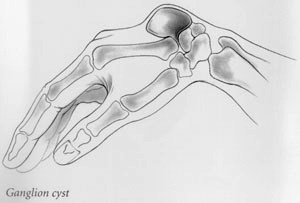
Ganglion cysts are the most common mass or lump in the hand. They are most common on the back of the wrist. These non-cancerous, fluid-filled cysts arise from the ligaments, joint linings, or tendon sheaths when they are irritated or inflamed.

They may disappear or change size quickly. Many ganglion cysts do not require treatment.

However, if the cyst is painful, interferes with function or the patient does not like the appearance, your orthopaedic surgeon can remove the fluid with a needle (aspiration) or remove it surgically.

Carpal Tunnel Syndrome
cons1_9_8
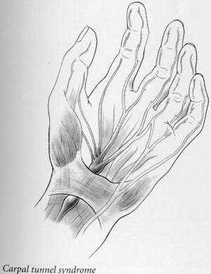
Common symptoms of carpal tunnel syndrome are numbness and tingling in the hand, especially at night; pain with prolonged gripping such as holding a steering wheel; or clumsiness in handling objects. Sometimes the pain can go all the way up to the shoulder.

These symptoms are caused by pressure on the median nerve as it enters the hand through a tunnel in the wrist. The tendons that bend your fingers and thumb also travel in this tunnel.

Mild cases can be treated with a splint or brace to rest the wrist. Steroid injections into the carpal canal to decrease swelling may be used in addition to splinting.

Those cases that do not respond to nonsurgical treatment and those that are diagnosed late often require surgery. This is generally done in an outpatient setting under local anesthesia.

Tendonitis of the Wrist
cons1_10_8
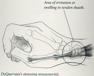
DeQuervain’s stenosing tenosynovitis is an irritation and swelling of the sheath or tunnel which surrounds the thumb tendons as they pass from the wrist to the thumb. Pain when grasping or pinching and tenderness over the tunnel are the most common symptoms.

Sometimes a lump or thickening can be felt in this area. If the hand is made into a fist with the thumb “tucked in” and bent towards the little finger, the pain gets worse (Finkelstein test).

Tendonitis may be caused by overuse and also can be seen in association with pregnancy or inflammatory arthritis such as rheumatoid disease.

If treated early, many cases improve with short periods of rest in a splint, followed by stretching exercises designed to get the tendons gliding. Injection with steroids and/or taking anti-inflammatory medications. More severe cases or those that do not respond to other treatment may require surgery.

Modification of the activities which caused the symptoms initially also may be required.

“Wear and Tear” Arthritis of the Hand
cons1_11_8
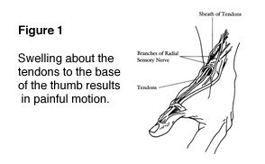
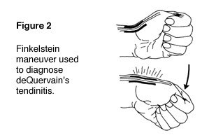
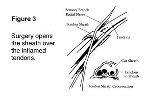
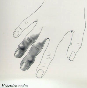
Wear and tear arthritis is very common at the base of the thumb. Pain localized to the base of the thumb, particularly with use, is a very common early symptom.

Early disease can be treated with anti-inflammatory medication, steroid injections into the joint, or splinting.
As the wear and deformity progress, surgery is frequently required. There are many procedures to relieve pain and improve function.

Heberden nodes are “bumps” which occur at the last joint of the finger or thumb due to wear and tear arthritis (osteoarthritis). As the joints deteriorate, small bone spurs form over the back of the joints and make them appear “lumpy.”

Since most Heberden nodes are not painful and seldom interfere with function, no specific treatment is usually required. Patients with pain can be treated with anti-inflammatory medications. All patients should continue moving their hands; disuse frequently results in stiffness.

Dupuytren’s Contracture
cons1_12_8
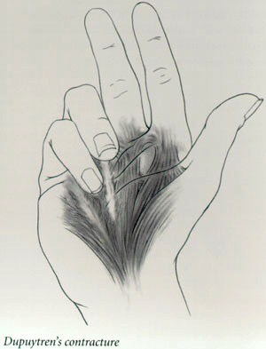
Dupuytren’s contracture is a hereditary thickening of the tough tissue called fascia that lies just below the skin of your palm.

This condition may vary from small lumps or bands to very thick bands which may eventually pull the fingers into the palm.

Dupuytren’s disease is familial, and may be associated with cigarette smoking, vascular disease, epilepsy, and diabetes.

The mainstay of treatment is surgical and is recommended if there is progressive contracture drawing the fingers into the hand. Sometimes a steriod injection will be used in a painful nodule. Small nodules or lumps in the palm do not need treatment until they are very large and interfere with hand function. Even with successful surgical removal, the bands may reappear or occur in other fingers.

Trigger Finger
cons1_13_8Trigger finger is an irritation of the digital sheath which surrounds the flexor tendons. When the tendon sheath becomes thickened or swollen it pinches the tendon and prevents it from gliding smoothly.

In some cases the tendon catches and then suddenly releases as though a “trigger” were released.

Sometimes the swelling can be treated with rest, activity modification, oral anti-inflammatories, or steroid injections. The tendon sheath will then return to its normal, pain-free condition. More severe cases may require surgery to release the tendon.

This can be done as an outpatient procedure. Normal activity can be resumed as pain allows.

Arthritis of the Wrist

Arthritis affects millions of people in the United States. Often, arthritis strikes at the weightbearing joints of the body, such as the knees and the shoulders. But a significant number of people suffer from arthritis in their wrists and hands that make it difficult for them to perform the activities of daily living.

Although there are hundreds of kinds of arthritis, most wrist pain is caused by just two types:

  • Osteoarthritis (OA) is a progressive condition that destroys the smooth articular cartilage covering the ends of bones. The bare bones rub against each other, resulting in pain, stiffness and weakness. OA can develop due to normal “wear-and-tear” on the wrist or as a result of a traumatic injury to the forearm, wrist or ligaments.
  • Rheumatoid arthritis (RA) is a systemic inflammatory disease that affects the joint linings and destroys bones, tissues, and joints. Rheumatoid arthritis often starts in smaller joints, like those found in the hand and wrist, and is symmetrical, meaning that it usually affects the same joint on both sides of the body.

Signs and symptoms

  • OA of the wrist joint manifests with swelling, pain, limited motion and weakness. These symptoms are usually limited to the wrist joint itself.
  • RA of the wrist joint usually manifests will swelling, tenderness, limited motion and decreased grip strength. In addition, hand function may be impaired and there may be pain in the knuckle joints (metacarpophalangeal or MP joints).
  • Joint swelling may also put pressure on the nerves that travel through the wrist. This can cause a lesion to develop (compression neuropathy) or lead to carpal tunnel syndrome.

Diagnosis and treatment
Six bones make up the wrist joint: the two bones of the lower arm (the radius and the ulna) and four wrist bones (the carpals). Your physician will use a combination of physical examination, patient history, and tests to diagnose arthritis of the wrist. X-rays can help distinguish among various forms of arthritis. Some, but not all, forms of RA can be confirmed by a laboratory blood test.

In general, early treatment is nonsurgical and designed to help relieve pain and swelling. Several therapies can be used to treat arthritis, including:

  • Modifying your activities.
  • Immobilizing the wrist for a short time in a splint.
  • Taking anti-inflammatory medications such as aspirin or ibuprofen.
  • Following a prescribed exercise program.
  • Getting a steroid injection into the joint.

Your physician may prescribe other therapies, depending on the type of arthritis you have. For example, additional therapies for patients with rheumatoid arthritis include antimalarial drugs, antimetabolites, gold, immunosuppresive drugs (both non-steroidal and corticosteroids) and newer genetically-engineered medications.

When such conservative methods are no longer effective, or if hand function decreases, surgery is an option. The goal of surgery is to relieve pain; depending on the type of surgery, joint function may also be affected. Surgical options include removing the arthritic bones, joint fusion (making the joint solid and preventing any movement at the wrist) and joint replacement.

You and your physician should discuss the options and select the one that is best for you.