Boutonnière Deformity

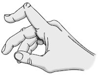
a00004f01Boutonnière deformity is an injury to the tendons in your fingers that usually prevents the finger from fully straightening. The result is that the middle joint of the injured finger bends down, while the fingertip bends back. This is the characteristic shape of a boutonnière deformity. Unless this injury is treated promptly, the deformity may progress, resulting in permanent deformity and impaired functioning.

There are several tendons in your fingers that work together to bend and straighten the finger. These tendons run along the side and top of the finger. The tendon on the top of the finger attaches to the middle bone of the finger (the central slip of tendon). When this tendon is injured, the finger is not able to be fully straightened.

Boutonnière deformity is generally caused by a forceful blow to the bent finger.

It also can be caused by a cut on the top of the finger, which can sever the central slip from its attachment to the bone. The tear looks like a buttonhole (“boutonnière” in French). In some cases, the bone actually can pop through the opening.

Boutonnière deformities may also be caused by arthritis. About one third of all people with rheumatoid arthritis also have fingers with boutonnière deformities.

Arthritis of the Hand

Description
The hand and wrist have multiple small joints that work together to produce motion. This gives you the fine motion needed to thread a needle or tie a shoelace. When the joints become affected with arthritis, activities of daily living can be difficult. Arthritis can occur in multiple areas of the hand and wrist. It can have multiple causes.

All arthritic joints lose cartilage, which works as nature’s “shock absorber.” Cartilage provides a smooth gliding surface for the joint. When the cartilage becomes worn or damaged, or is lost due to disease or trauma, the joint no longer has a painless, mobile area of motion.

The body attempts to make up for the lost cartilage. It produces fluid in the joint lining (synovium), which tries to act like a cushion, like water in a waterbed. But it also causes the joint to swell. This restricts motion. The swelling causes stretching of the joint covering (capsule), which causes pain.

Over time, if the arthritis is not treated, the bones that make up the joint can lose their normal shape. This causes more pain and further limits motion.

Risk Factors/Prevention
It is estimated that one out of every five people living in the United States has at least one joint with signs or symptoms of arthritis. About half of arthritis sufferers are under age 50. Arthritis is the leading cause of disability in the United States. It typically occurs from either disease or trauma. The exact number of people with arthritis in the hand and wrist is not known.

When arthritis occurs due to disease, the onset of symptoms is gradual and the cartilage decreases slowly. The two most common forms of arthritis from disease are osteoarthritis and rheumatoid arthritis. Osteoarthritis is much more common and generally affects older people.

It appears in a predictable pattern in certain joints. Rheumatoid arthritis has other system-wide symptoms and may be passed from parent to child (genetically).

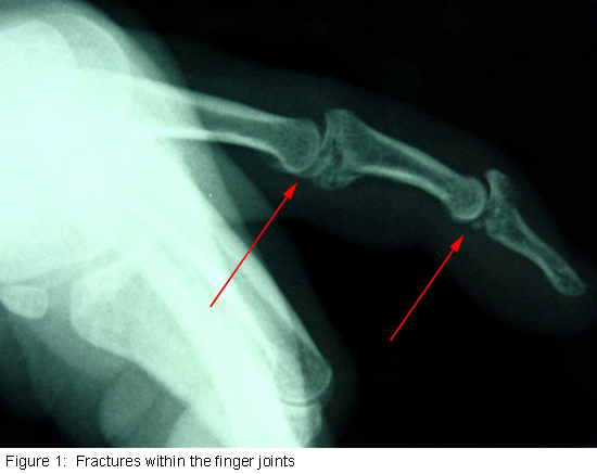
When arthritis is due to trauma, the cartilage is damaged. People of any age can be affected. Fractures–particularly those that damage the joint surface–and dislocations are the most common injuries that lead to arthritis (see Figure 1). An injured joint is about seven times more likely to become arthritic, even if the injury is properly treated.

Arthritis does not have to result in a painful or sedentary life. It is important to seek help early so that treatment can begin and you can return to doing what matters most to you.
cons1_407_307

Diagnosis
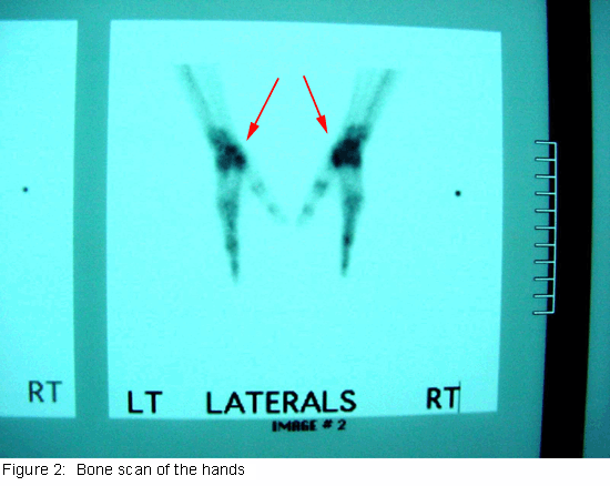
A doctor can diagnose arthritis of the hand by examining you and taking X-rays. Specialized studies such as MRI (magnetic resonance imaging) scans are usually not needed. Sometimes a bone scan is helpful (see Figure 2).
cons1_408_307

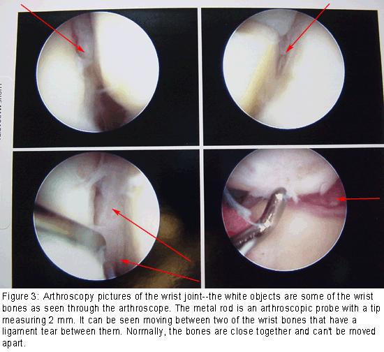
A bone scan may help the doctor diagnose arthritis when it is in an early stage, even if X-rays look normal. Arthroscopy is another way to look at the joint by direct inspection. During an arthroscopic procedure, the surgeon inserts a small camera into the joint to look inside.

It provides the clearest picture of the joint without having to make a large incision. However, this is an invasive procedure and should not be used as a routine diagnostic tool (see Figure 3).
cons1_409_307

Symptoms
Early symptoms of arthritis of the hand include joint pain that may feel “dull,” or a “burning” sensation. The pain often occurs after periods of increased joint use, such as heavy gripping or grasping. The pain may not be present immediately, but may show up hours later or even the following day.

Morning pain and stiffness are typical. As the cartilage wears away and there is less material to provide shock absorption, the symptoms occur even with less use. In advanced disease, the joint pain may wake you up at night.

When the affected joint is subject to greater stress than it can bear, it may swell in an attempt prevent further joint use. Your pain might be made worse with use and relieved by rest. Many people with arthritis complain of increased joint pain with rainy weather. Activities that once were easy, such as opening a jar or starting the car, become difficult due to pain.

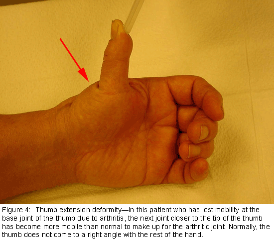
To prevent pain at the arthritic joint, you might adapt the way you use your hand. In patients with advanced thumb base arthritis, the neighboring joints may become more mobile than normal (see Figure 4).
cons1_410_307

The arthritic joint may feel warm to touch. This is due to the body’s inflammatory response. There may be a sensation of grating or grinding in the affected joint (crepitation). This is caused by damaged cartilage surfaces rubbing against one another. If arthritis is due to damaged ligaments, the support structures of the joint may be unstable or “loose.”

In advanced cases, the joint may appear larger than normal (hypertrophic). This is usually due to a combination of bone changes, loss of cartilage and joint swelling.

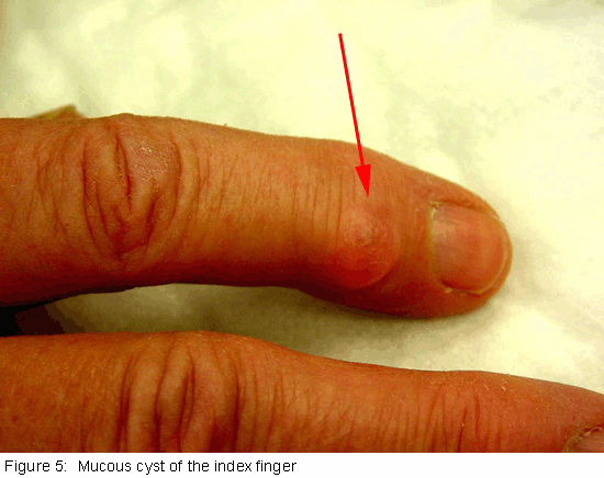
When arthritis affects the end joints of the fingers (DIP joints), small cysts (mucous cysts) may develop (see Figure 5). The cysts may then cause ridging or dents in the nail plate of the affected finger.
cons1_411_307

Treatment Options: Nonsurgical
Treatment options for arthritis of the hand and wrist include medication, splinting, injections and surgery. Treatment depends on many factors:

  • How far the arthritis has progressed
  • How many joints are involved
  • Your age, activity level and other medical conditions
  • If the dominant or non-dominant hand is affected

Your personal goals, home support structure, and ability to understand the treatment and comply with a therapy program

Medications: Medications treat symptoms but cannot restore joint cartilage or reverse joint damage. The most common medications for arthritis are anti-inflammatories, which stop the body from producing chemicals that cause joint swelling and pain.

Examples of anti-inflammatory drugs include over-the-counter medications such as Tylenol® and Advil® and prescription drugs such as Celebrex®. Glucosamine and chondroitin are widely advertised “neutraceuticals.”

Neutraceuticals are not drugs. Rather, they are compounds that are the “building blocks” of cartilage. They were originally used by veterinarians to treat arthritic hips in dogs. However, neutraceuticals have not yet been studied as a treatment of hand and wrist arthritis.

Injections: When first-line treatment with anti-inflammatory medication is not appropriate, injections may be used. These typically contain a long-acting anesthetic, similar to novacaine but longer lasting, and a steroid that can provide pain relief for weeks to months.

The injections can be repeated, but only a limited number of times, due to possible side effects, such as lightening of the skin, weakening of the tendons and ligaments and infection.

Splinting: Injections are usually combined with splinting of the affected joint. The splint helps support the affected joint to ease the stress placed on it by activities. Splints are typically worn during periods when the joints hurt.

They should be small enough to allow functional use of the hand when they are worn. Wearing the splint for too long can lead to muscle wasting (atrophy). Muscles can assist in stabilizing injured joints, so atrophy should be prevented.

Treatment Options: Surgical
If non-operative treatment fails to give you relief, surgery is usually discussed. There are many surgical options. The option chosen should be one that has a reasonable chance of providing long-term pain relief and return to function. It should be tailored to your individual needs. It is important that the treating physician is well versed in current surgical techniques. If there is any way the joint can be preserved or reconstructed, this option is usually chosen.

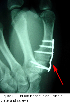
When the damage has progressed to a point that the surfaces will no longer work, a joint replacement or fusion (arthrodesis) is performed (see Figure 6). Joint replacements attempt to provide pain relief and functional joint motion. Joint fusions provide pain relief but stop joint motion. The fused joint no longer moves; the damaged joint surfaces are gone, so they cannot cause symptoms.
cons1_412_307

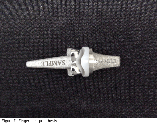
As with hip and knee replacements, there have been significant improvements in joint replacements in the hand and wrist. The replacement joints are made of materials similar to those used in weight-bearing joints, such as ceramics or long wearing metal and plastic parts (see Figure 7). The goal is to improve the function and longevity of the replaced joint. Most of the major joints of the hand and wrist can be replaced.

A surgeon often needs additional training to perform the surgery. As with any evolving technology, the long-term results of the hand or wrist joint replacements are not yet known. Early results have been promising. Talk with your doctor to find out if these implants are right for you.
cons1_413_307

After any type of joint reconstruction surgery, there is a period of recovery. Often, you will be referred to a trained hand therapist, who can help you maximize your recovery. You may need to use a postoperative splint or cast for awhile after surgery. This helps protect the hand while it heals.

During this postoperative period, you may need to modify activities to let the joint reconstruction heal properly. Typically, pain medication you take by mouth is also used to reduce discomfort.

It is important to discuss your pain with your doctor so it can be adequately treated. Length of recovery time varies widely and depends on the extent of the surgery performed and multiple individual factors. However, people usually can return to most if not all of their desired activities in about three months after most major joint reconstructions.

Research on the Horizon/What’s New?
Increasingly, doctors are focusing on how to preserve the damaged joint. This includes getting an earlier diagnosis and repairing joint components before the entire surface becomes damaged. Arthroscopy of the small joints of the hand and wrist is now possible because the equipment has been downsized.

There have been encouraging results in cartilage repair and replacement in the larger joints such as the knee, and some of these techniques have been applied to the smaller joints of the hand and arm. In addition, stem cell research may be an option to regenerate damaged joint surfaces.
Animal Bites

Each year millions of people in the United States-most of them children—are bitten by animals. Most animal bites are from dogs; cat bites are second most common. However, the risk of infection from a cat bite is much higher than that from a dog bite. Most bites occur on the fingers of the dominant hand, but children may also be bitten about the head and neck area.

A major concern about an animal bite is the possibility of rabies. Because most pets in the U.S. are vaccinated, most cases of rabies result from the bite of a wild animal such as a skunk, bat or raccoon. However, in other countries, dog bites are the most common source of rabies. If you are bitten by a dog outside the U.S., consult a doctor immediately.

Signs and Symptoms of Animal Bites

In some cases, the bite will not break the skin but may cause damage to underlying tendons and joints. If the skin is broken, there is the additional possibility of infection as well as injury to tendons and nerves. Dogs have powerful jaws and can cause crushing injuries to bone, muscles, tendons, ligaments and nerves.

Signs of an infection include:

  • Warmth around the wound
  • Swelling
  • Pain
  • A pus discharge
  • Redness around the puncture wound

Signs of damage to tendons or nerves include:

  • An inability to bend or straighten the finger
  • A loss of sensation over the tip of the finger

First aid

  • Don’t put the bitten area in your mouth! You will just be adding the bacteria in your mouth to that already in the wound.
  • If the wound is superficial, wash the area thoroughly. Use soap and water or an antiseptic such as hydrogen peroxide or alcohol. Apply an antibiotic ointment and cover with a non-stick bandage. Watch the area carefully to see if there are signs of damaged nerves or tendons. Some bruising may develop, but the wound should heal within a week to 10 days. If it does not, or if you see signs of infection or damage to nerves and tendons, seek medical help.
  • If there is bleeding, apply direct pressure with a clean dry cloth. Elevate the area. Do not clean a wound that is actively bleeding. Cover the wound with a clean sterile dressing and always seek medical help.
  • If the wound is to the face and/or head and neck area, seek medical help immediately.
  • Contact your physician to see whether additional treatment is needed.
  • Report the incident to your public health department. They may ask your assistance in locating the animal so that it can be confined and observed for symptoms of rabies.

Medical assistance

Tell your doctor how you got the bite. Your physician will wash the wound area thoroughly and check for signs of nerve or tendon damage. The doctor may examine your arm to see if there are signs of a spreading infection. Your physician will probably leave the wound open (without stitches), unless you have a facial wound. You may need to get X-rays and a blood test. You may also need to get a tetanus shot and a prescription for antibiotics. If the tendons or nerves have been injured, you may need to see a specialist for additional treatment.

More about Rabies

Rabies is a disease that affects only mammals (such as raccoons, bats, dogs, horses, and humans). It is caused by a virus that attacks the nervous system. Without treatment, it is 100 percent fatal. Rabies develops in two stages.

During the first stage, which can last up to 10 days, the individual may have a headache, fever, decreased appetite, vomiting and general malaise, along with pain, itching, and tingling at the wound site. Symptoms of stage two include difficulty in swallowing, agitation, disorientation, paralysis, and coma. At this point, there is no known, effective treatment.

If rabies is identified early, a series of highly effective vaccinations can be administered. That’s why it’s important to capture and observe the animal that bit you. If the animal cannot be captured, but must be killed, the head should be kept intact so the brain can be examined for signs of rabies.

Preventing Animal Bites

Follow these recommendations to prevent animal bites and rabies.

  • Do not try to separate fighting animals.
  • Avoid animals that appear sick or act strangely. Call animal control.
  • Leave animals, even pets or other animals you know, alone when they are eating or sleeping.
  • Keep pets on a leash when out in public.
  • Never leave a young child alone with a pet. Don’t allow children to tease an animal by waving sticks, throwing stones, or pulling a tail.
  • Be sure your pet is vaccinated.
  • Do not approach or play with any kind of wild animal. Teach children not to pet strange animals, even pets on a leash, without asking permission of the owner first.

Rotator Cuff Tears – Dr. Franklin

From orthoinfo.aaos.org

From orthoinfo.aaos.org

Anatomy
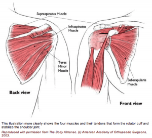
The rotator cuff is made up of 4 muscles surrounding the shoulder.

Three muscles on the back of the shoulder (supraspinatus, infraspinatus and teres minor) converge as a tendon to insert on the outer edge of the humeral head.

They act to elevate and externally rotate the shoulder. The fourth muscle (subscapularis) is on the front of the shoulder and helps internally rotate the shoulder.

Injury Mechanism
Rotator cuff tears can occur after an acute injury such as a fall or catching a heavy falling object, or they can occur over time as a gradual wearing of a hole in the rotator cuff from ongoing rubbing on the acromion such as overuse with overhead or throwing sports.

Symptoms
With acute complete tears of the rotator cuff, patients often describe a burning or tearing sensation at the time of injury. Early along, it is difficult to raise the arm overhead. Patients describe pain and weakness. The pain is usually located over the outer (lateral) aspect of the upper arm. Rotator cuff tears are often painful at night. Older patients may not recall an injury, but may just describe a gradual aching in the shoulder that has progressed over several months or years.

Diagnosis
The physician’s work-up will start with a careful history and exam.

X-rays are often obtained to see that no fractures have occurred with the injury and to help assess the overall condition of the shoulder joint. An MRI scan is often obtained to confirm the diagnosis and to evaluate any associated injuries to the labrum, rotator cuff or damage to the joint surfaces. Often the radiologist will inject contrast into the shoulder joint with a small needle to coat the undersurface of the rotator cuff and to see if the contrast leaks through the rotator cuff suggesting a complete tear. An MRI with contrast is called an arthro/MRI.

Plain x-rays show the bones of the shoulder, while MRI scans reveal the soft tissues around the shoulder including the labrum (lip of cartilage around the socket) and the rotator cuff tendons.

Treatment
Your physician will discuss treatment options with you. Treatment decisions are based on age, activity level and the severity of symptoms. In older patients with less activity demands and less severe symptoms, one will usually start with non-operative treatment including rest, ice, anti-inflammatories, stretching and occasional injections to see if the symptoms become tolerable.

In younger, more active patients, surgery is almost always recommended when a full thickness rotator cuff tear has occurred, since the rotator cuff has poor blood supply, therefore poor healing potential. Rotator cuff surgery is usually done on an outpatient basis. In most cases, the orthopedist will start with an exam under anesthesia to see that full motion of the shoulder is present and to see that the joint is stable. Next, one usually looks into the joint with a small arthroscope (a small lens and camera) so the surgeon can see and probe all of the structures in the joint. Once the complete rotator cuff tear is confirmed, the torn tendon is repaired back to the bony attachment site.

Because there is poor blood supply at the attachment site to the bone, one usually creates a small groove in the bone and pulls sutures through drill holes or uses anchors in the bone for fixation.

What to expect after surgery
Rotator cuff surgery is performed as an outpatient procedure. The surgery takes about 60 minutes. Patients go home in a sling that they use for 3 or 4 weeks. Most patients are uncomfortable for the first 2 or 3 days, but prescription medication is used to help alleviate the pain. Patients are seen back in the office one week after surgery to check their incisions and to start their exercise routine.

For the first 4 to 6 weeks, patients avoid any active elevation of their arm or lifting with that arm. At that point, formal physical therapy is started to improve range of motion and strength. It takes about 3 months from surgery before most of the strength and use of the shoulder returns. Full recovery may take 4 to 6 months.

Shoulder Impingement

Shoulder Impingement (Rotator Cuff Tendinitis)

The rotator cuff is made up of tendons and muscles that allow for a great range of motion of your arm. This is a frequent source of pain for athletes and an area that is at risk for injury during sporting activities. Shoulder impingement is often referred to as rotator cuff tendinitis and is one of the most common causes of shoulder pain.

What causes rotator cuff tendinitis?
When you raise your arm to shoulder height, the space between the bone and rotator cuff narrows. The bone can rub against (or impinge on the tendon and the bursa, causing irritation and pain when the arm is used repeatedly. Young athletes who use their arms for overhead action are particularly vulnerable. This includes those who play tennis, softball and baseball, and swimmers.

What are the symptoms of shoulder impingement?
When the rotator cuff is irritated this can lead to local swelling and tenderness in the front aspect of the shoulder. You may also have pain and stiffness when you lift your arm. There is also a sensation of tenderness when the arm is lowered from an elevated position. Other symptoms include sudden pain when reaching or lifting, pain radiating from the front of the shoulder to the side of the arm, minor pain at rest, and pain when throwing or using the arm.

How is rotator cuff tendinitis treated without surgery?
Your orthopedic specialist wants to reduce your pain and restore function of your shoulder. He will consider your activity level, your age, and your general state of health. Many times shoulder impingement can be treated with medications and rest. It is not uncommon for athletes to be ordered physical therapy to help restore normal motion of the shoulder. Your therapist will teach you specific stretching and strengthening exercises to relieve your shoulder pain and help you get back to normal activities.

What is involved with surgical treatment?
The goal of surgery is to create more space for the rotator cuff and this involves removing a portion of the inflamed bursa. Your orthopedic specialist will perform an anterior acromioplasty, where part of the bone is removed to allow for movement of the rotator cuff. Many times, the surgeon opts to perform this procedure by way of arthroscope.

The arthroscopic technique allows for use of small thin surgical instruments to be inserted around puncture wounds around the shoulder. The doctor can see inside the shoulder through a small camera inserted into the joint that displays images onto a computer TV monitor.