What is foot fracture surgery?

Foot Fractures

There are 26 bones in each of your feet, all of which can break. There are several different types of fractures:

  • Non-displaced: The bone breaks but stays in place
  • Displaced: The bone breaks into two pieces that move apart from one another
  • Comminuted: The bone is broken in multiple places
  • Open fracture: The bone breaks through the skin after fracturing

If you injure your foot, your foot and ankle orthopedic surgeon will take X-rays to see if you have a broken bone. X-rays will identify most fractures but some smaller and more subtle fractures may require CT or MRI scans. Not all fractures require surgery, and your surgeon will help determine how your fracture should be treated.

If you need surgery for your foot fracture, the goals are to restore the fractured bone to its correct position, stabilize the bone in this position, encourage healing, restore function, and reduce the risk of future problems such as persistent pain, loss of motion, and arthritis.

Diagnosis

All foot fractures are different, but generally speaking if a fracture is significantly displaced it is likely to benefit from surgery. This is especially true if a fracture enters a joint and the joint surface is disrupted and displaced. Restoring the alignment and stabilizing the fracture in its correct position will decrease the risk of future problems, such as pain, swelling, deformity, and arthritis.

In some cases, surgery may be appropriate for non-displaced or minimally displaced fractures if the broken bones are likely to be unstable. In such cases surgery can maintain the alignment and encourage healing in the right position.

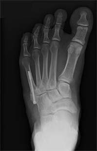
Even some non-displaced and stable fractures may benefit from surgery. One such fracture, called a Jones fracture, often is treated surgically in active and athletic individuals because it is likely to get them back to their activities more quickly than non-surgical treatment.

When the risks of surgery outweigh potential benefits, your foot and ankle orthopedic surgeon may recommend non-surgical treatment. This decision is based on an understanding of your entire body and pre-existing conditions. For example, if you have a history of heart problems, your surgeon may recommend non-surgical treatment or that you see your cardiologist to determine if surgery is safe before proceeding.

Treatment

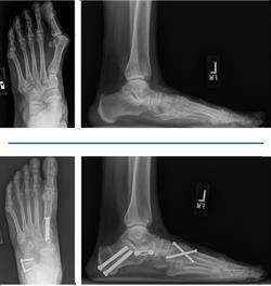
Foot fracture surgery involves making an incision in the skin centered over the fractured bone. The bone is then exposed so the orthopedic surgeon can see the fracture. The fractured bone fragments are realigned as well as possible and secured with implants such as pins, wires, screws, and plates. After stabilizing the fracture your surgeon will stitch the incision closed, apply a sterile bandage, and place your foot in a cast, splint, boot, or post-operative shoe.

With some fractures, the surgeon can restore the alignment of the fractured bone without a large incision. In this method, the fracture is fixed with appropriate implants through one or more small incisions. This is called a percutaneous fracture fixation. The advantages of this technique include smaller incisions, less trauma to the tissues, less disruption of the blood supply to the bone, and less pain after surgery.

Recovery

After surgery your foot and ankle orthopedic surgeon will place your foot into a cast, splint, boot, or post-op shoe. It is important to keep your foot elevated as much as possible to reduce pain and swelling. In most cases your doctor also will want you to stay off your foot completely for 1-3 months, depending on the injury.

A few weeks after surgery your surgeon will check the wound and remove the stitches. Your surgeon may have you begin working on range-of-motion exercises of the ankle, foot, and toes. They may also refer you to a physical therapist.

Your doctor will see you at regular intervals and check X-rays to see how well the fracture is healing. Based on your fracture type and this evaluation, your surgeon will decide when you can begin to bear weight on the injured foot. They may have you begin weightbearing with a special boot and allow you to advance slowly to your normal footwear as symptoms allow. In most cases you can expect 3-6 months or more before you return to full activity without restrictions, and up to a year before you reach maximum improvement.

Risks and Complications

All surgeries come with possible complications, including the risks associated with anesthesia, infection, damage to nerves and blood vessels, and bleeding or blood clots. To minimize the risk for blood clots, you may be placed on aspirin or a blood thinning medication for several weeks after surgery. Unfortunately, even when on these medications, blood clots can occur post-operatively.

Potential complications of foot fracture surgery include wound breakdown, failure of the fracture to heal (nonunion), fracture healing in a bad position (malunion), loss of fracture alignment prior to healing, implant failure, persistent pain, loss of motion, and arthritis. Smoking is a significant risk factor for multiple complications. If you are a smoker, your surgeon will discuss this with you before operating.

Foot and ankle orthopedic surgeons are uniquely qualified to identify and treat fractures of the foot and should be your first resource when you experience a foot injury.

What is foot drop treatment?

Foot Drop

Foot drop occurs when the muscles and tendons that flex the foot up are no longer working. Commonly, it is the result of a nerve injury, stroke, or nerve disease (neuropathy). It also can occur after an injury to a muscle or tendon. If a person is unable to flex the foot up when walking, the foot or toes can drag on the ground. This can make walking difficult and lead to frequent falls.

The goal of a foot drop procedure is to improve a patient’s ability to actively flex the foot and ankle up in situations where this function is weak or lost completely.

Symptoms

When the muscles that flex the foot up are not working, but other muscles around the foot and ankle still function, tendon transfer surgery can be helpful. If there is no muscle function at the ankle, this procedure cannot be performed. A patient must have some working muscles for a tendon transfer procedure to be successful.

Treatment

Usually, initial treatment involves use of an off-the-shelf or custom brace called an AFO (ankle foot orthosis), which helps to improve the position of the foot while a person is walking. When this brace is no longer helpful, surgery can be considered. You should discuss your options with your foot and ankle orthopedic surgeon before proceeding with surgery.

The surgical procedure for a foot drop is called a tendon transfer. In general, a tendon transfer is a procedure in which a tendon (and attached muscle) that is still working is taken from one part of the foot and moved to another part of the foot to try to replace the missing muscle function. The most common tendon transferred is the posterior tibial tendon.

Specific Technique

A posterior tibial tendon transfer procedure can involve the posterior tibial tendon alone or transfer with two other tendons, the peroneus longus and the anterior tibialis. When all three tendons are used it is called a Bridle procedure.

Multiple incisions are needed to move the tendon from one position to another. The posterior tibial tendon is taken off its insertion on the navicular bone on the inner side of the foot. This is the first incision. A second incision is made above the ankle and the tendon and muscle are identified. The tendon is pulled into this second incision and then transferred in between the tibia and fibula bones to the front of the ankle.

Another incision is made on the top of the foot at the bone to which the tendon is going to be transferred. The tendon is routed under the skin to this bone and fixed into a tunnel in the bone.

If a Bridle procedure is performed, a second tendon, the peroneus longus, is cut above the level of the ankle on the outer side of the leg. This tendon is then routed to the front of the ankle and the free end is attached to the posterior tibial tendon and the anterior tibial tendon in a bridle configuration. With this construct the posterior tibial muscle pulls on all three tendons to pull the foot up. The posterior tibial tendon is routed to the top of the foot as described above.

Occasionally, the ankle can be very stiff from long-standing weakness. If it is not possible to pull the ankle up for attachment of the tendon transfer, the Achilles tendon is lengthened to help bring the foot and ankle up. This is done either through an incision on the calf or an incision right over the Achilles tendon. The location of the incision is based on which portion of the Achilles is too tight.

Recovery

The ankle is placed in a splint in the operating room to hold the position of the foot and ankle and protect the newly transferred tendon(s). Strict elevation and non-weightbearing are enforced over the first 10 to 14 days. Stitches are removed in about two weeks, after which the foot will be in a cast for about six weeks. The patient usually is non-weightbearing during this time.

Once the cast is removed, the patient will be allowed to walk in a special boot. Physical therapy to retrain the tendon(s) in its new position continues for 8-12 weeks. A night splint is worn for three months after surgery to prevent premature stretching of the tendon transfer.

As swelling improves a custom-molded brace can be worn in an athletic shoe and the walker boot is discontinued. Once the patient’s strength and motion are improved with physical therapy, the brace may be discontinued. The goal of the surgery is for the patient to walk in a regular shoe without the need for a brace.

Risks and Complications

Potential complications of this treatment can include wound infection, deep infection that can compromise the tendon transfer, and failure of the tendon or tearing of the repair.

FAQs

Will I have “normal” ankle movement after this procedure?

A foot drop procedure changes the function of the ankle and allows a patient to walk without a brace. However, it is not possible to restore normal strength and full range of motion with this procedure.

Will I be able to pull up my toes?

Often a nerve injury that causes a foot drop also limits the ability to pull up the toes. The tendon transfer will not restore this function. If this is a concern, make sure that you mention it to your doctor.

Will I be able to walk without a brace?

The purpose of the procedure is to try to improve function of the ankle so that a patient can walk without a brace. However, with severe nerve injury it is not always possible to guarantee that a patient will be brace-free all the time.

Can foot drop surgery be performed if I have no working muscles at all?

The foot drop tendon transfer surgery only works if some of the muscles around the ankle are still working.

Is there a “best time” to have the procedure after a foot drop occurs? Is sooner better than later?

Nerve injuries can improve very slowly over time. At the initial time of injury, it is possible that over months to a year, the foot drop will actually improve and strength will return. By about one year, if no function has returned, a foot drop procedure is reasonable to consider.

For a long-standing foot drop (that is, the injury happened many years ago) a foot drop procedure can still work if the other muscles around the ankle are still working. So no matter how long ago it happened, it is reasonable to seek the opinion of a foot and ankle orthopedic surgeon to see if you are a candidate for this procedure.

Will I need physical therapy?

It is helpful to have formal physical therapy to help retrain the muscle for its new function. Therapy continues until progress is made in both strength and motion and you are able to transition out of the boot or brace.

What is flatfoot surgical correction?

Adult Acquired Flatfoot Deformity

Adult acquired flatfoot deformity (AAFD) is a collapse of the arch of the foot. Flatfoot surgery addresses the bones, ligaments, and tendons that support the arch, often through a combination of procedures. The goals of the surgery are to improve the alignment of the foot and restore more normal pressure during standing and walking. This surgery can also reduce pain and improve walking ability.

Diagnosis

Patients with a painful flatfoot frequently mention ankle and/or foot pain and difficulty with daily activities. A foot and ankle orthopedic surgeon should do a complete evaluation of the foot, including a medical history, physical exam, and X-rays. Non-surgical treatments such as rest, immobilization, shoe inserts, braces, and physical therapy should be tried first. If these are unsuccessful, then surgery may be considered.

Patients who have diabetes or take oral steroids should be evaluated by their primary care physician to determine if surgery is safe. Obese patients and smokers are at higher risk for blood clots and wound problems. Full recovery from flatfoot surgery can take up to a year. Patients who are unable or unwilling to complete this process should not have this surgery.

Treatment

Surgery can be performed under regional anesthesia, which is numbing of the foot and ankle with a nerve or spinal block, or general anesthesia, which may require a breathing tube. A nerve block often is placed behind the knee to reduce pain after surgery.

Comprehensive surgical treatment for AAFD usually involves a combination of several procedures. Your foot and ankle orthopedic surgeon will develop a treatment plan based on your deformity and the surgeon’s preferences. The following procedures may be considered.

Achilles Lengthening

In AAFD, the Achilles tendon becomes tight and contracted. Almost every surgical procedure for AAFD includes some kind of Achilles tendon lengthening. There are multiple types, each with different benefits. The most commonly performed types are gastrocnemius recession and triple-cut/percutaneous Achilles tendon lengthening.

Medializing Calcaneal Osteotomy

Also called a heel slide, this procedure involves cutting the heel bone to shift it back into correct alignment under the leg. The bone is then held in place with screws, staples, or a plate.

Tendon Transfers

Typically the flexor digitorum longus (FDL) tendon, which flexes your toes, is transferred to help bring some strength back to the posterior tibial tendon. It is cut in the foot and transferred to the navicular bone. If the posterior tibial tendon is severely damaged, your surgeon may remove it altogether. Sometimes, tendon transfers on the outside of the foot are also done to help realign the forces working on the foot.

Ligament Repairs

The spring ligament and the deltoid ligament are two ligaments that help hold the correct alignment of the foot and ankle. In patients with severe disease, one or both ligaments may be torn. In some cases, your surgeon may recommend repair or reconstruction of one or both of these ligaments.

Lateral Column Lengthening

In this procedure, the calcaneus bone is cut on the outside of the foot and “lengthened” to help correct the foot deformity. This is typically done by inserting either a cadaver bone or a metal wedge into the cut bone to lengthen it. Often, screws or a plate are used to help hold the bones in position while they heal.

Cotton (Medial Cuneiform) Osteotomy

In this procedure, the medial cuneiform bone is cut through an incision on the top of your foot. Spreading the cut bone apart with a bone or metal wedge helps recreate an arch.

Midfoot Fusion

Some patients with arthritis and/or deformity of their midfoot may require a midfoot fusion. This may involve one or more of the multiple midfoot joints, including the tarsometatarsal joints or the naviculocuneiform joint. This procedure is also useful for restoring the arch.

Subtalar Fusion

This procedure is done for more severe deformities. The talus and the calcaneus bones are fused together, which allows the surgeon to correct more of the deformity.

Double or Triple Arthrodesis

This procedure is done for the most severe deformities or ones with arthritis. In a triple arthrodesis, three joints are fused: the subtalar, talonavicular, and calcaneocuboid joints. Often, just the subtalar and talonavicular joints are fused (double arthrodesis). The foot will be stiff after this surgery, but usually pain and alignment are improved and the foot feels more stable for walking.

X ray views of a flatfoot before and after
X-ray views of a flatfoot before and after

Recovery

Patients may go home the day of surgery or they may require an overnight hospital stay. The leg will be placed in a splint or cast and should be kept elevated for the first two weeks. At that point, sutures are removed. A new cast or a removable boot is then placed. It is important that patients do not put any weight on the corrected foot for 6-8 weeks following the operation. In most cases, patients may begin bearing weight after the first 6-8 weeks and progress to full weightbearing by 10-12 weeks. For some patients, weightbearing requires additional time. After 12 weeks, patients usually can transition to wearing a shoe. Inserts and ankle braces often are used. Physical therapy may be recommended. Swelling and discomfort can last for months after surgery, and full recovery can take 1-2 years.

Risks and Complications

All surgeries come with possible complications, including the risks associated with anesthesia, infection, damage to nerves and blood vessels, and bleeding or blood clots.

Complications following flatfoot surgery may include wound breakdown or incomplete healing of the bones (nonunion). These complications often can be prevented with proper wound care and rehabilitation. Occasionally, patients may notice some discomfort due to prominent hardware. Removal of hardware can be done at a later time if this is an issue. The overall complication rates for flatfoot surgery are low.

FAQs

Will surgical correction of my flatfoot improve the cosmetic appearance of my foot?

Surgical correction of flatfoot is aimed primarily at reducing pain and restoring function. Although surgery likely will improve the cosmetic appearance of the foot, it is not a primary goal of treatment.

What activities will I be able to do following flatfoot surgery?

With proper correction and rehabilitation, many patients return to active lifestyles. Activities such as walking, biking, driving, and even golfing are well tolerated. It is less likely, however, that patients will be able to participate in very strenuous high impact activities requiring running, cutting, or jumping.

What is fifth metatarsal fracture surgery?

Fifth Metatarsal Fracture

The metatarsal bones are the long bones in the middle of the foot. Each metatarsal bone has a base, a shaft, a neck, and a head. The fifth metatarsal is the last bone at the outside of the foot, and most breaks of the fifth metatarsal occur at the base.

The majority of fifth metatarsal fractures are treated without surgery. However, certain situations may require surgical treatment. Surgery can be performed to help the bone heal in a correct position and return the patient to full function. Surgery may reduce the time needed for immobilization and improve the chance of healing compared to non-surgical treatment.

Diagnosis

The base of the fifth metatarsal is divided into three fracture zones.

  • Zone 1 fractures are avulsion or chip fractures that occur at the tip of the base of the fifth metatarsal. These fractures typically are treated without surgery using a cast, boot, or hard-soled shoe and tend to heal within 6-8 weeks.
  • Zone 2 fractures are known as Jones fractures. They occur at the intersection between the base and the shaft of the fifth metatarsal. These fractures are known to have a higher chance of not healing (nonunion). They also are at risk of refracture even after healing. Surgery is common for these fractures due to the risk for delayed healing or reinjury.
  • Zone 3 fractures happen at the junction of the wide portion of the bone with the shaft of the fifth metatarsal. These typically are stress fractures, and may be associated with symptoms prior to the fracture, or with minimal trauma that still results in a fracture. Lengthy healing times and risk of refracture may be reasons for surgical repair in these fractures.

Surgery is not indicated in a fracture where there is an infection or severely damaged soft tissue. Zone 1 fractures that are not displaced do not require surgery, and most fifth metatarsal shaft fractures without significant displacement do not require surgery.

Treatments

There are many surgical options for fifth metatarsal fractures. One popular technique (for zone 2 and 3 fractures) is a surgery where a screw is placed lengthwise within the bone (intramedullary screw fixation). Fractures of the shaft of the metatarsal are fixed with a plate and screws. These procedures can be performed under general or regional anesthesia with the patient going home the same day.

Specific Techniques

The surgical incision for an intramedullary screw is typically no more than a stab incision at the base of the fifth metatarsal. An X-ray machine is used to guide the screw placement. The screw threads cross the fracture site and allow for the fracture ends to be squeezed together. If bone grafting is needed, such as in a chronic fracture that has failed non-operative treatment, then a separate incision may be needed over the fracture to insert the bone graft or bone graft substitute.

Screw fixation of a fifth metatarsal base fracture

Recovery

For the first 7-14 days after surgery, you may be allowed to weight bear through the heel but typically not through the front of the foot. This could last as long as six weeks (sometimes longer based on healing and other factors such as diabetes). Weight bearing in a removable walking boot is allowed after that. Patients can expect to return to full activity 3-4 months after a typical fracture.

This includes returning to sports. Some fractures may require bone grafting and have longer recoveries. The screw is not usually removed unless it causes discomfort.

Risks and Complications

All surgeries come with possible complications, including the risks associated with anesthesia, infection, damage to nerves and blood vessels, and bleeding or blood clots.

Some complications can result in the need for repeat surgery. The metatarsal fracture may not heal and can become a nonunion. Another rare but serious complication is a re-fracture after fixation. Some patients may be at greater risk for poor healing or re-fracture due to the shape of their foot. A high-arched foot or a heel that turns in can put extra pressure on the fifth metatarsal and may require other surgical procedures to change the architecture of the foot.

FAQs

If I have a zone 2 fracture of the base of the fifth metatarsal, will I jeopardize my chance of bone healing if I try non-operative treatment first?

Most of these breaks will go on to heal after 12 weeks with appropriate treatment. The nonunion rate of these fractures may still be as high as 15 to 20 percent. A fracture that fails to heal and is painful may require surgical repair. The surgery can be more difficult at that point and may require a bone graft.

If I have a fracture of the neck or shaft of the metatarsal, is there a need for surgery?

The need for surgery in these fracture types depends on the degree of deformity. Rotational deformity of the little toe, angulation of the shaft of the metatarsal with a change in the shape of the foot and shortening as a result of the fracture are a few of the reasons for surgical repair.

If I choose not to have surgery and use a walker boot for six weeks, are there any other treatments that can help accelerate healing?

There is some evidence that electromagnetic bone stimulation may be useful in increasing healing rates and reducing time for healing in zone 2 fractures of the base of the fifth metatarsal. However, more evidence is needed before these therapies can be recommended to all patients with such fractures.

Is there any role for PRP or stem cell injections?

This is a relatively new and not well researched area currently. No recommendation for these injections can currently be made; they are not typically covered by insurance and can be quite expensive without data to suggest that they improve results. However, you should be aware that even if the biology can be improved with injections, these will not alter the mechanics of the foot. An increasing number of orthopedic practices are providing these injections. You should speak with your foot and ankle orthopedic specialist who is best educated to evaluate the full spectrum of care including non-surgical and surgical options.

What is extracorporeal shock wave therapy?

Shock Wave Therapy

Shock wave therapy is a non-invasive method that uses pressure waves to treat various musculoskeletal conditions. High-energy acoustic waves (shock waves) deliver a mechanical force to the body’s tissues.

Diagnosis

Shock wave therapy may treat conditions such as degenerated tendons (Achilles tendinitis), heel pain (plantar fasciitis), and tennis elbow (lateral epicondylitis).

Complications are rare with shock wave therapy. People who have poor sensation (neuropathy) or hypersensitivity in the target area should not have this procedure. Open sores also are a reason to avoid shock wave therapy. Shock wave therapy is not used in patients with heart conditions or seizures. It should not be used during pregnancy.

Treatment

A non-invasive probe is applied to the skin. An electrical charge creates an energy wave that is focused on the area of concern. The shock waves create a force on the tissues that may induce healing. It’s not clear why this approach to healing works for some people, but it may be that shock waves cause inflammation and improve blood flow to encourage the body to repair and heal itself.

Specific Technique

Shock wave therapy is an outpatient procedure. A probe is placed on the skin after a gel is applied to help conduct the shock waves. Therapy is more successful with active patient participation where the patient tells the therapist whether or not the probe is at the area of pain. One or more treatment sessions may be needed.

Recovery

Patients typically bear weight after treatment. Patients are advised to reduce the level of physical activity for 1-2 weeks after treatment. Shock wave therapy may give good outcomes for some tendon problems or chronic degenerative conditions. Examples include Achilles tendinitis and plantar fasciitis.

Risks and Complications

The main complications are pain and hypersensitivity at the site of treatment. These problems typically resolve with time. Pain and disability may persist when shock wave therapy is not successful.