Wrist Fracture

Other common names: Distal Radius Fracture, Fracture of the end of the Radius, Broken Wrist, Wrist FractureWhat is a Broken Wrist?Though there are 10 bones that make up the wrist, and each bone has the potential to fracture (medical term for broken bone), by far the most common broken bone in the wrist is a fracture of the end of the radius bone (Figure 1). Other common injuries include a wrist sprain, scaphoid fracture, Ulna fracture, fracture of the triquetrum.

Typically, a patient sustains a broken wrist from one of two scenarios; one is that they were in an accident, or fell with such force on their hand that the otherwise normal wrist crushed under significant forces. The second scenario involves one with less than ideal bone density who falls on an outstretched hand in a way that causes the bone to break. The difference between the two is the difference in the quality of the bone and the amount of force required to break the bone. The distinction between the two mechanisms is significant as the patient with higher quality bone often has multiple injuries due to the significant force of the injury, and in the patient with poorer bone quality, a thorough bone quality investigation should be considered.

How are Fractures to the end of the Radius evaluated?
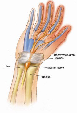
X-rays and a physical exam can usually give the information needed to evaluate a distal radius fracture. Occasionally, further imaging such as a CT or MRI may be considered, but this is rare. In addition to the bone, there is certainly injury to the surrounding soft structures such as tendons, ligaments, muscles and nerves which will affect your outcome.

How is my broken wrist going to be treated? Do I need surgery?
Many factors go into deciding the best treatment for your wrist fracture, but typically I choose the treatment regimen which will allow the best recovery potential, both in the short term and in the long term. This usually initially involves an attempt at manipulating the fracture into the most ideal healing position and holding that position with a splint or cast. If the wrist fracture falls into an unacceptable position for healing despite maximal non-operative care, then surgical intervention is considered. With experience, training and careful review of current research, I can often predict which fractures can be treated without surgery and which ones will need further surgical stabilization.

How long is the recovery from my broken wrist?
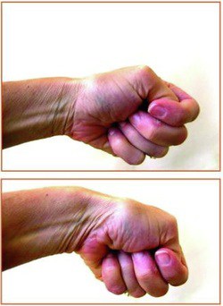
Most patients, regardless of the type of break, have very good long term results. Most fractures treated without surgery will require 6 weeks in a cast, and another 6 weeks to regain most of the strength that was lost. Distal Radius fractures which require surgical stabilization often only need 2 weeks of splinting, followed by 4 weeks of exercise to regain range of motion and another 6 weeks to regain strength. Most patients are able to do most activities by three months after the injury, but individual circumstances certainly vary.

Thumb Base Arthitis

Arthritis at the Base of the Thumb

Other names: Basal Joint arthritis, 1st CMC arthritis

6871969What is it?
The thumb is made up of three highly mobile joints which allow for a high degree of motion. These joints are normally quite mobile and consist of the cartilage covered bone ends, allowing for shock absorption and smooth gliding coordinated movement. Over time, the cartilage can wear down in these joints, causing a painful gliding surface and less shock absorption, creating a condition commonly known as arthritis. The most common joint in to wear down in the hand is the highly mobile joint at the base of the thumb known as the thumb basal joint, other wise known as the carpometacarpal (CMC) joint. (see figure) You can feel for pain in this joint by palpating it in the fleshy part of the thumb side of the palm, about a centimeter above the wrist joint.

Who gets it?
The common causes for arthritis at the base of the thumb include age, female gender, joint laxity and previous injury to the joint. Genetics probably has the greatest influence on the condition as the exact cause is unknown.

This type of arthritis is generally called osteoarthritis, which is the progressive wearing down of a joint over time due to age. The other common form of arthritis is rheumatoid arthritis, which is an autoimmune inflammatory disease that rapidly destroys joints throughout the body.How do I know if I have it?Pain at the base of the thumb is the hallmark of the condition, though there are other conditions that can also cause similar symptoms which should be ruled out such as carpal tunnel syndrome or wrist arthritis. Pain occurs with pinching, grasping, buttoning buttons or opening a jar and eventually the destruction in the joint surfaces alters the thumb mechanics causing deformity and weakness.

To fully evaluate the joint, routine x-rays are helpful in identifying the condition and quantifying the amount of joint destruction.

What can be done to treat the arthritis at the base of the thumb?
First, this is a common condition and should be generally considered a normal part of the aging process. In addition, there are no cures for this this type of arthritis (osteoarthritis) so treatments are generally aimed at symptom relief and functional recovery. Perhaps someday we will have treatments that can address the cause of this arthritis.

Many people with this form of arthritis need no treatment as the symptoms remain tolerable. Often anti-inflamatory medication such as ibuprofen or Tylenol can decrease symptoms to a tolerable level, especially before significant activity. Thumb splints are also helpful in immobilizing the joint while allowing hand motion. These can be custom made by a hand therapist for optimal fit and function. Some patients find relief with steroid injections into the joint, which can be repeated up to 3 times a year for symptomatic treatment. Eventually, some patients continue to have pain and elect to proceed to surgical management which consists of removing the offending joint and reconstructing the thumb ligaments to eliminate the bone-on-bone pain and restore function.

DeQuervain’s Tenosynovitis

Finklestein’s Test above, causing intense pain
By Scott Ruhlman MD 1/31/2011

5416710

Do you experience pain at the base of the thumb after lifting your new child? Does the back of your thumb hurt after typing? Do repetitive activities such as knitting, gardening, or sports cause severe wrist pain? You may be experiencing DeQuervain’s Tenosynovitis , a common treatable condition resulting from inflammation of the thumb tendons.

The following are activities that commonly result in Dequervain’s tenosynovitis:

  • Knitting
  • Gardening
  • Playing a musical instrument
  • Improperly holding your child (lift with your shoulders and fixed wrists, not by flexing your wrists)
  • Typing
  • Carpentry
  • Walking your pet on a leash
  • Sporting activity

What is DeQuervain’s Tenosynovitis?
DeQuervain’s Tenosynovitis is a condition where synovial tissue surrounding the thumb extensor tendons become inflamed as they pass through a tight pulley. As the synovium becomes inflamed, the process amplifies, exacerbating the painful symptoms. Eventually all thumb movement become painful.

How is DeQuervain’s Tenosynovitis diagnosed?
The most sensitive test is Finklestein’s test. This is a test which causes exquisite pain at base of the thumb when the thumb is placed in the palm and the wrist ulnarly deviated.

It is important to visit a hand surgeon to help differentiate DeQuervain’s tenosynovitis from other common conditions such as carpal tunnel syndrome, arthritis, nerve injury, or even fracture.

How is DeQuervain’s Tenosynovitis Treated?
The first step is proper identification of the condition and your particular reasons for the inflammation. Activity modifications such properly lifting of your baby can help alleviate symptoms. Bracing, anti-inflammatory medications and steroid injections can also dramatically decrease the inflammation.

Occasionally surgical release of the affected tendons are required to relieve the symptoms of Dequervain’s tenosynovitis, but this is usually reserved for persistent cases. The vast majority of cases I see do not require surgery.

Carpal Tunnel Surgery

Carpal Tunnel Syndrome

596967

What are the treatment options?
Treatment usually begins with wrist splints, especially at night, anti-inflammatory medications, and activity modifications. Steroid injections into the carpal tunnel may also provide relief of symptoms, but unfortunately no interventions short of surgery give adequate space for the nerve in the carpal tunnel once there is too much pressure. Ultimately, surgery may be necessary to relieve the pressure on the median nerve in the carpal tunnel.

What is involved in surgery?
Carpal tunnel surgery involves an incision in the base of the palm to gain access to the carpal tunnel. The goal of surgery is to provide more space to for the constricted nerve, which the 30 minute procedure reliably accomplishes. I perform the procedure typically in an outpatient setting and most patients have only a few days of discomfort from the incision are able to use their hand fully by 2 weeks after the sutures are removed. Many patients are able to return to work within 1-3 days, and the dressing stays in place until the sutures are removed.

It is important to note that in severe cases, the nerve has sustained permanent injury from years of compression. Though the surgery reliably relieves pressure on the nerve, sometimes the damage is irreversible and persistent numbness or weakness remain.Interested in learning more about your symtoms? Contact Dr. Scott Ruhlman’s office at 206-633-8100 or submit your question here

Arthroplasty

The wrist is a more complicated joint than the hip or the knee. At the base of the hand are two rows of bones, with four bones in each row. These are called the carpals. The long thin bones of the hand radiate out from one row of carpals toward the fingers and thumb. The two bones of the lower arm (radius and ulna) form a joint with the other row of carpals. All the bone ends are covered with a slick, elastic tissue called cartilage, which enables the bones to move smoothly against each other. However, if the cartilage is worn away or damaged by injury, infection or disease, the bones will rub against each other causing pain. During any total joint replacement, the worn-out bone ends are removed and replaced by an artificial joint (prosthesis).