Knee Replacement Surgery

Knee replacement surgery is also known as knee arthroplasty. This is a procedure that can help relieve pain of the knee and restore function of the knee joint. The knees develop osteoarthritis and other disorders that prevent them from bending appropriately.

During this knee surgery, the surgeon will remove damaged bone and cartilage from the thighbone, shinbone, kneecap, and surrounding areas and replace what is removed with an artificial prosthetic joint.

Today there are many good alternatives to the old crude hinges of yesteryear. You can have a metal alloy knee or one made with high-grade plastic and polymer. The surgeon performing knee replacement surgery in Seattle will decide which one is best for you based on your age, activity level, weight, and overall health.

What is arthritis?

Arthritis is means “inflammation of the joint.” Most people think of arthritis as the wearing away of joint cartilage. This causes severe inflammation and pain within the joint. When most of this cartilage is lost and the bone is exposed, we consider this osteoarthritis.

It is the “wear-and-tear” that occurs with age or athletic activities. Other types of arthritis are rheumatoid arthritis (a more severe type), gouty arthritis (more painful and less common), and lupus arthritis (uncommon).

What are the risks of knee replacement arthroplasty?

The risks of a knee replacement include: infection, knee stiffness, heart attack, stroke, nerve damage, or blood clots in the leg vein or lungs. Only around 2 percent of those undergoing this procedure will have serious complications, according to the American Association of Orthopedic Surgeons.

Who is a candidate for a knee replacement?

The most frequent reason for knee replacement surgery in Seattle is for the repair of joint damage caused by osteoarthritis and rheumatoid arthritis. You may be an applicant for knee replacement if:

You have disabling pain. Individuals who need knee replacement surgery commonly have problems walking, stooping, climbing stairs, and getting in and out of chairs. These people also experience moderate or severe knee pain at rest. Surgery may be an option in this case.

You have a knee deformity. Knee replacement surgery can be particularly helpful for people who have a knee that bows in or out or one that has lost function and shape from rheumatoid arthritis.

You’re 55 or older. Knee replacement is normally performed in older adults, but it may be considered for adults of any ages. Young physically active people are more likely to wear out their new knees prematurely, so doctors try not to replace young knee joints.

You have failed on other treatments. More conservative treatments are weight loss, physical therapy, a cane or other walker, medications, and braces. If these don’t help you, you may be a candidate for a knee replacement.

Your general health is good. Conditions such as restricted blood flow, cardiovascular disease, diabetes, serious lung disease, cancer, or infections can complicate surgery and recovery. The ideal candidate will not be in poor health.

What are the alternatives to knee replacement surgery?

Knee replacement is typically reserved for patients who have tried all of the other treatments and failed with them. Some of these individuals are still left with significant pain during normal activities, regardless of what medication or treatment they have taken.

Patients who only have occasional pain, are who are able to participate in athletic activities may not need a knee replacement. Others who have not tried non-operative treatments are probably not ready for a knee arthroplasty, either.

Non-operative treatment options include: arthrocentesis, arthroscopy, cortisone injections, Synavisc injections, physical therapy, heat therapy, massage therapy, cartilage transplantation, specialized knee braces, and arthrodesis with knee fusion, weight loss, and oral medications.

What are the Contraindications of knee arthroplasty?

There are a few reasons your doctor would not want you to undergo knee replacement surgery in Seattle. These include but are not limited to: knee sepsis, severe vascular disease, recurvatum deformity with muscle weakness, extensor mechanism dysfunction, a remote source of ongoing infection, and the presence of a well-functioning joint.

There are also relative contraindications where the medical condition doesn’t make the procedure safe or effective. These include: obesity, neuropathic joint, past history of osteomyelitis of the knee, and skin conditions that affect the knee (like psoriasis).

Remember, total knee replacement is an elective and life-enhancing surgery. It is not a life-saving surgery. The decision to undergo total knee surgery is one you must make once you are informed and well-educated on the alternatives, risks, and complications. It is important that you be aware of your options and be realistic with your expectations.

Meniscus Tears and Repairs

Meniscus tears occur on the C-shaped disc that supports and cushions the knee. When this structure is damaged or torn, there may be pain, swelling, stiffness, and limited range of motion. Twisting or turning incorrectly can bring on a meniscus tear or injury. Knee arthroscopy is a safe procedure the orthopedic specialist may perform to resect or repair a meniscus tear and diagnose the extent of the injury to the knee.

What is a Meniscus Tear?

The meniscus is a rubbery, C-shaped disc that supports and cushions the knee. Injury to this part of the knee is common. There are two menisci in each knee. One is at the outer, or lateral side of the knee and the other is at the inner, or medial, side. These structures keep the knee steady by allowing for balance of weight across the knee. If one of these menisci is torn, the knee does not function properly and the torn meniscus can scuff and damage the surfaces of the knee resulting in arthritis.

What are the symptoms of a Meniscus Tear?

The symptoms associated with meniscus tears vary greatly depending on the severity. Minor tears may result in slight pain and swelling. If there are no mechanical symptoms, such as catching or locking, these tears may resolve on their own in around 2 or 3 weeks. More moderate tears can lead to pain at the side and back of the knee. The swelling of a moderate tear slowly gets worse over a 2 or 3 day period.

The knee will feel stiff with this type of injury and there will be limitations to how far the knee can be bent. The symptoms may go away after a week or two but can come back anytime there is re-injury or overuse of the knee. The pain of a moderate tear could go on for years if the tear is not treated properly.

The third type of tear is a severe tear. With these, pieces of the meniscus are torn and can displace into the joint space. This will make the knee pop, catch or lock without notice. It will be difficult to straighten the knee as well. The knee may be described as “wobbly” and give way without any warning. Most people who suffer a severe tear have pain, swelling, and stiffness immediately following the injury and it gets worse over the next few days.

What is the Cause of a Meniscus Tear?

Twisting or turning quickly can lead to a meniscus tear. Oftentimes, the foot is planted while the knee is bent. These types of tears occur when the person is lifting something really heavy or playing sports. As people get older, the likelihood of meniscus wear and tear increases.

How is a Meniscus Tear Diagnosed?

Most of the time, the orthopedic specialist inquires with the patient regarding past injuries and accidents. The doctor will also perform a physical examination to help find out if the meniscus is torn and causes pain. Testing may involve X-Rays and/or an MRI so the doctor can see if the meniscus is torn and how serious the injury actually is.

How is a Meniscus Tear Treated?

The orthopedic specialist will treat the tear based on the severity of symptoms, where the tear is located, how serious the tear is, your age, and how active you are. Treatment could involve rest, ice therapy, non-steroidal anti-inflammatories, elastic bandage wrapping, and elevating the leg up on pillows. The doctor may order physical therapy, too.

Sometimes, surgery is necessary to repair the meniscus or remove parts of the torn tissue. Surgical repair is usually the best choice for younger people who need to continue working and participating in sports.

What is Knee Arthroscopy?

Knee arthroscopy is one of the most commonly performed surgical interventions for repair of the meniscus. The orthopedic specialist inserts a small lens into the knee area through a tiny incision that is hooked up to a sterile camera and light source. This allows him a clear view of inside of the knee. Then, the doctor can use miniature surgical instruments to trim and repair the meniscus tear.

After the surgery, the doctor may put a brace on the knee to allow it to be immobilized to heal if the meniscus tear has been repaired rather than removed. If the tear is removed, patients generally are able to fully weight bear immediately following surgery. If necessary, a prescribed rehabilitation program will help you get back on your feet after the procedure.

Ulnar Nerve Entrapment

master_74Description
Ulnar nerve entrapment occurs when one of the nerves in the arm (the ulnar nerve) becomes compressed and can’t function normally. This can give symptoms of “falling asleep” in the ring finger and little finger, especially when the elbow is bent. You may have aching pain on the inside of the elbow.

In some cases, you may have trouble moving the fingers in and out, or manipulating objects. Carpal tunnel syndrome has similar symptoms but involves a different nerve (the median nerve). Carpal tunnel syndrome typically causes tingling in the thumb, index finger and long finger.

The ulnar nerve is one of the three main nerves in the arm. It travels from under the collarbone and along the inside of the upper arm. It passes through a tunnel (the cubital tunnel) behind the inside of the elbow. Here you can feel the nerve through the skin. It is commonly called the “funny bone.”

Beyond the elbow, the nerve travels under muscles on the inside of the arm, and into the hand on the pinky side of the palm. When the nerve goes into the hand, it travels through another tunnel (Guyon’s canal). The most common place where the nerve gets compressed is behind the elbow. Sometimes it gets compressed at the wrist, beneath the collarbone, or as it comes out of the spinal cord in the neck.

The nerve functions to give sensation to the little finger and the half of the ring finger that is near the little finger. It also controls most of the little muscles in the hand that help with fine movements, and some of the bigger muscles in the forearm that help to make a strong grip.

cons1_352_143
cons2_352_143

Risk Factors/Prevention
It is not known exactly what causes compression of the ulnar nerve. Some factors can make it more likely that the nerve will be compressed. These include prior fractures of the elbow, bone spurs, swelling of the elbow joint, or cysts. A direct blow to the inside of the elbow, leaning on the elbow for prolonged periods, or repetitive activity that requires a bent elbow can irritate the nerve if it is already compressed. If the ulnar nerve is compressed at the wrist, the cause is more likely to be a cyst in Guyon’s canal.

Symptoms
Numbness and tingling in the ring finger and little finger are common symptoms of ulnar nerve entrapment. Often these symptoms come and go. They happen more often when the elbow is bent, such as when you are driving or talking on the phone. Some people wake up at night because their fingers are numb. You may also have weakness of grip and difficulty with finger coordination (such as typing or playing an instrument). If the nerve is very compressed or has been compressed for a long time, muscle wasting in the hand can occur. Once this happens, muscle wasting cannot be reversed. For this reason, it is important to see the doctor as soon as you experience any of the symptoms.

Diagnosis
Always see an orthopaedist if you are having symptoms of ulnar nerve entrapment that interfere with normal activities or last more than a few weeks.
The doctor will examine the arm to check the nerve, and try to determine where the nerve is compressed. If the nerve is irritated, tapping over the nerve at the “funny bone” can cause a shock into the little finger and ring finger, although this can happen when the nerve is normal as well. The doctor will probably move the shoulder, elbow and wrist to see if any of these cause symptoms. The doctor will test the sensation in the fingers.

Although most causes of compression of the ulnar nerve cannot be seen on X-ray, the doctor may take an X-ray of the elbow or wrist to look for bone spurs, arthritis or other places that the bone may be compressing the nerve. If the doctor thinks that the nerve is compressed at the wrist, a CT scan (computed tomography) or MRI (magnetic resonance image) may be recommended to see if a cyst or other structure is the cause of the compression.

The doctor may recommend nerve conduction studies. These are special tests to determine how well the nerve is working and to help localize the area of compression. Nerves work like wires; when the nerve is not working well, it takes too long for the nerve to conduct. During this test, the nerve is stimulated in one place; the amount of time it takes for the response to be conducted to another place is determined. The area where the nerve conduction takes too long is likely to be the place where the nerve is compressed. Sometimes, a small needle is put into some of the muscles that the ulnar nerve controls. This can determine if there is any evidence of muscle wasting.

cons1_354_143Treatment Options
Unless you have a lot of muscle wasting, your doctor will probably recommend nonsurgical treatment initially. The following treatments may help to improve the symptoms. They may be all the treatment you need.

  • Avoid frequent use of the arm with the elbow bent. If you use a computer frequently, make sure that your chair is not too low. Do not rest the elbow on the armrest.
  • Avoid leaning on the elbow or putting pressure on the inside of the arm. For example, do not drive with the arm resting on the open window.
  • Keep the elbow straight at night when you are sleeping. This can be done by wrapping a towel around the straight elbow, wearing an elbow pad backwards, or using a special brace.

If symptoms are acute, the doctor may recommend that you take an anti-inflammatory medicine such as ibuprofen to help reduce swelling around the nerve. Steroid (cortisone) injections around the ulnar nerve are not generally used because there is a risk of damage to the nerve.

cons1_355_143

Some doctors think that exercises to help the nerve slide through the tunnels can improve the symptoms. These exercises can help keep the arm and wrist from getting stiff.

cons1_356_143
cons2_356_143

Treatment Options: Surgical
If you are not improving with the strategies listed above, if the nerve is very compressed, or if you have muscle wasting, the doctor may recommend surgery to take pressure off of the nerve. Most often, the surgery is done around the elbow, but it can be done at the wrist if that is the place of the compression. Sometimes, the nerve is compressed in both places, so surgery is done at both the elbow and the wrist.

Surgeons use various ways to relieve compression from the nerve around the elbow. All of the operations involve making an incision around the elbow. In one operation, only the “roof” is taken off of the cubital tunnel. This tends to work best when the nerve compression is mild. More commonly, the nerve is moved from its place behind the elbow to a new place in front of the elbow. This is called an anterior transposition of the ulnar nerve. The nerve can be moved to lie under the skin and fat but on top of the muscle (subcutaneous transposition), within the muscle (intermuscular transposition) or under the muscle (submuscular transposition). There are many factors that go into deciding where the nerve is moved. The doctor will recommend the best option for you.

cons1_357_143

If the nerve is compressed at the wrist, a zigzag incision will be made at the base of the palm on the pinky side. The surgeon will open the roof of Guyon’s canal to take the pressure off the ulnar nerve. If there is a cyst or another reason for the compression, the surgeon will remove that at the same time.
The surgery is usually done on an outpatient basis or with an overnight stay in the hospital. Depending on the type of surgery, you may need to wear a splint for a few weeks after the operation. A submuscular transposition usually requires a longer time (3-6 weeks) in a splint. The surgeon may recommend physical therapy to help you regain strength and motion in the arm.

cons1_358_143

The results of the surgery are generally good. If the nerve is very badly compressed or if you have muscle wasting, the nerve may not be able to get back to normal and you may have some symptoms even after the surgery. Nerves recover slowly, and it can take a long time to know how well the nerve will do after the operation.

Throwing Injuries in the Elbow

Description
With the start of the baseball season each spring, doctors frequently see an increase in elbow problems in young baseball players. A common elbow problem is Little Leaguer’s Elbow.

The elbow is the joint where the upper arm bone (humerus) meets the two bones of the lower arm (ulna and radius). The elbow is a combination hinge and pivot joint. The hinge part of the joint lets the arm bend like the hinge of a door; the pivot part lets the lower arm twist and rotate. The rounded ends of the upper arm bone give the elbow its two “knobs” or bumps (epicondyle). Several muscles, nerves and tendons (connective tissues between muscles and bones) cross at the elbow.

master_66

Injury occurs when the repetitive throwing creates an excessively strong pull on elbow tendons and ligaments. The young player feels pain at the knobby bump on the inside of the elbow.

Little Leaguer’s Elbow can be serious if it becomes aggravated. Repeated pulling can tear the ligament and tendon away from the bone. The tearing may pull tiny bone fragments with it in the same way a plant takes soil with it when it is uprooted. This can disrupt normal bone growth, resulting in deformity.

Osteochondrosis dissecans is a less common condition that is also caused by excessive throwing and may be the source of the pain on the outside of the elbow.
Muscles work in pairs. In the elbow, if there is pulling on one side, there is pushing on the other side. As the elbow is compressed, the joint smashes immature bones together. This can loosen or fragment the bone and cartilage. The resulting condition is called osteochondrosis dissecans.

Risk Factors / Prevention
Little Leaguer’s Elbow affects pitchers and other players who throw repetitively. Continuing to throw may lead to major complications and jeopardize a youngster’s ability to remain active in a sport that requires throwing.

Symptoms
Little Leaguer’s Elbow may cause pain on the inside of the elbow. A child should stop throwing if any of the following symptoms appear:

  • Elbow pain
  • Restricted range of motion
  • Locking of the elbow joint

Treatment Options
If left untreated, osteochondrosis dissecans can become a complicated condition. Younger children tend to respond better to nonsurgical treatments.

  • Rest the affected area.
  • Apply ice packs to bring down any swelling.
  • If pain persists after a few days of complete rest of the affected area or if pain recurs when throwing is resumed, stop the activity again until the youngster gets treatment.
  • Return to throwing.

Treatment Options: Surgical
Surgery may be necessary, especially in girls more than 12 years old and boys more than 14 years old.

Tennis Elbow (Lateral Epicondylitis)

master_71Description
Tennis elbow is a degenerative condition of the tendon fibers that attach on the bony prominence (epicondyle) on the outside (lateral side) of the elbow. The tendons involved are responsible for anchoring the muscles that extend or lift the wrist and hand (see Figure 1).

Risk Factors/Prevention
Tennis elbow happens mostly in patients between the ages of 30 years to 50 years. It can occur in any age group. Tennis elbow can affect as many as half of athletes in racquet sports. However, most patients with tennis elbow are not active in racquet sports. Most of the time, there is not a specific traumatic injury before symptoms start. Many individuals with tennis elbow are involved in work or recreational activities that require repetitive and vigorous use of the forearm muscles (see Table 1). Some patients develop tennis elbow without any specific recognizable activity leading to symptoms.

Symptoms
Patients often complain of severe, burning pain on the outside part of the elbow. In most cases, the pain starts in a mild and slow fashion. It gradually worsens over weeks or months. The pain can be made worse by pressing on the outside part of the elbow or by gripping or lifting objects. Lifting even very light objects (such as a small book or a cup of coffee) can lead to significant discomfort. In more severe cases, pain can occur with simple motion of the elbow joint. Pain can radiate to the forearm.
To diagnose tennis elbow, tell the doctor your complete medical history. He or she will perform a physical examination.

  • The doctor may press directly on the bony prominence on the outside part of the elbow to see if it causes pain.
  • The doctor may also ask you to lift the wrist or fingers against pressure to see if that causes pain.

cons1_304_132X-rays are not necessary. Rarely, MRI (magnetic resonance imaging) scans may be used to show changes in the tendon at the site of attachment onto the bone.

Treatment Options
In most cases, nonoperative treatment should be tried before surgery. Pain relief is the main goal in the first phase of treatment. The doctor may tell you to stop any activities that cause symptoms. You may need to apply ice to the outside part of the elbow. You may need to take acetaminophen or an anti-inflammatory medication for pain relief.
Orthotics can help diminish symptoms of tennis elbow. The doctor may want you to use counterforce braces and wrist splints. These can reduce symptoms by resting the muscles and tendons (see Figure 2).

cons1_305_132

Symptoms should improve significantly within four weeks to six weeks. If not, the next step is a corticosteroid injection around the outside of the elbow. This can be very helpful in reducing pain. Corticosteroids are relatively safe medications. Most of their side effects (i.e., further degeneration of the tendon and wasting of the fatty tissue below the skin) occur after multiple injections. Avoid repeated injections (more than two or three in a specific site).

cons1_306_132
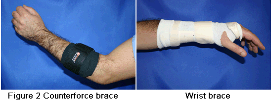
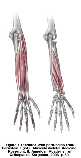
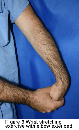
After pain is relieved, the next phase of treatment starts. Modifying activities can help make sure that symptoms do not come back. The doctor may want you to do physical therapy. This may include stretching and range of motion exercises and gradual strengthening of the affected muscles and tendons (see Figure 3). Physical therapy can help complete recovery and give you back a painless and normally functioning elbow. Nonoperative treatment is successful in approximately 85 percent to 90 percent of patients with tennis elbow.

Treatment Options: Surgical
Surgery is considered only in patients who have incapacitating pain that does not get better after at least six months of nonoperative treatment.
The surgical procedure involves removing diseased tendon tissue and reattaching normal tendon tissue to bone (see Figure 4). The procedure is an outpatient surgery; you do not need to stay in the hospital overnight. It can be performed under regional or general anesthesia.

Technique for surgical treatment of lateral epicondylitis. A, Skin incision over the lateral epicondyle. B, Distal reflection of the extensor mechanism exposing the lateral compartment of the elbow. C, Excision of pathologic tissue from the underside of the extensor mechanism. D, Decortication of the lateral epicondyle. E, Drilling of two V-shaped tunnels within the lateral epicondyle. F, Reattachment of the extensor mechanism to the lateral epicondyle. G, Side-to-side repair of the extensor tendon mechanism.

cons1_307_132

Most commonly, the surgery is performed through a small incision over the bony prominence on the outside of the elbow. Recently, an arthroscopic surgery method has been developed.

So far, no significant benefits have been found to using the arthroscopic method over the more traditional open incision.

After surgery, the elbow is placed in a small brace and the patient is sent home. About one week later, the sutures and splint are removed. Then exercises are started to stretch the elbow and restore range of motion. Light, gradual strengthening exercises are started two months after surgery. The doctor will tell you when you can return to athletic activity. This is usually approximately four months to six months after surgery. Tennis elbow surgery is considered successful in approximately 90 percent of patients.

Tell us how we did!
close slider