mm

About Orthopedic Specialists

Orthopedic Specialists of Seattle provides new and advanced procedures including endoscopic carpel tunnel release surgery for carpal tunnel syrome, complex joint restoration procedures, anterior approach hip replacement surgery, and more.

What happens if bunion surgery does not work?

Bunions

Most bunions can be treated without an operation, but sometimes surgical procedures are needed to relieve pain and deformity. Unfortunately, in some cases bunion repairs fail and the pain or deformity returns. There are multiple factors that can contribute to this challenging scenario. Bunions can recur because of medical conditions or non-healing of the bone after surgery. In some cases, there are technical aspects that can be improved to achieve the desired result with additional surgery.

The goal of correcting a failed bunion repair, using a procedure called revision surgery, is to relieve pain and deformity of the first toe that remains after the initial surgery. Sometimes arthritis develops after bunion surgery. This may require a different procedure than the first. It is important to figure out why the first surgery failed to prevent another failure.

Symptoms

Revision surgery is for those with deformity and pain after bunion surgery. The pain may be the same or different from that experienced before the first surgery. Your foot and ankle orthopedic surgeon will examine deformities of bones and joints and their various angles around the first toe.

Revision bunion surgery is not advised for patients with poor blood flow or certain nerve conditions. Bunions should not be revised if they are painless and do not cause problems. No bunion surgery should be performed solely to make the foot look better.

Treatment

Revision surgery typically takes a little longer to perform than the first procedure because it can be more complicated due to scar tissue and altered anatomy. Incisions are made around the first toe and by the arch of the foot. Special instructions may include wrapping, protection with boots or braces, and limited activity. Usually this is an outpatient procedure, meaning you can go home the same day as surgery.

There are numerous accepted methods of correcting a bunion. Some are treated with a bone cut of the first toe, while others require a fusion. Hardware in the form of plates and/or screws may be used to maintain corrections or hold fusions solid.

Recovery

Recovery will take at least as long as the first surgery. After surgery, you will be placed in a brace or shoe. You will have to avoid putting weight on the foot or only put weight on the heel for a period of time determined by your surgeon. Sutures usually are removed two weeks after surgery.

What are Sesamoids?

Sesamoids

Sesamoids are bones that develop within a tendon. The one most people are familiar with is in the kneecap, however they most commonly occur in the foot and hand. Two sesamoids, each about the size of a corn kernel, typically are found near the underside of the big toe.

Symptoms

Pain from a sesamoid injury is focused under the big toe on the ball of the foot. With sesamoiditis or a stress fracture, pain may develop gradually, whereas with a fracture, the pain will be immediate after trauma. Swelling and bruising may or may not be present. There may be difficulty and pain when bending and straightening the big toe.

Causes

Sesamoids act like pulleys, increasing the ability of the tendons to transmit muscle forces. The sesamoids in the forefoot also assist with weight bearing and help elevate the bones of the big toe. Like other bones, sesamoids can break in a traumatic injury. They also can develop a stress fracture from overuse. In addition, the tendons surrounding the sesamoids can become irritated or inflamed. This is called sesamoiditis and is a form of tendinitis or tendinosis. It is common among ballet dancers, runners, and professional athletes.

Diagnosis

During your examination, your foot and ankle orthopedic surgeon will look for tenderness at the sesamoid bones. Your doctor may manipulate the bone slightly or ask you to bend and straighten the toe. He or she also may bend the big toe up toward the top of the foot to see if the pain intensifies.

Your surgeon will request X-rays of the forefoot to ensure a proper diagnosis. In many people, the sesamoid bone nearer the center of the foot (the medial sesamoid) has two parts (bipartite). Because the edges of a bipartite medial sesamoid are generally smooth, and the edges of a fractured sesamoid are generally jagged, an X-ray is useful in making an appropriate diagnosis. Your physician also may request X-rays of the other foot to compare the bone structure. If the X-rays appear normal, the physician may suggest additional tests such as an MRI or CT scan.

Treatment

Treatment for sesamoiditis usually is nonoperative and successful, but can be frustrating in how long it takes for symptoms to resolve. If conservative measures fail, your physician may recommend surgery to remove the sesamoid bone. First, your specialist will recommend the following:

  • Stop the activity that causes the pain.
  • Take acetaminophen or ibuprofen to relieve the pain.
  • Rest and ice the sole of your foot. Do not apply ice directly to the skin; use an ice pack or wrap the ice in a towel.
  • Wear soft-soled, low-heeled shoes.
  • Use a felt cushioning pad around the sesamoid to relieve stress.
  • Return to activity gradually and continue to wear a cushioning pad of dense foam rubber under the sesamoids to support them. Avoid activities that put your weight on the balls of the feet.
  • Tape the big toe so that it remains bent slightly downward.

In rare occasions, a steroid injection may be appropriate.

If symptoms persist, you may need to wear a removable boot or a cast for 4-6 weeks. Sesamoids tend to heal slowly.

If you have fractured a sesamoid bone, your foot and ankle orthopedic surgeon may recommend conservative treatments before resorting to surgery. You will need to wear a stiff-soled shoe, a boot, or possibly a cast, and your physician may tape the joint to limit movement of the big toe. You also may have to wear a J-shaped pad around the area of the sesamoid to relieve pressure as the fracture heals. Pain relievers such as acetaminophen or ibuprofen may be recommended as well, but know that it may take several months for the discomfort to subside.

In some cases, a fractured sesamoid requires surgery. In this case, repair sometimes can be performed, but often removal of part or all of the sesamoid will be needed.

Recovery

Healing of the sesamoid typically is slow and can take up to six months. The process can be frustrating but is usually successful.

Risks

Failure of healing, avascular necrosis, development of arthritis at the joint between the sesamoid and the first metatarsal, and continued pain are the risks associated with sesamoid injuries. If these should develop, excision of part or all of the sesamoid can usually resolve symptoms.

What are Osteochondral Lesions?

Osteochondral Lesions

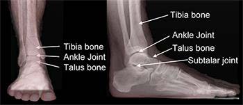
Osteochondral lesions are injuries to the talus (the bottom bone of the ankle joint) that involve both the bone and the overlying cartilage. They may also be called osteochondritis dessicans or osteochondral fractures. These injuries may include softening of the cartilage layers, cyst-like lesions within the bone below the cartilage, or fracture of the cartilage and bone layers. Throughout this article, these injuries will be referred to as osteochondral lesions of the talus (OLT).

Anatomy

The talus is the bottom bone of the ankle joint. Much of this bone is covered with cartilage. The tibia and fibula bones sit above and to the sides of the talus, forming the ankle joint. This joint permits much of the up (dorsiflexion) and down (plantarflexion) motion of the foot and ankle. The blood supply to the talus is not as rich as many other bones in the body, and as a result, injuries to the talus sometimes are more difficult to heal than similar injuries in other bones.

Symptoms

OLTs usually occur after an injury to the ankle, either a single traumatic injury or as a result of repeated trauma. Common symptoms include prolonged pain, swelling, catching, and/or instability of the ankle joint. Symptoms can be vague. After an injury such as an ankle sprain, the initial pain and swelling should decrease with appropriate attention (rest, elevation). Persistent pain in spite of appropriate treatment after several months may raise concern for an OLT.

You may feel pain primarily at the lateral (outside) or medial (inside) point of the ankle joint. Severe locking or catching symptoms, where the ankle freezes up and will not bend, may indicate that there is a large osteochondral lesion or even a loose piece of cartilage or free bone within the joint.

Causes

The majority of OLTs, as many as 85%, occur after a traumatic injury to the ankle joint. Ankle sprains are a common cause of OLTs. With this type of injury, a section of the talus surface may impact another part of the ankle joint (tibia or fibula) and injure the talus. Some patients, however, have no history of an injury to their ankle.

Diagnosis

Foot and ankle orthopedic surgeons diagnose OLTs with a combination of clinical and special studies. Your surgeon may have a suspicion that you have this type of injury from the history you provide and their physical examination. Imaging is necessary to confirm the diagnosis. Occasionally, regular X-rays can show an OLT but frequently additional imaging is needed, such as a CT scan or an MRI.

Treatments

Once the diagnosis has been confirmed, treatment may be surgical or non-surgical, depending on the nature of the OLT, presence of other injuries, and patient characteristics.

Non-surgical Treatment

Non-surgical treatment is appropriate for certain lesions and usually involves immobilization and restricted weightbearing. This may be followed with gradual progression of weightbearing and physical therapy. The goal of non-surgical treatment is to allow the injured cartilage and bone to heal. Patients may have an OLT that is present and doesn’t cause pain or limitations or a lesion that becomes painful but improves. In these cases, no additional treatment is necessary. It was once believed that all OLTs progress and worsen with time. This has been shown to no longer be true.

Surgical Treatment

Other lesions may be more appropriately treated with surgery. The goals of surgery are to restore the normal shape and gliding surface of the talus in order to re-establish normal mechanics and joint forces. The hope is to minimize symptoms and limit the risk of developing arthritis.

Depending on the characteristics and location of the OLT, surgery may done arthroscopically or by opening the skin. Arthroscopy uses a camera and small instruments to view and work within the joint through small incisions. It may not be possible to properly treat certain lesions arthroscopically due to the size or location of the lesion. Treatments may include debridement (removing injured cartilage and bone), fixation of the injured fragment, microfracture or drilling of the lesion, bone grafting the bone cyst below the cartilage, and/or transfer or grafting of bone and cartilage. You and your foot and ankle orthopedic surgeon can discuss these treatment options and decide which one is best. Often, there may be several treatment options.

If you have any underlying conditions that may predispose to an OLT such as ankle instability, ankle impingement, a high arched (cavovarus) foot, or tight calf muscles, it may be necessary to correct these problems at the time of surgery as well.

Recovery

Recovery after OLT treatment varies depending upon the nature of the lesion and the treatment. Most treatments require a period of immobilization and restricted weightbearing that can range from several weeks to several months. More involved procedures that include bone grafting or cartilage transfer may require a longer period of recovery.

The results of non-surgical treatment of OLTs have been disappointing. Most studies show that full resolution of the pain from an OLT occurs in less than half of cases. Studies examining the outcomes after surgical debridement and microfracture (drilling) of OLTs have shown that more than 70% of patients have a good or excellent outcome. Procedures that transfer bone or cartilage to an OLT also have good outcomes. In general, the best results can be expected for smaller lesions.

Risks and Complications

All surgeries come with possible complications, including the risks associated with anesthesia, infection, damage to nerves and blood vessels, and bleeding or blood clots. Complications, such as infection or wound healing problems, are uncommon after arthroscopic ankle surgery. More complex procedures with an open surgical approach or bone or cartilage transfer may have additional risks. In addition to standard surgical risks, additional complications may include the failure of any transplanted tissue (bone or cartilage). Despite surgery going as well as possible, there is still a chance the pain will persist requiring additional treatment in the future.

What are Biologics?

Biologics

Biologics refers to a group of substances that your surgeon may inject in the office or use during your surgery to help you heal. They contain specific material or cells that have an effect on other nearby cells and processes in your body. Depending on the contents, they potentially can help stimulate your body to form new bone, build new blood vessels, or limit damaging inflammation.

Where do biologics come from?

Some biologics are harvested directly from you! By taking some of your own blood, bone marrow, or fat cells, your surgeon can isolate certain types of your own stem cells and growth factors. Examples of biologics that can be harvested from your own tissue include platelet-rich plasma (PRP) and bone marrow aspirate concentrate (BMAC).

Other biologics are harvested from other sources, usually from human donors. These commercially available biologics have many different brand names, which can be confusing. On individual product websites, you can find more information about what the biologics actually contain. However, the following are some examples of the substances found in commercially available biologics:

  • Stem cells and/or bone cells
  • Bone morphogenetic protein (BMP) – a signal that helps new bone to form
  • Platelet-derived growth factor (PDGF) – a signal involved in the healing process after injury
  • Vascular endothelial growth factor (VEGF) – a signal that stimulates new blood vessels to form
  • Amniotic membrane

Treatment

Your foot and ankle orthopedic surgeon may recommend a biologic injection to treat conditions in the office. For example, PRP injections may be used for Achilles tendinitis, plantar fasciitis, or ankle arthritis. There is still debate in the scientific community about the effectiveness of these injections, and research is continuing.

Your surgeon also may recommend using biologics during your surgery. In foot and ankle surgery, they are most commonly used to help bone healing. Many foot and ankle surgeries involve fusions, or trying to get two or more separate bones to become one. In certain situations, it can be more difficult for bones in the foot to join together, and using biologics can help increase the chance of the bones healing properly.

Risks and Complications

Though uncommon, your immune system may react negatively to the biologics. Using biologics from your own body lowers the chance of this happening, and commercially-available biologics are tested for disease and treated to minimize reactions.

You may experience local pain or changes from obtaining the material from your own body, and you may be limited in the amount available in your body. Biologics from donor sources can be significantly more expensive and vary in quality. If the biologics come from cadaver sources, there is a small risk of transmission of infection. Talk to your foot and ankle orthopedic surgeon to discuss the pros and cons of each option for your specific situation.

FAQs

Can biologics help me avoid surgery?

Because so much depends on your specific situation, you should discuss your goals with your foot and ankle orthopedic surgeon before considering biologics. We know that some biologics can help your bones heal together when used during surgery. However, there is debate about how helpful they are for other conditions.

For patients with arthritis, for example, these injections are not going to regenerate lost cartilage or substitute for surgery, but they may decrease inflammation and improve the symptoms. As long as you are making an informed choice, and have agreed upon reasonable expectations with your surgeon, biologics can be a useful tool in your treatment plan.

A Final Note

This is a very exciting field with new products being developed and new studies being reported almost every year. The above is a current summary of this area of medicine, but given the rate of change, it is quite possible to be different in the future as this field develops.

Post-operative Instructions for Hip Arthroscopy

Wound Care

  • You will have a sterile gauze dressing covered with tape. Please keep the dressing clean and dry. You may take a sponge bath, or shower with waterproof plastic wrap over the surgical area (use tape at the edges to prevent leaks).
  • Remove the dressing 3 days after surgery to inspect the incisions. Some clear, yellow, or bloody drainage from the incision is normal. If this happens, keep the incision covered with gauze and change the dressing daily until there is no further drainage. If there is no drainage you may leave the hip open to air. You may get the incision wet 5 days after surgery, but do not submerge in water. Sutures will be removed at your follow up appointment.
  • Occasionally there is excessive bloody drainage; please change the dressing when it becomes completely saturated. Sterile gauze is available at the pharmacy. If you continue to have saturated dressings beyond the first few dressing changes, please call the office.
  • If the incisions are draining pus (opaque, thick, white fluid), or if there is redness that worsens over the next 1-2 days, call the office immediately. Do not apply any ointments or creams.

Activity

You may be weight bearing as tolerated with the use of crutches to assist your operative leg. Please continue to use both crutches at all times for the first 2-4 weeks after surgery. You may gradually increase the amount of time you spend standing and walking. Formal outpatient physical therapy is typically not required.

Avoid heavy lifting, exercising, stretching, running, climbing, squatting, and any jarring activities. Please use pain as your guide; any activity that causes severe pain should be avoided.

Controlling your pain and inflammation

Some pain, swelling, and bruising is expected after surgery. It is usually most severe for the first 2-3 days. The following strategies are especially important during this time.

  • Rest — Take things easy for the first few days, try to rest and avoid prolonged walking or standing.
  • Ice – Apply an ice pack (or a cold therapy machine if you have one) to your operative hip to reduce pain and inflammation. Take care not to put ice directly on the skin. Ice for 30 minutes at a time, and remove for 30 minutes in between sessions. You should continue this for the first 2-3 days or longer if you still have pain and swelling.
  • Elevate – Put pillows under your operative leg, or lie on your opposite hip to elevate. This will help to drain fluid from the leg and reduce swelling.
  • Medication — You may have received a prescription for narcotic and/or anti-inflammatory medication. Please take them as instructed. The medication is most helpful if taken 30-45 minutes prior to any planned activity.

Follow up appointment
If an appointment has not already been scheduled, please call the office at 206-633-8100 and schedule an appointment for 7-10 days after your surgery. During this visit we will examine the surgical incisions, remove sutures if necessary, and take xrays.

Returning to work
You may return to work when it is safe to do so within the above activity restrictions. Please note that your employer may prohibit narcotics while at work. Please continue to rest and ice while at work. You may need to ask for frequent breaks in order to avoid prolonged standing or walking. A doctor’s note or a Duty Status form can be provided during your follow up appointment.

Driving
For those who had LEFT hip surgery, you may drive an automatic transmission once it is comfortable to do so and you are no longer taking narcotic medication. For the RIGHT hip, or those with manual transmission, it may take anywhere from 2-4 weeks depending on your pain level, strength, etc. Please wait to drive until after your follow up appointment so that we can assess your progress.

Medications and common side effects:

  • Narcotics (oxycodone, hydrocodone, etc.) – prescription medication for reducing pain. They may cause drowsiness, confusion, nausea, and constipation. To avoid constipation, increase your intake of fiber, fruits, and vegetables, and stay hydrated. Over the counter laxatives can be taken to treat constipation while on narcotics; please see separate handout or ask your pharmacist.
  • Anti-inflammatories (Ibuprofen, Naproxen, etc.) – available over-the-counter to reduce pain and inflammation. Avoid them if you have diagnosed kidney disease or active ulcers. This medication can cause upset stomach; please take them with food. To treat an upset stomach, take an over-the-counter antacid or proton-pump inhibitor (ask your pharmacist for assistance).
  • Acetaminophen (Tylenol) – Used to reduce pain and decrease fever. Avoid taking this medication if you have liver dis-ease. Taking more than the recommended dose can lead to liver damage. For an adult, it is safe to take up to 3-4,000 milligrams each day (24 hour period). Avoid taking with Percocet, Vicodin, Norco; these prescription narcotics already have acetaminophen in them. It is safe to take Tylenol and an anti-inflammatory at the same time.
  • Antihistamines (e.g., benadryl, hydroxyzine) – Used to treat some side effects from narcotic use, such as itching and nausea. Can cause drowsiness and confusion.

Please call the office if you have the following:

  • Fever above 101°, pus draining from wound, worsening redness or rash
  • Difficulty breathing
  • Continuous bleeding from wound (see “wound care” above)
  • Numbness or weakness of the leg
  • Intolerable pain when the above strategies for pain control have failed.