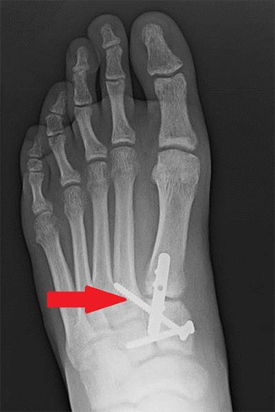
mm

About Orthopedic Specialists

Orthopedic Specialists of Seattle provides new and advanced procedures including endoscopic carpel tunnel release surgery for carpal tunnel syrome, complex joint restoration procedures, anterior approach hip replacement surgery, and more.

What is plantar fasciitis?

Plantar Fasciitis

If your first few steps out of bed in the morning cause severe pain in the heel of your foot, you may have plantar fasciitis, an overuse injury that affects the sole of the foot. A diagnosis of plantar fasciitis means you have inflamed the tough, fibrous band of tissue (fascia) connecting your heel bone to the base of your toes.

Causes

You’re more likely to develop the condition if you’re female, overweight, or have a job that requires a lot of walking or standing on hard surfaces. You’re also at risk if you walk or run for exercise, especially if you have tight calf muscles that limit how far you can flex your ankles. People with very flat feet or very high arches also are more prone to plantar fasciitis.

Symptoms

Plantar fasciitis typically starts gradually with mild pain at the heel bone often referred to as a stone bruise. You’re more likely to feel it after (not during) exercise. The pain classically occurs right after getting up in the morning and after a period of sitting. If you don’t treat plantar fasciitis, it may become a chronic condition.

Treatments

Stretching is the best treatment for plantar fasciitis. It may help to try to keep weight off your foot until the initial inflammation goes away. You can also apply ice to the sore area for 20 minutes 3-4 times a day to relieve your symptoms. Rolling a frozen water bottle on the bottom of your foot can be beneficial.

Your foot and ankle orthopedic surgeon may prescribe a nonsteroidal anti-inflammatory medication such as ibuprofen or naproxen. Home exercises to stretch your Achilles tendon and plantar fascia are the mainstay of treatment and reduce the chance of recurrence.

In one exercise, you lean forward against a wall with one knee straight and heel on the ground. Your other knee is bent. Your heel cord and foot arch stretch as you lean. Hold for 10 seconds, relax and straighten up. Repeat 20 times for each sore heel. It is important to keep the knee fully extended on the side being stretched.

Stretch for Plantar Fasciitis

What is peroneal tendinosis?

Peroneal Tendons

The peroneal tendons are on the outside of the ankle just behind the bone called the fibula. Peroneal tendinosis is the name for the enlargement, thickening, and swelling of these tendons. This usually occurs with overuse, such as a repetitive activity that irritates the tendon over long periods of time.

Symptoms

People with peroneal tendinosis typically have tried a new exercise or markedly increased their activities. Characteristic activities include marathon running or others that require repetitive use of the ankle. Patients usually have pain around the back and outside of the ankle. There often is no history of a specific injury.

Causes

Improper training or rapid increases in training and poorly fitting shoes can lead to peroneal tendinosis. Also, patients who have high arches may be more susceptible because their heel is turned inwards slightly, which requires the peroneal tendons to work harder to turn the ankle to the outside. The harder the tendons work, the more likely patients are to develop tendinosis.

Anatomy

Tendons connect muscle to bone and allow them to exert their force across the joints that separate bones. Ligaments, on the other hand, connect bone to bone. There are two peroneal tendons that run along the back of the fibula. The first is called the peroneus brevis. It runs down around the back of the bone called the fibula on the outside of the leg and connects to the fifth metatarsal on the side of the foot.

The peroneus longus takes its name because it has a longer course. It runs all the way underneath the foot to connect to the first metatarsal on the other side. Both tendons share the major job of turning the ankle to the outside. The tendons are held in a groove behind the back of the fibula and are covered by a ligament-type tissue called a retinaculum.

What is percutaneous Achilles tendon lengthening?

Percutaneous Achilles Tendon Lengthening

Percutaneous Achilles tendon lengthening in a procedure used to stretch a tight Achilles tendon and increase motion at the ankle joint.

Diagnosis

People with a tight Achilles tendon tend to walk on their toes. The tight tendon prevents the foot from sitting flat on the floor, and ankle motion can be limited. When this tightness cannot be treated with non-surgical stretching or physical therapy, surgery is needed.

This procedure may be only one part of a surgery to help correct the position of the foot. Patients that develop ulcers in the front part of the foot may require an Achilles lengthening to decrease pressure on the front part of the foot and allow the ulcers to heal.

This procedure is not recommended when there is active infection or redness around the Achilles tendon. A very tight tendon may require a more complex lengthening surgery.

Treatment

The procedure is minimally invasive. It may be done alone or with other procedures in order to improve the overall position of the foot. It is typically an outpatient procedure, and general or regional anesthesia is typically used. It only takes a few minutes to perform this procedure.

The foot and ankle orthopedic surgeon makes three small incisions at the back of the ankle along the Achilles tendon. The tendon is cut approximately 50 percent at each of these three sites in an alternating pattern. The surgeon does this while an assistant is holding the ankle and stretching the tendon. The tendon stretches as the fibers are cut.

Recovery

Healing time for tendons is approximately 6-8 weeks. You may be in a protective cast, splint, or walking boot initially while the tendon heals. Physical therapy and rehabilitation often are needed after the initial healing period to help with strength and range of motion.

Risks and Complications

All surgeries come with possible complications, including the risks associated with anesthesia, infection, damage to nerves and blood vessels, and bleeding or blood clots. With a percutaneous Achilles tendon lengthening, specific complications are rare. Wound problems such as nonhealing incisions or infection can occur. The Achilles tendon can remain tight after surgery. The Achilles tendon also can completely rupture during surgery or recovery.

FAQs

When will I be able to walk again after this procedure?

Generally, the Achilles tendon will take about 6-8 weeks to heal, so weight bearing and therapy will begin at that point. It may take several months to be able to walk normally. The routine post-operative course may be altered if other procedures are performed.

What is os trigonum syndrome?

Os Trigonum

An os trigonum is a small extra (accessory) piece of bone in the back of the ankle. While up to 15% of people have this extra bone, it usually does not cause any symptoms. It may be present only on one side but can be found in both feet. An os trigonum does not move as it attached by thick tissue to the talus bone just behind the ankle joint.

Os trigonum syndrome is the term used when it becomes painful. This is due to the piece of bone and surrounding tissue becoming irritated and inflamed. It may happen as a result of a single injury or due to repetitive/overuse injury such as pointing the toes in dancers, downhill running, or frequent kicking seen in soccer players. It has been termed a “nutcracker injury” as the os trigonum is compressed like a nut when the toes are pointed down.

Symptoms

Frequently, people experience a deep achy pain at the back of the ankle and pain when pointing the toes. It also can be associated with pain when simply moving the big toe because the big toe tendon runs close to the os trigonum at the back of the ankle. Often symptoms are worse when patients are active and improve with rest. As it is quite deep at the back of the ankle, swelling is not common.

Treatments

Patients are usually successfully treated with rest, ice, anti-inflammatory medication, and occasionally immobilization in a walking boot. Sometimes an injection of steroid medications will be considered to relieve the pain and settle the inflammation. If pain persists despite these therapies, surgery can be considered. This simply involves removal of the bone and inflamed tissue which can be performed either by an open procedure or arthroscopically using two small incisions.

Recovery

Complete healing usually takes 4-6 weeks but depending on their activity level some people find they can have discomfort for several months. If surgery is performed, patients usually wear a surgical boot for 2-4 weeks to let the tissues heal. Activities can be resumed as symptoms allow, although the ability to run and perform jumping activities comfortably may take 3-6 months.

Risks and Complications

All surgeries come with possible complications, including the risks associated with anesthesia, infection, damage to nerves and blood vessels, and bleeding or blood clots. Also, there are two main sensory nerves on either side at the back of the ankle which are at slight risk, but the surgical approach generally avoids them.

FAQs

Did this occur because I broke a bone in my ankle?

No. The os trigonum is a naturally occurring bone in up to 15 percent of people who are walking around with no symptoms in this area. It is joined to the rest of the ankle bone by a thick but slightly flexible cartilage. Some people can develop pain if they damage this attachment. As noted above, this damage can occur either with repetitive small injuries or by one big injury. There can be a broken bone in this same area, but generally it looks quite different on X-ray.

Does the removal of this bone affect my athletic ability?

No studies to date show an impact on athletic ability. Most people improve because they no longer have the pain, and most people get back to full athletic activities. It can take several months to get there to allow for slow healing.

What is naviculocuneiform fusion?

Naviculocuneiform Joint

The naviculocuneiform (NC) joint is located in the middle of the foot. It consists of four bones: the tarsal navicular and the medial, middle, and lateral cuneiforms.

The main reason to perform NC joint fusion is to relieve pain related to arthritis. Arthritic joints in the midfoot typically occur after trauma to that region or as part of a collapsing foot arch. When a joint is arthritic, the cartilage has worn away and the bony surfaces rub together, which causes pain. The goal of the fusion is to get the bones to heal together so the pain goes away. Sometimes patients whose arches have collapsed will have a deformity that requires NC fusion to correct it.

Diagnosis

Patients will notice pain on top of the foot with tight shoes or weightbearing activities. This pain can prevent them from being able to walk for any length of time. The pain usually is worst after the patient gets up from resting or sitting. It can improve slightly with walking but then gets worse with continued walking.

Surgery should be avoided if you have any signs or symptoms of a bone or skin infection to the same foot or ankle as the planned procedure. An infection can prevent the bones from healing together and lead to more surgery in the future.

Smoking increases the risk of blood clots, wound healing problems, and the possibility the fusion won’t heal. You should completely stop nicotine use at least one month before surgery and abstain until the fusion has healed. Inability or unwillingness to follow the treatment plan may mean surgery is not for you.

Treatment

Depending on the general health of the patient and what other procedures are planned to take place with the NC fusion, the procedure often is done on an outpatient basis. Patients are put to sleep with general anesthesia. A nerve block may also be offered. This will make the operative foot and ankle numb and help with pain control after surgery.

Generally, a single incision along the inside of the foot is used to gain access to the joint. After the joint is identified, the cartilage that remains on the bones is removed and the bones are then positioned back in their correct location and held in place with screws or a combination of plates and screws. The wounds are then closed with stitches or staples and the patient is placed into a well-padded splint. Patients stay off the operative foot for six to eight weeks.

Specific Techniques

During surgery, the lining of the joint is opened and the joint surfaces are evaluated. More often than not, the top of the joint surface has been worn away while the bottom of the joint still has cartilage in place. After all of the cartilage has been removed, holes are placed in the underlying bone to allow bleeding at the joint, which helps the bones heal together. Bone graft also may be used to help the bones heal together.

Recovery

Normally, patients are placed into well-padded splints while in the operating room. Patients are asked to keep their dressings clean and dry at all times and to not remove the dressing unless instructed to do so by their foot and ankle orthopedic surgeon.

The first two weeks after surgery usually are spent with the foot elevated to help decrease swelling. At around two weeks the stitches/staples are removed and X-rays are taken. The patient typically will be placed into a cast or boot and remain non-weightbearing for another 4-6 weeks.

Six to eight weeks after surgery, the patient will come out of the cast or boot and X-rays will be taken to assess healing. If all looks good the patient can begin weightbearing slowly. Physical therapy may be recommended to improve strength and range of motion of the foot and ankle. Some patients may have residual swelling and periodic discomfort in the year following surgery, but the majority of patients are back to normal activities in four to six months.

Risks and Complications

All surgeries come with possible complications, including the risks associated with anesthesia, infection, damage to nerves and blood vessels, and bleeding or blood clots.

Complications from this procedure can include delayed wound healing, infection and delayed bone healing or no bone healing at all. All of these complications generally require further surgery to try and correct the problems. These complications are rare but happen more frequently in diabetic patients and those who smoke.

FAQs

After the bones grow together, will I still be able to walk or run?

In a normal foot, there is limited motion at these joints, so removing painful motion generally will not have any negative effect on your ability to walk or run. A successful naviculocuneiform fusion should allow you to walk or run pain-free once you have recovered fully from the surgery.

Why do I need to be non-weightbearing for so long?

Typically, in a healthy non-smoking patient without diabetes, bones take six to eight weeks to heal. Patients are asked to remain non-weightbearing for that period to prevent motion between the bones that are trying to heal together. If there is too much motion between the bones, it can take longer for them to heal or they may not heal at all.

What if my bones do not heal together?

The term for this is nonunion. It is more common in patients who are diabetic or who smoke. Additionally, patients who put weight on the foot prior to the bones healing can cause this. If this happens the patient will continue to have pain just like before surgery. A nonunion requires another surgery. More or bigger plates and screws can be tried the second time, and usually some form of bone graft is used to try and help the bones heal.