mm

About Orthopedic Specialists

Orthopedic Specialists of Seattle provides new and advanced procedures including endoscopic carpel tunnel release surgery for carpal tunnel syrome, complex joint restoration procedures, anterior approach hip replacement surgery, and more.

Animal Bites

Each year millions of people in the United States-most of them children—are bitten by animals. Most animal bites are from dogs; cat bites are second most common. However, the risk of infection from a cat bite is much higher than that from a dog bite. Most bites occur on the fingers of the dominant hand, but children may also be bitten about the head and neck area.

A major concern about an animal bite is the possibility of rabies. Because most pets in the U.S. are vaccinated, most cases of rabies result from the bite of a wild animal such as a skunk, bat or raccoon. However, in other countries, dog bites are the most common source of rabies. If you are bitten by a dog outside the U.S., consult a doctor immediately.

Signs and Symptoms of Animal Bites

In some cases, the bite will not break the skin but may cause damage to underlying tendons and joints. If the skin is broken, there is the additional possibility of infection as well as injury to tendons and nerves. Dogs have powerful jaws and can cause crushing injuries to bone, muscles, tendons, ligaments and nerves.

Signs of an infection include:

  • Warmth around the wound
  • Swelling
  • Pain
  • A pus discharge
  • Redness around the puncture wound

Signs of damage to tendons or nerves include:

  • An inability to bend or straighten the finger
  • A loss of sensation over the tip of the finger

First aid

  • Don’t put the bitten area in your mouth! You will just be adding the bacteria in your mouth to that already in the wound.
  • If the wound is superficial, wash the area thoroughly. Use soap and water or an antiseptic such as hydrogen peroxide or alcohol. Apply an antibiotic ointment and cover with a non-stick bandage. Watch the area carefully to see if there are signs of damaged nerves or tendons. Some bruising may develop, but the wound should heal within a week to 10 days. If it does not, or if you see signs of infection or damage to nerves and tendons, seek medical help.
  • If there is bleeding, apply direct pressure with a clean dry cloth. Elevate the area. Do not clean a wound that is actively bleeding. Cover the wound with a clean sterile dressing and always seek medical help.
  • If the wound is to the face and/or head and neck area, seek medical help immediately.
  • Contact your physician to see whether additional treatment is needed.
  • Report the incident to your public health department. They may ask your assistance in locating the animal so that it can be confined and observed for symptoms of rabies.

Medical assistance

Tell your doctor how you got the bite. Your physician will wash the wound area thoroughly and check for signs of nerve or tendon damage. The doctor may examine your arm to see if there are signs of a spreading infection. Your physician will probably leave the wound open (without stitches), unless you have a facial wound. You may need to get X-rays and a blood test. You may also need to get a tetanus shot and a prescription for antibiotics. If the tendons or nerves have been injured, you may need to see a specialist for additional treatment.

More about Rabies

Rabies is a disease that affects only mammals (such as raccoons, bats, dogs, horses, and humans). It is caused by a virus that attacks the nervous system. Without treatment, it is 100 percent fatal. Rabies develops in two stages.

During the first stage, which can last up to 10 days, the individual may have a headache, fever, decreased appetite, vomiting and general malaise, along with pain, itching, and tingling at the wound site. Symptoms of stage two include difficulty in swallowing, agitation, disorientation, paralysis, and coma. At this point, there is no known, effective treatment.

If rabies is identified early, a series of highly effective vaccinations can be administered. That’s why it’s important to capture and observe the animal that bit you. If the animal cannot be captured, but must be killed, the head should be kept intact so the brain can be examined for signs of rabies.

Preventing Animal Bites

Follow these recommendations to prevent animal bites and rabies.

  • Do not try to separate fighting animals.
  • Avoid animals that appear sick or act strangely. Call animal control.
  • Leave animals, even pets or other animals you know, alone when they are eating or sleeping.
  • Keep pets on a leash when out in public.
  • Never leave a young child alone with a pet. Don’t allow children to tease an animal by waving sticks, throwing stones, or pulling a tail.
  • Be sure your pet is vaccinated.
  • Do not approach or play with any kind of wild animal. Teach children not to pet strange animals, even pets on a leash, without asking permission of the owner first.

Rotator Cuff Tears – Dr. Franklin

From orthoinfo.aaos.org

From orthoinfo.aaos.org

Anatomy
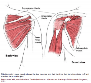
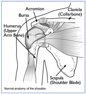
The rotator cuff is made up of 4 muscles surrounding the shoulder.

Three muscles on the back of the shoulder (supraspinatus, infraspinatus and teres minor) converge as a tendon to insert on the outer edge of the humeral head.

They act to elevate and externally rotate the shoulder. The fourth muscle (subscapularis) is on the front of the shoulder and helps internally rotate the shoulder.

Injury Mechanism
Rotator cuff tears can occur after an acute injury such as a fall or catching a heavy falling object, or they can occur over time as a gradual wearing of a hole in the rotator cuff from ongoing rubbing on the acromion such as overuse with overhead or throwing sports.

Symptoms
With acute complete tears of the rotator cuff, patients often describe a burning or tearing sensation at the time of injury. Early along, it is difficult to raise the arm overhead. Patients describe pain and weakness. The pain is usually located over the outer (lateral) aspect of the upper arm. Rotator cuff tears are often painful at night. Older patients may not recall an injury, but may just describe a gradual aching in the shoulder that has progressed over several months or years.

Diagnosis
The physician’s work-up will start with a careful history and exam.

X-rays are often obtained to see that no fractures have occurred with the injury and to help assess the overall condition of the shoulder joint. An MRI scan is often obtained to confirm the diagnosis and to evaluate any associated injuries to the labrum, rotator cuff or damage to the joint surfaces. Often the radiologist will inject contrast into the shoulder joint with a small needle to coat the undersurface of the rotator cuff and to see if the contrast leaks through the rotator cuff suggesting a complete tear. An MRI with contrast is called an arthro/MRI.

Plain x-rays show the bones of the shoulder, while MRI scans reveal the soft tissues around the shoulder including the labrum (lip of cartilage around the socket) and the rotator cuff tendons.

Treatment
Your physician will discuss treatment options with you. Treatment decisions are based on age, activity level and the severity of symptoms. In older patients with less activity demands and less severe symptoms, one will usually start with non-operative treatment including rest, ice, anti-inflammatories, stretching and occasional injections to see if the symptoms become tolerable.

In younger, more active patients, surgery is almost always recommended when a full thickness rotator cuff tear has occurred, since the rotator cuff has poor blood supply, therefore poor healing potential. Rotator cuff surgery is usually done on an outpatient basis. In most cases, the orthopedist will start with an exam under anesthesia to see that full motion of the shoulder is present and to see that the joint is stable. Next, one usually looks into the joint with a small arthroscope (a small lens and camera) so the surgeon can see and probe all of the structures in the joint. Once the complete rotator cuff tear is confirmed, the torn tendon is repaired back to the bony attachment site.

Because there is poor blood supply at the attachment site to the bone, one usually creates a small groove in the bone and pulls sutures through drill holes or uses anchors in the bone for fixation.

What to expect after surgery
Rotator cuff surgery is performed as an outpatient procedure. The surgery takes about 60 minutes. Patients go home in a sling that they use for 3 or 4 weeks. Most patients are uncomfortable for the first 2 or 3 days, but prescription medication is used to help alleviate the pain. Patients are seen back in the office one week after surgery to check their incisions and to start their exercise routine.

For the first 4 to 6 weeks, patients avoid any active elevation of their arm or lifting with that arm. At that point, formal physical therapy is started to improve range of motion and strength. It takes about 3 months from surgery before most of the strength and use of the shoulder returns. Full recovery may take 4 to 6 months.

Shoulder Impingement

Shoulder Impingement (Rotator Cuff Tendinitis)

The rotator cuff is made up of tendons and muscles that allow for a great range of motion of your arm. This is a frequent source of pain for athletes and an area that is at risk for injury during sporting activities. Shoulder impingement is often referred to as rotator cuff tendinitis and is one of the most common causes of shoulder pain.

What causes rotator cuff tendinitis?
When you raise your arm to shoulder height, the space between the bone and rotator cuff narrows. The bone can rub against (or impinge on the tendon and the bursa, causing irritation and pain when the arm is used repeatedly. Young athletes who use their arms for overhead action are particularly vulnerable. This includes those who play tennis, softball and baseball, and swimmers.

What are the symptoms of shoulder impingement?
When the rotator cuff is irritated this can lead to local swelling and tenderness in the front aspect of the shoulder. You may also have pain and stiffness when you lift your arm. There is also a sensation of tenderness when the arm is lowered from an elevated position. Other symptoms include sudden pain when reaching or lifting, pain radiating from the front of the shoulder to the side of the arm, minor pain at rest, and pain when throwing or using the arm.

How is rotator cuff tendinitis treated without surgery?
Your orthopedic specialist wants to reduce your pain and restore function of your shoulder. He will consider your activity level, your age, and your general state of health. Many times shoulder impingement can be treated with medications and rest. It is not uncommon for athletes to be ordered physical therapy to help restore normal motion of the shoulder. Your therapist will teach you specific stretching and strengthening exercises to relieve your shoulder pain and help you get back to normal activities.

What is involved with surgical treatment?
The goal of surgery is to create more space for the rotator cuff and this involves removing a portion of the inflamed bursa. Your orthopedic specialist will perform an anterior acromioplasty, where part of the bone is removed to allow for movement of the rotator cuff. Many times, the surgeon opts to perform this procedure by way of arthroscope.

The arthroscopic technique allows for use of small thin surgical instruments to be inserted around puncture wounds around the shoulder. The doctor can see inside the shoulder through a small camera inserted into the joint that displays images onto a computer TV monitor.

Biceps Tendon Tears

From orthoinfo.aaos.org

From orthoinfo.aaos.org

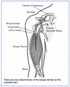
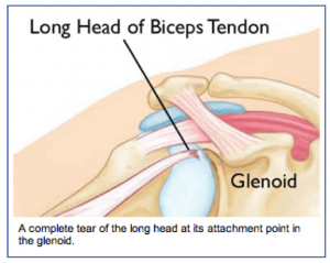
Anatomy: The biceps is a large muscle in the front of the shoulder. The upper end of the biceps has to heads (attachment sites). The long head of the biceps or its attachment to the labrum (cartilage lip at the top of the shoulder socket) is commonly injured.

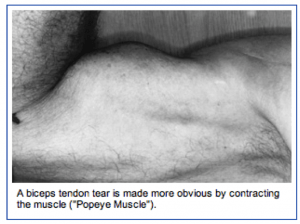
When it is torn at the upper end or at the labral attachment, there is often pain in the front of the shoulder. When the long head of the biceps is completely torn, the biceps muscle has a typical bulging appearance, but oftentimes does not cause much discomfort of weakness.

Injury Mechanism: Acute injuries to the long head of the biceps occur with heavy lifting or by grabbing the railing to support one’s body weight during a fall. Tears of the biceps can also occur with repetitive use of the shoulder in sports or work.

Symptoms: Acute complete tears of the long head of the biceps usually present as a sharp pain in the front of the shoulder, followed by bruising and the “Popeye muscle” deformity of the biceps (bulging in the middle of the upper arm where the muscle has retracted). Chronic tears or fraying of the biceps usually presents as aching in the front of the shoulder with use of the biceps.

From orthoinfo.aaos.orgDiagnosis: The physician’s work-up will start with a careful history and exam. X-rays will be normal as they show the bones and joints, not the muscles and tendons. An MRI scan can be obtained to confirm the diagnosis, especially if the diagnosis is not obvious by history and exam.
Treatment: Your physician will discuss treatment options with you. Most complete tears of the rotator cuff can be treated without surgery. Rarely patients notice weakness or cramping in the biceps. Stretching, strengthening, anti-inflammatories, and time will usually settle down symptoms to a tolerable level.

The partial tears are usually more painful. Injections can sometimes manage the discomfort, but occasionally surgery is necessary to help alleviate symptoms.

From orthoinfo.aaos.orgSurgery is usually accomplished through the arthroscope to remove the frayed tissue.
If the partial tear is nearly complete, the tendon is released from its attachment to the socket (labrum) and attached further down on the upper arm bone (humerus). This procedure is called a biceps tenodesis. Sometimes a small incision is necessary to re-attach the tendon to the bone.

What to expect after surgery: If the frayed tendon is just removed through the arthroscope, the recovery is rapid. Usually, after a few days of rest, gentle exercise is started. A full recovery is expected after 4 to 6 weeks. If the tendon is re-attached to bone, a sling is used with the elbow bent for 3 or 4 weeks to protect the repair. No resisted use of the biceps is started until 6 to 8 weeks after the repair.

Bursitis and Tendinitis

From orthoinfo.aaos.org

From orthoinfo.aaos.org

Both bursitis and tendinitis are forms of impingement syndrome. Bursitis occurs when the bursa (normally thin fluid filled sack that allows the rotator cuff to glide smoothly under the acromion) becomes swollen or inflamed.

Tendinitis occurs when the tendons of the rotator cuff or the long head of the biceps becomes swollen or inflamed (normally thin fluid filled sack that allows the rotator cuff to glide smoothly under the acromion) becomes swollen or inflamed.

Injury Mechanism: Bursitis and tendinitis are commonly seen in throwing athletes, those doing overhead lifting, or with repetitive motions and overuse of the shoulder. Often, one does not recall a specific injury, but awakens with pain.

Symptoms: When patients have bursitis, they describe pain over the lateral aspect of the shoulder with overhead reaching or laying on that shoulder at night. With tendinitis, pain occurs when using the rotator cuff or biceps in addition to the pain with overhead reaching or laying on that shoulder at night.

Diagnosis: The physician’s work-up will start with a careful history and exam. With tendinitis, the physician can isolate the tendons involved by eliciting pain with use of those tendons plus the impingement test is positive. With bursitis, pain cannot be elicited with use of specific tendons, but the impingement test is still positive.

X-rays are often normal with bursitis or tendinitis, but occasionally a spur is identified on a special “outlet view” that can be obtained in the physician’s office. An MRI scan is sometimes obtained to confirm that the rotator cuff is not torn, especially when patients do not respond to initial treatment.

Treatment: The mainstay of treatment for bursitis and tendinitis is non-surgical. Most patients respond to a short course of anti-inflammatories, stretching, and gentle strengthening of the external rotators of the shoulder. This can be accomplished at home with rubber tubing. If symptoms persist, one can inject the bursa with cortisone to cause the swollen tissues to shrink so they will no longer rub, or impinge.

If the symptoms respond temporarily to the injections, but keep recurring, one can consider surgery to shave down the undersurface of the acromion (acromioplasty) to make more clearance for the rotator cuff and bursa to slide underneath without rubbing. This type of surgery is performed through the arthroscope, using 2 or 3 small incisions, and looking inside the shoulder with a small lens and camera.”

What to expect after surgery: Most shoulder surgery is now performed as an outpatient procedure. In the rare event that an acromioplasty is needed to stop the impingement process, most patients find that they do best by resting their shoulder in a sling for 3 or 4 days following surgery.

They are then encouraged to come out of the sling for gentle stretching exercises. Once comfortable (usually 1 or 2 weeks following surgery), gentle strengthening is initiated with rubber tubing. Once the patient is off of his/her medication, they can resume driving and returning to office work. Most patients can return to full activities in 6 to 8 weeks.