Shoulder Anatomy

from orthoinfo.aaos.org

from orthoinfo.aaos.org

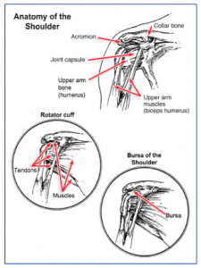
The shoulder joint is a complicated joint.

It is made up of three bones: the scapula (shoulder blade), the clavicle (collarbone) and the humerus (upper arm bone). The true shoulder joint is where the shoulder blade meets the upper arm bone.

A second joint exists where the collarbone meets the shoulder blade on the top and front of the shoulder and is called the AC or acromioclavicular joint. When people refer to a shoulder separation, it is really the AC joint that has been injured, not the true ball and socket (glenohumeral) joint of the shoulder. A third joint exists where the shoulder blade sits on the back of the ribcage called the scapulothoracic joint.

The shoulder joint is unique in that it can move in almost every direction, unlike the knee that just straightens and bends. The large ball on the relatively flat socket is what allows the shoulder such unrestricted motion.

What keeps the humeral head (the ball) on the flat surface of the glenoid (socket) is the muscles, tendons, and ligaments that surround the head plus a lip of cartilage (labrum) that surrounds and extends the socket to help contain the head.

The rotator cuff is a group of 4 muscles that converge on the outside of the shoulder to help elevate and rotate the arm. The tendinous attachment of these tendons to the upper arm bone is called the rotator cuff. The long head of the biceps tendon also runs over the top of the shoulder and attaches to the upper part of the socket through the upper labrum.

Between the top of the shoulder blade (the acromion) and the rotator cuff and biceps tendons is a large fluid filled sack called the bursa. It is normally quite thin and helps allow these tissues to slide under the acromion without rubbing or abrading.

Arthritis of the Knee

arthritisknee1-300x164What is arthritis of the knee?
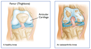
Arthritis of any joint is when there is inflammation and damage to the normal joint surface. The normal articular cartilage is smooth and firm. With arthritis, the surface of the joint becomes pitted, eroded, and irregular causing swelling and pain. There are many types of arthritis.

The most common type of arthritis is osteoarthritis, or wear and tear that is seen in older individuals. Another form of arthritis is rheumatoid arthritis. It is commonly involves multiple joints, especially in the hands and wrists.

It can occur at any age. One more common form of arthritis is post-traumatic arthritis. It follows an injury to the surfaces, the meniscus, or the ligaments, which makes the joint susceptible to arthritis often many years after the injury.

Symptoms
Patients with arthritis typically describe pain and stiffness, especially with the first few steps after getting up from a chair or first thing in the morning. In the early stages of arthritis, the knee is often swollen, and even warm to the touch. After years of having arthritis, the knee rarely swells, but it becomes more difficult to fully straighten or bend the knee. Arthritic knees are painful with activity.

Diagnosis
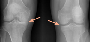
kneearthritis2-300x140The physician’s work-up will start with a careful history and exam. A history of prior injury, pain, stiffness and swelling may suggest arthritis. The exam often shows some swelling and decreased range of motion. Sometimes, patients develop deformities if one side of the knee wears out more than the other side.

The legs can then appear “knock-kneed” or “bow-legged.” X-rays are usually the key in determining the extent of the arthritis. Arthritic knees show loss of normal joint space, cysts and bone spurs on x-ray.

Treatment
Early treatment of arthritis may involve non-steroidal anti-inflammatories, ice, rest, injections and activity modification. If these methods are not satisfactory in keeping a patient from becoming sedentary, surgery is often discussed. Ideally for surgery, patients should be in their mid-60s (current knee replacements last about 20 years or more so are not ideal for those in their 40s or 50s), able to walk only a block or two (considered sedentary), and experience pain at night (effecting quality of life).

However, as techniques and materials become better, physicians are considering knee replacements in younger and younger patients.

Surgical options for arthritis range from arthroscopic debridement (cleaning out the loose pieces and smoothing the surfaces), osteotomies (cutting a wedge out of the bone above or below the knee to try to throw more of the weight onto the better half of the knee), cartilage replacement (transferring plugs of bone and cartilage to the involved areas), to partial or total knee replacements in which the arthritic surfaces are removed and replaced with metal and plastic surfaces.

Your orthopedist will have a thorough discussion with you about which surgical option is best for you, and discuss the risks and benefits of the surgery with you.

Patellofemoral Pain and Instability

patellar1-300x300Anatomy
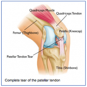
The kneecap (or patella) is a small bone that sits in front of the knee and provides mechanical advantage for our extensor mechanism (quadriceps muscles) in helping one straighten out there knee. The quadriceps tendon attaches to the upper pole of the kneecap and the patellar tendon attaches to the lower pole of the kneecap.

Normally the kneecap glides smoothly in a groove on the front of the femur (thigh bone). The Back of the kneecap and the front of the femoral groove are coated with smooth cartilage that allows the kneecap to glide smoothly.

What is patellofemoral pain?
Patellofemoral pain is a generic term for pain that occurs in the front of the knee. It can result from wearing or arthritis of the joint surface on the back of the kneecap, overuse, malalignment, muscle imbalance, flat feet (pronation) or trauma to the kneecap.

Symptoms
Most patients with patellofemoral pain complain of discomfort in the front of the knee that is worsened with stairs, inclines, sitting for long periods of time, squatting or kneeling, or even with prolonged standing. Occasionally patients report swelling, especially after rigorous activity or episodes in which the patellar has dislocated. Often patients describe grinding (crepitation) when they straighten the knee against resistance.

Diagnosis
The physician’s work-up will start with a careful history and exam. When pain is worse with squatting, kneeling, stairs, and prolonged sitting without a specific injury patellofemoral pain is suspected. The examination often reveals grinding under the kneecap, lateral tracking or malalignment of the kneecap, and often tenderness along either side of the kneecap. X-rays can help determine if the kneecap is tracking properly and if there is any wear starting behind the kneecap. MRI scans are usually not as helpful for patellofemoral pain, except to look for other pathology within the knee.

Treatment
Non-surgical treatment is helpful in the majority of patients with patellofemoral pain. Non-surgical treatment may involve formal physical therapy, cross-training and activity modification, weight loss and general fitness, braces. modification in training schedules and form, non-steroidal anti-inflammatories, taping of the kneecap, inserts for shoes, and other modalities. When non-surgical treatment fails, especially with abnormal anatomy (tight lateral restraints or poor alignment), surgery can be helpful.

Sometimes the tight lateral restraints that are tethering the kneecap on one side can be released through the arthroscope (lateral release). In more severe cases in which the kneecap is dislocating or wearing unevenly, more drastic steps are needed to help get the kneecap to track centrally, such as reconstructing the ligament on the inside of the knee or actually cutting the bony attachment of the patellar tendon and moving it to a more central position under the kneecap.

What to expect after surgery
On the rare occasions that surgery is performed for patellofemoral problems, the post-operative treatment depends on the extent of the surgical procedure. If a lateral release is all that is needed, the patient is usually placed in a straight let knee immobilizer for one week while weight bearing fully without crutches. After a week, the brace is removed and therapy is begun to regain motion and strength. It is often 6 to 8 weeks before returning to most normal activities.

If more extensive surgery is needed to re-align the patellar tracking by reconstructing a ligament or moving the bony insertion of the patellar tendon, crutches and bracing may be used initially and the return to full activities may be delayed until complete healing has occurred.

Cubital Tunnel

Cubital Tunnel – Numb hand from nerve pinched at the Elbow

Facts about Cubital Tunnel Syndrome / Ulnar Nerve Compression

6990160What is Cubital Tunnel Syndrome?
Cubital tunnel syndrome is a condition brought on by increased pressure on the ulnar nerve at the elbow. The ulnar nerve controls muscles used for gripping and the coordination of fine movements. The nerve passes through the cubital tunnel, a bony passageway. Cubital tunnel syndrome occurs when the ulnar nerve is compressed as it passes through this tunnel behind the elbow. When compressed, the ulnar nerve causes the sharp, tingling sensation that you feel when you hit your “funny bone.”

Cubital Tunnel Syndrome is the second most common peripheral nerve compression syndrome (after carpal tunnel syndrome).

What are the symptoms?
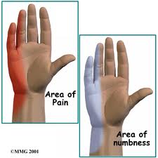
Common symptoms include pain and numbness in the hand, including the ring and small fingers. More severe cases may also lead to a weak grip and pain at the elbow. Often patients suffering from cubital tunnel syndrome have difficulty sleeping at night due to the pain and numbness.

What causes Cubital Tunnel Syndrome?
While the majority of cases of cubital tunnel syndrome are idiopathic, the syndrome has been termed “cell phone elbow,” as it can result from prolonged hyperflexion of the elbow (holding the elbow in a bent position for a long time). Bending the elbow increases the pressure on the ulnar nerve. Sustained bending of the elbow also tends to occur during sleep.

What are the treatment options?
Treatment usually begins with splinting the elbow, especially at night, and anti-inflammatory medications. Surgery may be necessary. Surgery involves either releasing the ulnar nerve from the compression, or actually moving the nerve (an ulnar nerve transposition) to allow more room for the nerve to move behind the elbow.

Carpal Tunnel Surgery

Carpal Tunnel Syndrome
Carpal Wrist

The Carpal Tunnel – The Median Nerve’s Path to the Hand

What is Carpal Tunnel Syndrome?

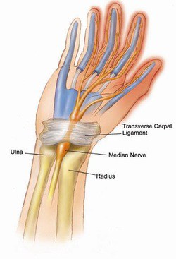
Carpal tunnel syndrome is a common condition in the adult hand, affecting nearly 3% of the population and is the most common peripheral nerve compression syndrome. The carpal tunnel is a bony passageway in the wrist, housing both the median nerve along with nine wrist flexor tendons.

Due to the nonconforming shape of the tunnel, any increase in the pressure of the tunnel causes compression on the median nerve, thereby decreasing its ability to function properly. The median nerve controls important muscles in the hand, giving sensation to many of the digits.

Carpal tunnel syndrome occurs when the median nerve is compressed as it passes through this tunnel in the hand. When compressed, the median nerve causes numbness, tingling, weakness and pain in the hand and wrist. The pain is often positional, meaning that extreme flexion or extension of the wrist exacerbate the symptoms.

What are the symptoms?
Compression of the median nerve reliably causes numbness in the thumb, index finger, middle finger and half of the ring finger. In addition, if the median nerve is compressed for a critical period of time, the nerve loses its ability to power key muscles of the thumb and hand, causing a loss of strength and dexterity for fine movements.
Other common symptoms include hand pain which is often worse at night or while driving, and a need to shake the hand to allow the hand to “wake up”.

What causes Carpal Tunnel Syndrome?

Contrary to much of the information readily available for patients, it is important to emphasize that the vast majority of patients have no known cause for their carpal tunnel syndrome. While it is tempting to attach blame to repetitive activity such as typing, or blame a particular injury, many well designed objective studies have consistently failed to show any effect of repetitive activity on the development or exacerbation of carpal tunnel syndrome.  

The best evidence suggests that some patients likely have a genetic predisposition to the development of carpal tunnel syndrome, despite intuitive interventions such as ergonomic workplace environments or other similar modifications.

Caveat: This is not to say that ergonomic improvements in the way we live and work are not positive improvements. Many significant advances have been made in workplace safety, and certainly in a patient with known carpal tunnel syndrome, minor modifications can be made which can decrease one’s symptoms. However it is important to underscore the fact that though these interventions may help symptomatic management, the lack of such interventions are not the cause of carpal tunnel syndrome.

What are the treatment options?
Treatment usually begins with wrist splints, especially at night, anti-inflammatory medications, and activity modifications.  Steroid injections into the carpal tunnel may also provide relief of symptoms, but unfortunately no interventions short of surgery give adequate space for the nerve in the carpal tunnel once there is too much pressure.  Ultimately, surgery may be necessary to relieve the pressure on the median nerve in the carpal tunnel.

What is involved in surgery?
Carpal tunnel surgery involves an incision in the base of the palm to gain access to the carpal tunnel. The goal of surgery is to provide more space to for the constricted nerve, which the 30 minute procedure reliably accomplishes. I perform the procedure typically in an outpatient setting and most patients have only a few days of discomfort from the incision are able to use their hand fully by 2 weeks after the sutures are removed. Many patients are able to return to work within 1-3 days, and the dressing stays in place until the sutures are removed.

It is important to note that in severe cases, the nerve has sustained permanent injury from years of compression. Though the surgery reliably relieves pressure on the nerve, sometimes the damage is irreversible and persistent numbness or weakness remain. Interested in learning more about your symptoms?  Contact Dr. Scott Ruhlman’s office at 206-633-8100.