deQuervain’s Tendinitis

What is deQuervain’s Tendinitis?

deQuervain’s tendinitis is a condition brought on by irritation or swelling of the tendons found along the thumb side of the wrist (Figure 1). The irritation causes the compartment (lining) around the tendon to swell, changing the shape of the compartment; this makes it difficult for the tendons to move as they should.

The swelling can cause pain and tenderness along the thumb side of the wrist, usually noticed when forming a fist, grasping or gripping things, or turning the wrist.

cons1_79_52

Signs and Symptoms
Pain over the thumb side of the wrist is the main symptom. The pain may appear either gradually or suddenly. It is felt in the wrist and can travel up the forearm. The pain is usually worse with use of the hand and thumb, especially when forcefully grasping things or twisting the wrist.

Swelling over the thumb side of the wrist is noticed and may be accompanied by a fluid-filled cyst in this region. There may be an occasional “catching” or “snapping” when moving the thumb. Because of the pain and swelling, it may be difficult to move the thumb and wrist, such as in pinching.

Irritation of the nerve lying on top of the tendon sheath may cause numbness on the back of the thumb and index finger.

cons1_80_52

Diagnosis
A Finkelstein test is generally performed. In this test, the patient makes a fist with the fingers over the thumb. The wrist is then bent in the direction of the little finger (Figure 2).

This test can be quite painful for the person with deQuervain’s tendinitis. Tenderness directly over the tendons on the thumb side of the wrist is the most common finding, however.

cons1_81_52

Treatment
The goal is to relieve the pain caused by the irritation and swelling. In some cases, your doctor may recommend resting the thumb and wrist by wearing a splint. Anti-inflammatory medication taken by mouth or injected into that tendon compartment may help reduce the swelling and relieve the pain.

In some cases, simply not doing the activities that cause pain and swelling may allow the symptoms to go away on their own. When symptoms are severe or do not improve, surgery may be recommended.

The surgery opens the compartment (covering) to make more room for the irritated tendons (Figure 3). Normal use of the hand can usually be resumed once comfort and strength have returned. Your hand surgeon can advise you on the best treatment for your situation.

Arthritis of the Wrist

Arthritis affects millions of people in the United States. Often, arthritis strikes at the weightbearing joints of the body, such as the knees and the shoulders. But a significant number of people suffer from arthritis in their wrists and hands that make it difficult for them to perform the activities of daily living.

Although there are hundreds of kinds of arthritis, most wrist pain is caused by just two types:

  • Osteoarthritis (OA) is a progressive condition that destroys the smooth articular cartilage covering the ends of bones. The bare bones rub against each other, resulting in pain, stiffness and weakness. OA can develop due to normal “wear-and-tear” on the wrist or as a result of a traumatic injury to the forearm, wrist or ligaments.
  • Rheumatoid arthritis (RA) is a systemic inflammatory disease that affects the joint linings and destroys bones, tissues, and joints. Rheumatoid arthritis often starts in smaller joints, like those found in the hand and wrist, and is symmetrical, meaning that it usually affects the same joint on both sides of the body.

Signs and symptoms

  • OA of the wrist joint manifests with swelling, pain, limited motion and weakness. These symptoms are usually limited to the wrist joint itself.
  • RA of the wrist joint usually manifests will swelling, tenderness, limited motion and decreased grip strength. In addition, hand function may be impaired and there may be pain in the knuckle joints (metacarpophalangeal or MP joints).
  • Joint swelling may also put pressure on the nerves that travel through the wrist. This can cause a lesion to develop (compression neuropathy) or lead to carpal tunnel syndrome.

Diagnosis and treatment
Six bones make up the wrist joint: the two bones of the lower arm (the radius and the ulna) and four wrist bones (the carpals). Your physician will use a combination of physical examination, patient history, and tests to diagnose arthritis of the wrist. X-rays can help distinguish among various forms of arthritis. Some, but not all, forms of RA can be confirmed by a laboratory blood test.

In general, early treatment is nonsurgical and designed to help relieve pain and swelling. Several therapies can be used to treat arthritis, including:

  • Modifying your activities.
  • Immobilizing the wrist for a short time in a splint.
  • Taking anti-inflammatory medications such as aspirin or ibuprofen.
  • Following a prescribed exercise program.
  • Getting a steroid injection into the joint.

Your physician may prescribe other therapies, depending on the type of arthritis you have. For example, additional therapies for patients with rheumatoid arthritis include antimalarial drugs, antimetabolites, gold, immunosuppresive drugs (both non-steroidal and corticosteroids) and newer genetically-engineered medications.

When such conservative methods are no longer effective, or if hand function decreases, surgery is an option. The goal of surgery is to relieve pain; depending on the type of surgery, joint function may also be affected. Surgical options include removing the arthritic bones, joint fusion (making the joint solid and preventing any movement at the wrist) and joint replacement.

You and your physician should discuss the options and select the one that is best for you.

Arthritis of the Hand

Description
The hand and wrist have multiple small joints that work together to produce motion. This gives you the fine motion needed to thread a needle or tie a shoelace. When the joints become affected with arthritis, activities of daily living can be difficult. Arthritis can occur in multiple areas of the hand and wrist. It can have multiple causes.

All arthritic joints lose cartilage, which works as nature’s “shock absorber.” Cartilage provides a smooth gliding surface for the joint. When the cartilage becomes worn or damaged, or is lost due to disease or trauma, the joint no longer has a painless, mobile area of motion.

The body attempts to make up for the lost cartilage. It produces fluid in the joint lining (synovium), which tries to act like a cushion, like water in a waterbed. But it also causes the joint to swell. This restricts motion. The swelling causes stretching of the joint covering (capsule), which causes pain.

Over time, if the arthritis is not treated, the bones that make up the joint can lose their normal shape. This causes more pain and further limits motion.

Risk Factors/Prevention
It is estimated that one out of every five people living in the United States has at least one joint with signs or symptoms of arthritis. About half of arthritis sufferers are under age 50. Arthritis is the leading cause of disability in the United States. It typically occurs from either disease or trauma. The exact number of people with arthritis in the hand and wrist is not known.

When arthritis occurs due to disease, the onset of symptoms is gradual and the cartilage decreases slowly. The two most common forms of arthritis from disease are osteoarthritis and rheumatoid arthritis. Osteoarthritis is much more common and generally affects older people.

It appears in a predictable pattern in certain joints. Rheumatoid arthritis has other system-wide symptoms and may be passed from parent to child (genetically).

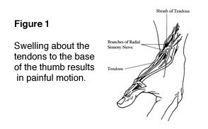
When arthritis is due to trauma, the cartilage is damaged. People of any age can be affected. Fractures–particularly those that damage the joint surface–and dislocations are the most common injuries that lead to arthritis (see Figure 1). An injured joint is about seven times more likely to become arthritic, even if the injury is properly treated.

Arthritis does not have to result in a painful or sedentary life. It is important to seek help early so that treatment can begin and you can return to doing what matters most to you.

cons1_407_307

Diagnosis
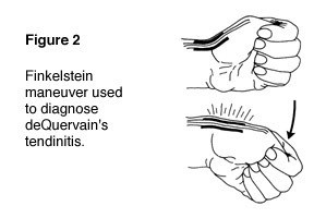
A doctor can diagnose arthritis of the hand by examining you and taking X-rays. Specialized studies such as MRI (magnetic resonance imaging) scans are usually not needed. Sometimes a bone scan is helpful (see Figure 2).

cons1_408_307

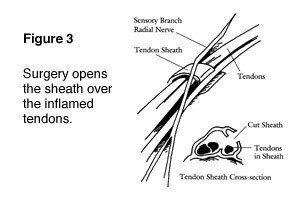
A bone scan may help the doctor diagnose arthritis when it is in an early stage, even if X-rays look normal. Arthroscopy is another way to look at the joint by direct inspection. During an arthroscopic procedure, the surgeon inserts a small camera into the joint to look inside.

It provides the clearest picture of the joint without having to make a large incision. However, this is an invasive procedure and should not be used as a routine diagnostic tool (see Figure 3).

cons1_409_307

Symptoms
Early symptoms of arthritis of the hand include joint pain that may feel “dull,” or a “burning” sensation. The pain often occurs after periods of increased joint use, such as heavy gripping or grasping. The pain may not be present immediately, but may show up hours later or even the following day.

Morning pain and stiffness are typical. As the cartilage wears away and there is less material to provide shock absorption, the symptoms occur even with less use. In advanced disease, the joint pain may wake you up at night.

When the affected joint is subject to greater stress than it can bear, it may swell in an attempt prevent further joint use. Your pain might be made worse with use and relieved by rest. Many people with arthritis complain of increased joint pain with rainy weather. Activities that once were easy, such as opening a jar or starting the car, become difficult due to pain.

To prevent pain at the arthritic joint, you might adapt the way you use your hand. In patients with advanced thumb base arthritis, the neighboring joints may become more mobile than normal (see Figure 4).

cons1_410_307

The arthritic joint may feel warm to touch. This is due to the body’s inflammatory response. There may be a sensation of grating or grinding in the affected joint (crepitation). This is caused by damaged cartilage surfaces rubbing against one another. If arthritis is due to damaged ligaments, the support structures of the joint may be unstable or “loose.”

In advanced cases, the joint may appear larger than normal (hypertrophic). This is usually due to a combination of bone changes, loss of cartilage and joint swelling.

When arthritis affects the end joints of the fingers (DIP joints), small cysts (mucous cysts) may develop (see Figure 5). The cysts may then cause ridging or dents in the nail plate of the affected finger.

cons1_411_307

Treatment Options: Nonsurgical
Treatment options for arthritis of the hand and wrist include medication, splinting, injections and surgery. Treatment depends on many factors:

  • How far the arthritis has progressed
  • How many joints are involved
  • Your age, activity level and other medical conditions
  • If the dominant or non-dominant hand is affected

Your personal goals, home support structure, and ability to understand the treatment and comply with a therapy program

Medications: Medications treat symptoms but cannot restore joint cartilage or reverse joint damage. The most common medications for arthritis are anti-inflammatories, which stop the body from producing chemicals that cause joint swelling and pain.

Examples of anti-inflammatory drugs include over-the-counter medications such as Tylenol® and Advil® and prescription drugs such as Celebrex®. Glucosamine and chondroitin are widely advertised “neutraceuticals.”

Neutraceuticals are not drugs. Rather, they are compounds that are the “building blocks” of cartilage. They were originally used by veterinarians to treat arthritic hips in dogs. However, neutraceuticals have not yet been studied as a treatment of hand and wrist arthritis.

Injections: When first-line treatment with anti-inflammatory medication is not appropriate, injections may be used. These typically contain a long-acting anesthetic, similar to novacaine but longer lasting, and a steroid that can provide pain relief for weeks to months.

The injections can be repeated, but only a limited number of times, due to possible side effects, such as lightening of the skin, weakening of the tendons and ligaments and infection.

Splinting: Injections are usually combined with splinting of the affected joint. The splint helps support the affected joint to ease the stress placed on it by activities. Splints are typically worn during periods when the joints hurt.

They should be small enough to allow functional use of the hand when they are worn. Wearing the splint for too long can lead to muscle wasting (atrophy). Muscles can assist in stabilizing injured joints, so atrophy should be prevented.

Treatment Options: Surgical
If non-operative treatment fails to give you relief, surgery is usually discussed. There are many surgical options. The option chosen should be one that has a reasonable chance of providing long-term pain relief and return to function. It should be tailored to your individual needs. It is important that the treating physician is well versed in current surgical techniques. If there is any way the joint can be preserved or reconstructed, this option is usually chosen.

When the damage has progressed to a point that the surfaces will no longer work, a joint replacement or fusion (arthrodesis) is performed (see Figure 6). Joint replacements attempt to provide pain relief and functional joint motion. Joint fusions provide pain relief but stop joint motion. The fused joint no longer moves; the damaged joint surfaces are gone, so they cannot cause symptoms.

cons1_412_307

As with hip and knee replacements, there have been significant improvements in joint replacements in the hand and wrist. The replacement joints are made of materials similar to those used in weight-bearing joints, such as ceramics or long wearing metal and plastic parts (see Figure 7). The goal is to improve the function and longevity of the replaced joint. Most of the major joints of the hand and wrist can be replaced.

A surgeon often needs additional training to perform the surgery. As with any evolving technology, the long-term results of the hand or wrist joint replacements are not yet known. Early results have been promising. Talk with your doctor to find out if these implants are right for you.

cons1_413_307

After any type of joint reconstruction surgery, there is a period of recovery. Often, you will be referred to a trained hand therapist, who can help you maximize your recovery. You may need to use a postoperative splint or cast for awhile after surgery. This helps protect the hand while it heals.

During this postoperative period, you may need to modify activities to let the joint reconstruction heal properly. Typically, pain medication you take by mouth is also used to reduce discomfort.

It is important to discuss your pain with your doctor so it can be adequately treated. Length of recovery time varies widely and depends on the extent of the surgery performed and multiple individual factors. However, people usually can return to most if not all of their desired activities in about three months after most major joint reconstructions.

Research on the Horizon/What’s New?
Increasingly, doctors are focusing on how to preserve the damaged joint. This includes getting an earlier diagnosis and repairing joint components before the entire surface becomes damaged. Arthroscopy of the small joints of the hand and wrist is now possible because the equipment has been downsized.

There have been encouraging results in cartilage repair and replacement in the larger joints such as the knee, and some of these techniques have been applied to the smaller joints of the hand and arm. In addition, stem cell research may be an option to regenerate damaged joint surfaces.

Bursitis and Tendinitis

From orthoinfo.aaos.org

From orthoinfo.aaos.org

Both bursitis and tendinitis are forms of impingement syndrome. Bursitis occurs when the bursa (normally thin fluid filled sack that allows the rotator cuff to glide smoothly under the acromion) becomes swollen or inflamed.

Tendinitis occurs when the tendons of the rotator cuff or the long head of the biceps becomes swollen or inflamed (normally thin fluid filled sack that allows the rotator cuff to glide smoothly under the acromion) becomes swollen or inflamed.

Injury Mechanism: Bursitis and tendinitis are commonly seen in throwing athletes, those doing overhead lifting, or with repetitive motions and overuse of the shoulder. Often, one does not recall a specific injury, but awakens with pain.

Symptoms: When patients have bursitis, they describe pain over the lateral aspect of the shoulder with overhead reaching or laying on that shoulder at night. With tendinitis, pain occurs when using the rotator cuff or biceps in addition to the pain with overhead reaching or laying on that shoulder at night.

Diagnosis: The physician’s work-up will start with a careful history and exam. With tendinitis, the physician can isolate the tendons involved by eliciting pain with use of those tendons plus the impingement test is positive. With bursitis, pain cannot be elicited with use of specific tendons, but the impingement test is still positive.

X-rays are often normal with bursitis or tendinitis, but occasionally a spur is identified on a special “outlet view” that can be obtained in the physician’s office. An MRI scan is sometimes obtained to confirm that the rotator cuff is not torn, especially when patients do not respond to initial treatment.

Treatment: The mainstay of treatment for bursitis and tendinitis is non-surgical. Most patients respond to a short course of anti-inflammatories, stretching, and gentle strengthening of the external rotators of the shoulder. This can be accomplished at home with rubber tubing. If symptoms persist, one can inject the bursa with cortisone to cause the swollen tissues to shrink so they will no longer rub, or impinge.

If the symptoms respond temporarily to the injections, but keep recurring, one can consider surgery to shave down the undersurface of the acromion (acromioplasty) to make more clearance for the rotator cuff and bursa to slide underneath without rubbing. This type of surgery is performed through the arthroscope, using 2 or 3 small incisions, and looking inside the shoulder with a small lens and camera.”

What to expect after surgery: Most shoulder surgery is now performed as an outpatient procedure. In the rare event that an acromioplasty is needed to stop the impingement process, most patients find that they do best by resting their shoulder in a sling for 3 or 4 days following surgery.

They are then encouraged to come out of the sling for gentle stretching exercises. Once comfortable (usually 1 or 2 weeks following surgery), gentle strengthening is initiated with rubber tubing. Once the patient is off of his/her medication, they can resume driving and returning to office work. Most patients can return to full activities in 6 to 8 weeks.

Reverse Total Shoulder Replacement

Dr. Peterson and Dr. Shapiro have been performing a relatively new procedure called reverse total shoulder replacement for the last several years.

This particular procedure is designed for people who have rotator cuff arthropathy or a large, irreparable rotator cuff tear. The rotator cuff is a group of muscles and tendons that surround the shoulder joint and allow you to lift your arm over your head. When this structure is severely torn, shoulder arthritis can set in and mobility is limited.

During this procedure, the surgeon removes damaged bone joint tissue.  A smooth,  polished, spherical alloy metal “glenosphere” is then fixed to the old bony “cup” of the shoulder, and a stemmed alloy and polymer cup to the shaft of the upper-arm bone.

Why is Reverse Total Shoulder Replacement Done?

This surgery was developed because traditional shoulder surgeries do not work well when patients also have a severe rotator cuff tear with arthritis. With reverse total shoulder replacement, the deltoid muscle powers the new prosthesis, allowing pain free motion overhead in many patients.

Who is a Candidate for Surgery?

Reverse total shoulder replacement may be recommended if you have:

  • A completely torn rotator cuff that cannot be repaired.
  • Cuff tear arthropathy (arthritis with a severe cuff tear).
  • A previous should replacement that was unsuccessful.
  • Severe shoulder pain and difficulty lifting your arm.
  • Tried other treatments that have not relieved your shoulder pain.

Reverse shoulder replacement may not be recommended for people who have:

  • Poor general health and may not tolerate anesthesia and surgery well.
  • An active infection or are at risk for infection.
  • Severe weakness of or damage to the deltoid muscle of the shoulder.
  • A shoulder problem deemed appropriate for more traditional replacement procedures.

How do I Prepare for this Procedure?

Anesthesia – This procedure can be performed under general or regional anesthesia, depending on what your orthopedic surgeon prefers.

Antibiotics – You will probably be prescribed antibiotics to take before and after the surgery to prevent infection.

Medications – Be sure you tell your orthopedic specialists about all the medications you are taking. He may advise you to stop certain medications before the procedure.

Home Planning – There are some things you should be aware of that will make your recovery period much easier. First of all, you will need to take several weeks off from work following the surgery. When you come home, you will need help for a few weeks with dressing, bathing, and simple household chores. Also, you may not be permitted to drive following the surgery and for a few weeks.

What Happens During the Surgery?

A reverse total shoulder replacement usually takes about 1.5 hours. The surgeon will make an incision at the top or front of your shoulder and remove the damaged bone. Then he will position the new components to restore function to your shoulder joint. The incision will then be closed with sutures.What Should I Expect After the Procedure?

After your procedure, the healthcare professionals will give you pain medication to keep you comfortable and several doses of antibiotics. Most patients are allowed to eat solid food and get out of bed the day after the surgery. You will go home on the first or second day following your procedure.

When you leave the surgical center, your arm will be in a sling to provide support. Your orthopedic specialist will instruct you on exercises to increase your mobility and endurance and plan a physical therapy program to strengthen your shoulder and improve your flexibility. Full recovery from this surgery usually occurs in 4-6 months.