
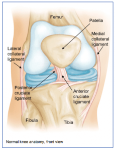
 The knee joint is one of the largest joints in the body. It is a complex joint with four bones: the femur (thigh bone), the tibia (main lower leg bone), the fibula (smaller lower leg bone), and the patella (kneecap). The bones are connected with four main ligaments: ACL (anterior cruciate ligament), PCL (posterior cruciate ligament), MCL (medial collateral ligament), and the LCL (lateral collateral ligament).
The ACL and PCL control the forward/backwards movement of the knee joint and prevent pivoting of the knee. The MCL and LCL prevent giving away on either side of the knee. The quadriceps is a group of 4 muscles that converge on the front of the thigh and together allow one to straighten their knee by pulling through the kneecap and patellar tendon, which attaches to the front of the lower leg bone (tibia).
The hamstrings are the muscles on the back of the thigh that help bending the knee by crossing the joint in the back of the knee and attaching to the lower leg bones. Between the femur and tibia, sitting centrally in the knee joint, are two C-shaped pads (the medial and lateral menisci) that act as cushions or shock absorbers between the two bones. The meniscal pads are made of cartilage.
There is also about a quarter of an inch of cartilage on the distal end of the thighbone and on the proximal end of the lower leg bone. Arthritis occurs when that joint cartilage becomes damaged or thin.