Reflex Sympathetic Dystrophy

What is Reflex Sympathetic Dystrophy?

Reflex sympathetic dystrophy, also known as RSD, is a condition of burning pain, stiffness, swelling, and discoloration of the hand. RSD includes other medical diagnoses such as casualgia, Sudeck’s atrophy, and shoulder-hand syndrome. RSD occurs from a disturbance in the sympathetic (unconscious) nervous system that controls the blood flow and sweat glands in the hand and arm. When the nervous system becomes overactive, burning pain is felt and swelling and warmth are left in the affected arm. If not treated, RSD can cause stiffness and loss of use of the affected part of the arm.

What causes Reflex Sympathetic Dystrophy?

In some cases, the cause of RSD is unknown. Often an injury can cause RSD, or the symptoms may appear after a surgery. Other causes include pressure on a nerve, infection, cancer, neck disorders, stroke, or heart attack. These conditions can cause pain, which sets off the sympathetic reflex causing RSD symptoms. Nerve injuries may change the way the nerve impulses are sent, causing a “short circuit” (Figure 2).

Signs and symptoms

The pain associated with reflex sympathetic dystrophy is often described as burning in nature. Swelling can cause painful joints and stiffness.

RSD has three stages:

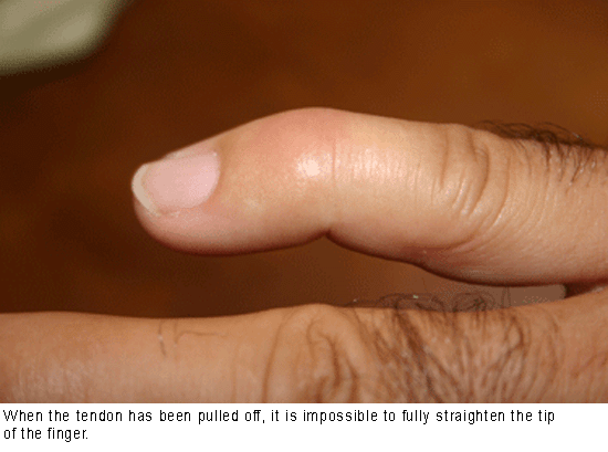
  • Stage I (acute) may last up to three months. During this stage the symptoms include pain and swelling, increased warmth in the affected part/limb, and excessive sweating. There may be faster-than-normal nail and hair growth and joint pain during movement of the affected area (Figure 1).
  • Stage II (dystrophic) can last three to 12 months. Swelling is more constant, skin wrinkles disappear, skin temperature becomes cooler, and fingernails become brittle. The pain is more widespread, stiffness increases, and the affected area becomes sensitive to touch.
  • Stage III (atrophic) occurs from one year on. The skin of the affected area is now pale, dry, tightly stretched, and shiny. The area is stiff, pain may decrease, and there is less hope of getting motion back.

Diagnosis

The diagnosis usually is made when at least three of the following symptoms are present: pain and tenderness, signs of changed blood flow (either increased or decreased), swelling with joint stiffness, or skin changes.

Treatment
Early diagnosis and treatment are important. Three forms of treatment may be combined: medication, physical therapy, and surgery. Medication taken by mouth can help decrease the symptoms. To reduce symptoms and provide long-term relief, local anesthetics may be injected into a nerve bundle at the base of the neck (stellate ganglion block). In some cases, a tourniquet is applied to the arm and medication can be injected into a vein along with an anesthetic.

Your hand surgeon may recommend therapy by a hand, occupational or physical therapist, or physician. Therapy is important to regain function and reduce discomfort caused by RSD. Successful treatment depends on the patient’s full and active effort in therapy. Occasionally, surgery is performed in the later stages, but the results can be disappointing. Your physician can advise you on the best treatment for your situation.

Mallet Finger (Baseball Finger)

Description
Mallet finger injuries occur when the tip of a finger or the thumb is forcefully bent. This condition is also known as baseball finger. It happens when a ball or other object strikes the tip of the digit. The force tears the thin tendon that allows you to straighten the finger. The force of the blow may even pull away a piece of bone along with the tendon.

Symptoms
A person with this injury has pain, swelling and bruising of the fingertip. The fingertip may droop noticeably. Occasionally, blood collects beneath the nail and the nail can even become detached from beneath the skin fold at the base of the nail.

cons1_397_151

Treatment Options
If you experience a mallet finger injury, immediately apply ice and elevate your hand above the level of your heart. Seek medical attention within the first week after this injury. It is very important to seek urgent attention if you have blood beneath the nail or if the nail is detached because you could also have a nail bed laceration or open (compound) fracture.

In most cases, the doctor will order X-rays to look for a major fracture or malalignment of the joint.

The majority of mallet finger injuries can be treated without surgery. The doctor may apply a splint to hold the fingertip straight (in extension) until it heals. Most of the time, you will have to wear the splint full-time for six weeks.

After six weeks, most patients gradually wean from the splint by wearing it less frequently over a three to four week period The finger usually regains acceptable function and appearance with this treatment plan; however, many patients may not regain full fingertip extension.

In children, mallet finger injuries may involve the cartilage that controls bone growth. A doctor must carefully evaluate and treat this type of injury, so the finger does not become stunted or deformed.

Treatment Options: Surgical

In certain cases, surgical repair may be considered. These cases include mallet finger injuries with:

  • Large fracture fragments
  • Joint malalignment

Surgical treatment in these cases often involves repair of the fracture using pins, pins and wire, or even small screws.

Surgery may also be considered if non-operative treatment fails. Repairing soft tissue mallet fingers is uncommon and usually reserved for patients with very severe deformities and functional problems. Surgical treatment can include tightening the stretched tendon tissue, using tendon grafts, or even fusing the joint straight. Consultation with an orthopaedic surgeon should be sought in these cases.

Kienböck’s Disease

Bone is living tissue that requires a regular supply of blood for nourishment. If the blood supply to a bone stops, the bone can die, a condition known as osteonecrosis. That’s what happens in Kienböck’s disease, which affects the lunate, one of the small bones of the hand near the wrist.

Kienböck’s Disease: Signs and Symptoms

The cause of Kienböck’s disease is unknown. Many people with Kienböck’s disease think they have a sprained wrist at first. They may have experienced some form of trauma to the wrist, such as a fall. This type of trauma can disrupt the blood flow to the lunate. In most people, two vessels supply blood to the lunate, but in some people there is only one source. This puts them at greater risk for developing the disease.

As the disease progresses, other signs and symptoms are noted, including:

  • A painful and sometimes swollen wrist
  • Limited range of motion in the affected wrist (stiffness)
  • Decreased grip strength in the hand
  • Tenderness directly over the bone (on the top of the hand at about the middle of the wrist)
  • Pain or difficulty in turning the hand upward

Progression

Kienböck’s disease follows a specific progressive pattern through four stages.

  • Stage 1: Symptoms are similar to those of a wrist sprain. X-rays may be normal or show a line indicating a possible fracture. Magnetic resonance imaging (MRI) may also be helpful in making the diagnosis in this early stage.
  • Stage 2: The lunate bone begins to harden. On an X-ray, it may appear brighter or whiter than the surrounding bones. These changes indicate that the bone is dying. Either an MRI or a computed tomography (CT) scan may be used to assess the condition of the bone. Recurrent pain, swelling and wrist tenderness are common.
  • Stage 3: The dead bone begins to collapse and break into pieces. The surrounding bones may begin to shift position. Patients experience increasing pain, weakness in gripping, and limited motion.
  • Stage 4: The surfaces of adjoining bones are affected, resulting in arthritis of the wrist.

Diagnosis and treatment
In its early stages, Kienböck’s disease may be difficult to diagnose because the symptoms are so similar to those of a sprained wrist. Even X-rays of the wrist may appear normal. At this point, the goal of treatment is to relieve the pressure on the lunate and help restore blood flow within the bone. Your physician may splint or cast your wrist for two to three weeks.

Anti-inflammatory medications such as aspirin or ibuprofen will help relieve any pain and reduce swelling. If the pain continues, your physician may refer you to an orthopaedic or hand surgeon for further evaluation.

Surgical options
Although there is no cure, there are several surgical options for treating the more advanced stages of Kienböck’s disease. The right procedure for you will depend on several factors, including disease progression, your personal activity levels and goals and your surgeon’s experience with various procedures.

Do not hesitate to discuss these options with your orthopaedic or hand surgeon and to ask why he or she is recommending a particular procedure.

In some cases, returning the blood supply to the bone (revascularization) may be possible. This procedure uses a bone graft from the inner bone of the lower arm. It may be combined with an external fixator, a metal device that helps relieve pressure on the lunate and preserve the spacing between bones.

If the bones of the lower arm are uneven in length, a joint leveling procedure may be recommended. Bones can be made longer with bone grafts, or shortened by removing a section of the bone.

This reduces the compressive forces on the lunate and seems to halt progression of the disease. If the lunate is severely collapsed or fragmented, it can be removed. The two bones on either side of it are also removed. This procedure is called a proximal row carpectomy and will relieve pain while maintaining partial wrist motion.

Another way to ease pressure on the bone is to fuse several of the small bones of the hand together. However, this will not improve range of motion. If the disease has progressed to severe arthritis of the wrist, fusing the bones will reduce pain and help maintain function, although motion is limited.

Hand Surgery

Our hands serve many purposes. Hands help us eat, dress, write, earn a living, create art and do many other activities. To accomplish these tasks and activities, our hands require sensation and movement, such as joint motion, tendon gliding and muscle contraction.

When a problem takes place in the hand, care must be given to all the different types of tissues that make function of the hand possible.

Hand surgeons are specifically trained to give that care:

  • Hand surgery is the field of medicine that deals with problems of the hand, wrist and forearm.
  • Hand surgeons care for these problems without surgery, and they are specially trained to operate when necessary.
  • Many hand surgeons are also experts in diagnosing and caring for shoulder and elbow problems.
  • Hand surgeons are orthopedic, plastic or general surgeons who have additional training in surgery of the hand.
  • To become a member of the American Society for Surgery of the Hand, a hand surgeon must complete a full year of additional training and must pass a rigorous certifying examination.

Dupuytren’s Contracture

Description
Dupuytren’s contracture is an abnormal thickening of tough tissue in the palm and fingers that can cause the fingers to curl. It is more common in men than in women and becomes more common as we grow older.

Risk Factors/Prevention
The cause of Dupuytren’s contracture is not known. It is not caused by an injury. It is not a cancer.

Dupuytren’s contracture is most common in people of Northern European or Scandinavian ancestry.
It is associated with smoking and drinking.
It is also associated with certain medical conditions such as diabetes, thyroid problems and seizures.

Symptoms
Dupuytren’s contracture usually occurs very gradually. It may begin as a small tender lump in the palm. Over time the pain usually goes away, but tough bands may form that cause the fingers to bend toward the palm. The ring and small fingers are most commonly affected.

Treatment Options
There is no way to stop or cure the problem. It is not dangerous. Dupuytren’s contracture usually progresses very slowly and may not be troublesome for years. If a painful lump is present, an injection may help diminish the pain. If the fingers become bent, they may interfere with use of your hand. Treatment is recommended when inability to straighten the fingers significantly limits your hand function. The treatment for Dupuytren’s Disease has advanced tremendously in the last few years. Treatments such as Needle Aponeurotomy and Xiaflex® injections have minimized the need for surgery for this condition.

Needle Aponeurotomy
Is a minimally invasive in office procedure that is performed under local anesthesia to disrupt the fascial bands formed by Dupuytren’s Disease and can correct significant deformity without the need for surgery. This procedure requires minimal recovery and no prolonged hand therapy.

XIAFLEX® Injections
Is an in office injection of Collagenase Clostridium Histolyticum which then enzymatically dissolves the Dupuytren’s cord and can also correct significant deformity with minimal recovery and downtime.

Surgical
Surgery for Dupuytren’s contracture divides or removes the thickened bands to help restore finger motion. Sometimes the wound is left open and allowed to heal gradually. Skin grafting may sometimes be needed. Risks of surgery include injury to nerves and blood vessels and infection. Some swelling and soreness are expected but severe problems are rare. Elevating your hand after surgery and gently moving the fingers helps minimize pain, swelling and stiffness. A physical therapist may be helpful during your recovery after surgery. Most people will have improved motion in the fingers after surgery.

Surgery does not cure the disease, which tends to progress gradually and recur over time.