Quadriceps Tendon Tears

patellar1-300x300Anatomy
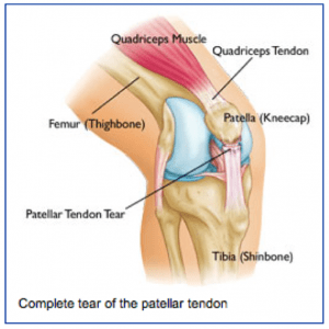
The quadriceps tendon attaches the main group of muscles in the front of the thigh (quadriceps) to the upper pole of the kneecap (patella).

The quadriceps muscle attaches to the upper pole of the kneecap through the quadriceps tendon to allow one to straighten out their leg or to support ones weight while squatting. When the quadriceps tendon is torn, one cannot support their weight when the knee is bent, such as getting up from a chair or going down the stairs.

Injury Mechanism
The quadriceps tendon is usually injured with a sudden high force across the tendon, such as landing from a jump. The quadriceps tendon can be injured by a direct blow or a sharp laceration across the tendon. Often, the quadriceps tendon is weak prior to the injury due to chronic illness or a period of inactivity.

Symptoms
Quadriceps tendon injuries present as significant pain across the front of the knee, just above the kneecap, and immediate weakness in supporting ones weight while squatting. There is often cramping of the thigh muscles. Patients usually cannot straighten their knee out fully with a complete tear of the quadriceps tendon. Rapid swelling occurs. Often, one can feel a gap above their kneecap at the site of the rupture.

Diagnosis
patellar2-300x235The physician’s work-up will start with a careful history and exam. The physician can often feel the defect in the quadriceps tendon and can appreciate the weakness on trying to straighten out the knee against resistance.

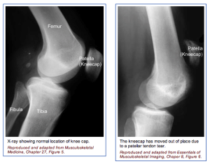
With a complete tear, x-rays reveal a low position of the patella, since the pull of the quadriceps is no longer pulling up on the patella.

An MRI scan is often obtained to confirm the diagnosis, especially if the physician is concerned that the injury may be just a partial tear that may be able to heal without surgical repair. Plain x-rays show the bones of the knee, while MRI scans reveal the soft tissues around the knee including the ligaments, menisci, muscles and tendons.

Treatment
Your physician will discuss treatment options with you. Treatment decisions are based on whether the injury is a complete tear or a partial tear. Partial tears are usually treated with a brace. The brace is initially locked out completely straight to take all of the pressure off of the injured tendon. As healing occurs, the hinges are unlocked and motion and strengthening are initiated. With complete tears, surgery is usually recommended.

Surgery involves sewing the two ends of the tendon together with strong suture material. If the tendon has pulled directly off of the kneecap, drill holes are made into the bone to repair the tendon directly to the bone. Risks and benefits of surgery are discussed thoroughly with the patient.

What to expect after surgery
Quadriceps tendon surgery is usually performed as an outpatient procedure. Patients are sent home with crutches and a knee brace with the knee fully straightened to protect the repair. Depending on the strength of the repair, patients start gentle range of motion and progressive weight bearing over 6-8 weeks.

Patients are using a stationary bike by 2 months, and an elliptical or stair climber shortly thereafter. Jogging is restricted until 4 months following surgery and full sports activities are not resumed until 6 months after surgery. Most patients can return to full activities, with no restrictions and no bracing at the 6th month point.

Posterior Cruciate Ligament Injuries and Multiple Ligament Injuries

Anatomy: The anterior cruciate ligament (ACL) runs from the back of the tibia (lower leg bone) to the front of the femur (thigh bone). It prevents the tibia from sliding backwards, especially when the knee is bent while going down stairs and inclines. Similar to the ACL, the PCL sits in the center of the joint and has poor blood supply that contributes to its poor healing potential.

The PCL is much thicker and stronger than the ACL, but when injured as an isolate injury, the PCL does not lead to as much instability as the ACL. However, often the PCL is injured in high energy accidents, such as motorcycle accidents, and often other ligaments are injured at the same time. With multiple ligament injuries, the combination often results in the knee being unstable for sports or daily activities, so multiple ligament injuries often require surgery to reconstruct the involved ligaments.

Injury Mechanism: The PCL is often injured when the lower leg bone sustains a direct blow from the front while the knee is bent, causing the lower leg bone to be pushed backwards. Occasionally, high-energy sporting injuries cause enough force to tear the PCL or several ligaments of the knee. PCLs can be an isolated injury, but often occur with associated injuries to the meniscus (cartilage pads) or other ligament combinations.

Symptoms: When the PCL or multiple ligaments are torn, usually rapid swelling occurs, usually within the first 24 hours. If multiple ligaments are involved, patients often describe a feeling that they “do not trust the knee.” The following day, one typically describes stiffness, swelling, and pain with weight bearing.

Diagnosis: The physician’s work-up will start with a careful history and exam. Often the description of sustaining a direct blow to the front of the lower leg, or often bruising over the front of the lower leg, can lead the physician to the suspected injury to the PCL. A history of immediate swelling of the knee (effusion) will further lead to this diagnosis. Once the patient can relax the muscles around the knee, the physician can feel the instability on the exam.

X-rays are often obtained to see that no fractures have occurred with the injury and to help assess the overall condition of the knee joint. An MRI scan is often obtained to confirm the diagnosis and to evaluate any associated injuries to the menisci, other ligaments, and damage to the joint surfaces. Plain x-rays show the bones of the knee, while MRI scans reveal the soft tissues around the knee including the ligaments, menisci, muscles and tendons.

Treatment: Your physician will discuss treatment options with you. Treatment decisions are based on age, activity level, degree of instability, and associated injuries to other structures about the knee. An isolated injury to the PCL can often be treated without surgery. The isolated PCL injury does not cause as much “giving way” as an ACL injury.

However, when the PCL is injured, one often develops arthritis in behind the kneecap and on the inside of the knee many years down the road from the original injury. Therefore, in a younger person, one considers reconstructing the PCL, even as an isolated injury, to prevent later arthritis. In an older individual, one can strengthen the muscles around the knee (especially the quadriceps) and expect to have a good long-term outcome from the PCL injury.

Occasionally, bracing can help for high-risk activities and sports. When the PCL is injured in addition to other ligaments in the knee, surgery is often required to re-establish stability in the knee. Surgery involves replacing the torn PCL and possibly the other injured ligaments with another tissue (a graft). Repairing the native PCL does not work, because of the poor blood supply mentioned above, so a substitute tissue must be used for the graft.

Tunnels are drilled in the femur and tibia at the attachment site of the normal PCL. The graft can then be passed through the tunnels to replicate the course of the normal PCL. The graft is fixed at both ends until the graft eventually heals into the tunnels and re-establishes a blood supply. Graft options will be discussed with one’s surgeon, but usually involves taking another tissue from elsewhere around the knee (such as the hamstrings or a portion of the patellar tendon) or taking tissue from a cadaver (another human being).

The cadaver tissue is often recommended because of the larger length and thickness of the PCL to provided an adequate graft. Cadaver tissue is often used for multiple ligament injuries as well because of the lack of enough tissue from ones own tendons to replace all of the damaged ligaments. Risks and benefits of each option will be discussed so a proper choice can be made for each patient.

What to expect after surgery: PCL and often even multiple ligament surgery is now performed as an outpatient procedure. Most patients find that they do best by resting their knee for 3 or 4 days following surgery with protected weight bearing with crutches. As swelling and pain subside, most patients are able to progress their weight bearing rapidly. Once fully weight bearing and not requiring pain medication, the patient can resume driving and returning to office work.

Formal physical therapy begins at one week following surgery and continues once or twice a week for six or eight weeks, depending on each individual’s progress. Patients are using a stationary bike by 3 weeks, and an elliptical or stair climber shortly thereafter. Jogging is restricted until 4 months following surgery and full sports activities are not resumed until 6 months after surgery. Most patients can return to full activities, with no restrictions and no bracing at the 6th month point.

Meniscal Tears

meniscal1-253x300Anatomy
Between the femur and tibia, sitting centrally in the knee joint, are two C-shaped pads (the medial and lateral menisci) that act as a cushion or shock absorber between the two bones.

The meniscal pads are made of cartilage. The meniscus has a poor blood supply except on the very periphery of the meniscus.

Peripheral tears, although rare, can be repaired, especially in the younger patients. With the inner portion of the meniscus having a poor blood supply, the meniscus has little healing potential, which leads many tears to surgery to help resolve the symptoms.

Injury Mechanism
meniscal2-300x184
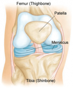
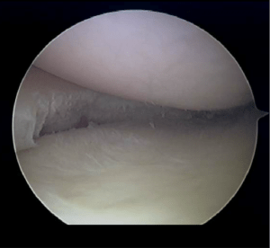
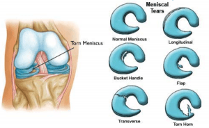
The meniscus is often injured with twisting the knee while in a squatting position. This creates a split or flap to occur while the meniscus is being compressed.
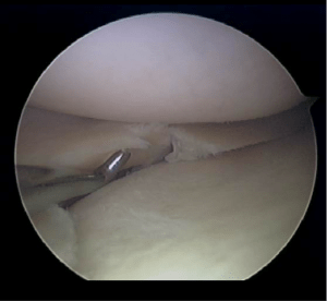
In younger patients, the meniscus is firm and is usually injured in sports activities. In older patient, the meniscus is often more brittle or friable and injuries can occur with daily activities such as squatting down to make the bed or working in the yard.

Symptoms
The “classic” meniscal injury creates pain on one side of the knee or in the back of the knee. The pain is usually worsened with more rigorous activities or with bending the knee fully. Bending the knee causes the meniscus to become pinched between the two bones. Meniscal tears can cause subtle mechanical symptoms like clicking.

However, sometimes patients will describe catching or actual “locking up” of the knee in which they cannot fully bend or straighten the knee. Swelling does not typically occur until several days after the injury and commonly with activities. Unlike with ACL injuries, the swelling will not usually subside until the meniscus tear is addressed surgically.

Diagnosis
The physician’s work-up will start with a careful history and exam. Sometimes patients recall a twisting injury when the pain began, but often patients just noticed that the knee began to hurt or swell with activities and cannot recall a specific event. On examination, the knee is often slightly swollen (effusion). Range of motion is often limited, and pain can usually be elicited with fully bending the knee. The knee is often tender on one side of the knee or the other specifically at the joint line.

X-rays are often obtained to see that no fractures have occurred with the injury and to help assess the overall condition of the knee joint. If significant arthritis is present, the outcome from surgery is less favorable even with a co-existing meniscus tear. An MRI scan is often obtained to confirm the diagnosis and to evaluate any associated injuries to the other meniscus, ligaments, or damage to the joint surfaces. Plain x-rays show the bones of the knee, while MRI scans reveal the soft tissues around the knee including the ligaments, menisci, muscles and tendons.

Treatment
Your physician will discuss treatment options with you. Treatment decisions are based on age, activity level, severity of symptoms, and associated arthritis within the knee. Often your surgeon will recommend surgery, since meniscus tears rarely heal without surgery, because of the poor blood supply mentioned above. In older patients with less activity demands and less severe symptoms, one can try nonoperative treatment including rest, ice, anti-inflammatories, occasional injections and see if the symptoms become tolerable.

In younger, more active patients, surgery is almost always recommended. Surgery involves removing or repairing the torn meniscal tissue. Most of the tears are in the central portion of the meniscus where there is no blood supply, therefore no healing potential. Those tears need to be removed, such as trimming off a hangnail. The surgeon tries to be conservative and only remove the damaged tissue so that adequate padding remains in the knee.

In the tears that are further out near the edge of the meniscus, where there is some healing potential, sutures can be placed through the arthroscope to repair the torn meniscus.

meniscal3-300x276
Probe shows tear in the meniscus
meniscus4-300x276
Same meniscus after removing the tear

What to expect after surgery
Meniscus surgery is performed as an outpatient procedure. The surgery takes about 30 minutes. Most patients are able to walk on their knee to the bathroom and meals even on the day of surgery without crutches, but most of the first day is spent resting with the leg elevated and iced. By the second or third day, most patients are able to drive and walk short distances and return to desk jobs.

By one week, patients are encouraged to start light, straight ahead exercising, such as using a stationary bike, an elliptical or stair climber, level walking or swimming. Activities are added during the next 2 to 3 weeks. By one month following surgery, most patients have returned to full activities. If the meniscus has been repaired rather than resected, a more prolonged recovery might become necessary to protect the repair.

Anterior Cruciate Ligament Injuries

anterior-240x300Anatomy
The anterior cruciate ligament (ACL) runs from the front of the tibia (lower leg bone) to the back of the femur (thigh bone). It prevents the tibia from sliding forward and keeps the knee from pivoting (instability). The ACL sits in the center of the joint and has poor blood supply that contributes to its poor healing potential.

Injury Mechanism
The ACL is often injured in sports with twisting type injuries or hyperextension injuries of the knee. ACL injuries can occur with rapid stopping while running and often as contact injuries. ACLs can be an isolated injury, but often occur with associated injuries to the meniscus (cartilage pads) or other ligament combinations.

Females are known to have a higher rate of ACL injuries than males in the same sports.

Arthroscopic view of the normal ACL

Symptoms
The “classic” ACL injury is described as a sudden “giving way” and hearing a “pop” at the time of the injury. Rapid swelling occurs, usually within the first 24 hours.

The following day, one typically describes stiffness, swelling, and pain with weight bearing.

Over the next 1 to 2 weeks, the swelling starts to subside and the range of motion of the knee improves, but patients may start to experience “giving way” or a sense that they cannot trust the knee.

Diagnosis
The physician’s work-up will start with a careful history and exam. Often the description of a sudden “giving way” episode and “pop” can lead the physician to the suspected injury to the ACL. A history of immediate swelling of the knee (effusion) will further lead to this diagnosis.

Once the patient can relax the muscles around the knee, the physician can feel the instability on the exam. X-rays are often obtained to see that no fractures have occurred with the injury and to help assess the overall condition of the knee joint.

An MRI scan is often obtained to confirm the diagnosis and to evaluate any associated injuries to the menisci, other ligaments, and damage to the joint surfaces. Plain x-rays show the bones of the knee, while MRI scans reveal the soft tissues around the knee including the ligaments, menisci, muscles and tendons.

Treatment
Your physician will discuss treatment options with you. Treatment decisions are based on age, activity level, degree of instability, and associated injuries to other structures about the knee. If a patient is older and does not participate in ACL dependent activities (soccer, basketball, court sports, or other twisting and pivoting sports) the patient may choose nonoperative treatment.

Nonoperative treatment does not mean “no treatment.” The patient is educated about the ACL injury and educated about high-risk activities. Often physical therapy is initiated to help regain full range of motion and strengthen the surrounding muscles that can help stabilize the knee. Occasionally, bracing can help for high-risk activities and sports. If patients in this group have “giving way” episodes after therapy or bracing, they may need to be considered for ACL surgery.

In the younger, more active patients, surgery is undertaken to help stabilize the joint to allow the patient to return to full activities. Surgery involves replacing the torn ACL with another tissue (a graft). Timing of surgery is important.

It has been found best to wait for at least 2 or 3 weeks after the injury before undertaking surgery to give the swelling time to resolve and to allow the patient to recover most of their range of motion before surgery. The chance of developing stiffness following surgery is decreased with better motion going into surgery.

Repairing the native ACL does not work, because of the poor blood supply mentioned above to the ACL, so a substitute tissue must be used for the graft. Tunnels are drilled in the femur and tibia at the attachment site of the normal ACL. The graft can then be passed through the tunnels to replicate the course of the normal ACL. The graft is fixed at both ends until the graft eventually heals into the tunnels and re-establishes a blood supply.

Graft options will be discussed with one’s surgeon, but usually involves taking another tissue from elsewhere around the knee (such as the hamstrings or a portion of the patellar tendon) or taking tissue from a cadaver (another human being). Risks and benefits of each option will be discussed so a proper choice can be made for each patient.

What to expect after surgery
ACL surgery is now performed as an outpatient procedure. Most patients find that they do best by resting their knee for 3 or 4 days following surgery with protected weight bearing with crutches. As swelling and pain subside, most patients are able to progress their weight bearing rapidly.

Once fully weight bearing and not requiring pain medication, the patient can resume driving and returning to office work. Formal physical therapy begins at one week following surgery and continues once or twice a week for six or eight weeks, depending on each individual’s progress.

Patients are using a stationary bike by 3 weeks, and an elliptical or stair climber shortly thereafter. Jogging is restricted until 4 months following surgery and full sports activities are not resumed until 6 months after surgery. Most patients can return to full activities, with no restrictions and no bracing at the 6th month point. Please see the complete ACL physical therapy protocol.

Common Knee Injuries Related to Sports

Common Knee Injuries

The knee is a complex structure with many components that make it vulnerable to a variety of sports injuries. Most people suffer a minor knee problem at one time or another. Knee injuries often occur during sporting activities, too. In 2009, knee injuries were the most common reason for visiting an orthopedic specialist.

Knee Anatomy and Function

The knee joint is the largest joint of the body and the one that is most easily injured. Two cushioning discs call menisci separate the upper and lower bones of the knee. The upper leg bone (the femur) and the bones of the lower legs are connected by ligaments, tendons, and muscles.

The surface of the bones is covered by cartilage, a substance that absorbs shock and provides a smooth, gliding surface. Knee injuries are the result of damage to one or more of these structures. Of the four major ligaments found in the knee, the three that are injured the most are the anterior cruciate ligament (ACL), the medial collateral ligament (MCL), and the posterior cruciate ligament (PCL).

ACL Injury

The ACL is injured when athletes are changing direction rapidly, slowing down when running, or landing from a jump. Individuals who play basketball, football, and soccer and those who ski are particularly at high risk for ACL injuries. If this structure is damaged, you could require surgery to regain full function of your knee. This will all depend on the severity of your injury and your activity level.

Most of the time ACL tears are too severe to be stitched back together. The orthopedic surgeon will have to surgically repair this structure by reconstructing the ligament. Most of the time the doctor will use a tissue graft to repair the ligament. This graft acts as scaffolding for new ligament to grow on. Most of the time grafts are taken from the patellar tendon or the hamstring tendons.

MCL Injury

A direct blow to the outer aspect of the knee commonly causes injuries to the MCL. Those athletes who play football and soccer are at the highest risk. When the knee is forced sideways, the MCL can tear and result in knee pain. Swelling will occur with a MCL injury as well and the knee will become unstable and give way.

When the MCL is torn severely and cannot heal correctly, surgery is necessary. This will involve grafting a piece of tendon to allow the portions of the torn ligament to connect to. Most of the time, however, these injuries can be treated without surgical intervention.

PCL Injury

The PCL is most commonly injured when an athlete receives a blow to the front of the knee. This structure can also be torn or injured if the individual makes a simple misstep on the playing field. Those who participate in football and soccer are at the greatest risk for a PCL injury. This ligament is located in the back of the knee and connects the femur to the shinbone (the tibia).

When an athlete suffers a PCL tear or injury, the orthopedic specialist will most likely recommend surgery. This is done to rebuild the ligament by replacing the torn structure with a tissue graft. During the procedure the doctor will rebuild the PCL. A tendon or other structure is used to replace the torn ligament.

Meniscus Injury

The menisci tear in different ways. The orthopedic specialist depending on how they look, where they occur, and how complex they are classes the tears. Common tears include the parrot-beak, flap, bucket handle, longitudinal, and mixed/complex. Sports-related tears of the meniscus often happen along with other knee injury. Sudden tears can occur when the athlete squats or twists the knee. Direct contact or a sharp blow can also cause meniscus injury.

When a meniscus tear is serious and the symptoms persist with nonsurgical treatment, your doctor may recommend an arthroscopic procedure of the knee. This is one of the most commonly performed surgical procedures where the orthopedic specialist inserts a miniature camera into the knee joint to trim and repair the tear to the meniscus. While he is doing this surgery, other torn structures can be repaired and treated as well.