Shoulder Injuries
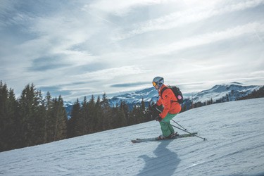
If you are a snowboarder or skiing fanatic, you know that injuries come with the territory. If you aren’t careful, you could end up with torn ligaments, sprained muscles, or broken bones.
Statistics tell us that less than 4 injuries happen for every 1,000 days of skiing or snowboarding.
Sometimes these injuries are minor and only will require home care. Other times, they can be serious, requiring you to seek medical help. This guide will help you to understand the most common snowboarding and skiing injuries.
Acromioclavicular Joint Damage
The Acromioclavicular Joint is often called the “AC joint” for short (pronounced ack-roe-my-oh-clah-vick-you-lar). This is basically a separation of the two bones that form this joint where the clavicle (collar bone) attaches to the scapula (shoulder blade). There is a ligament that attaches these two bones known as the AC ligament. The bony process that protrudes forward from the upper scapula is the acromion (pronounced ack-rome-ee-on). When these two main bones are separated, it is often referred to as a shoulder separation injury.
If you injure your AC joint skiing or snowboarding, expect to have pain at the end of your collarbone. This pain will spread throughout the shoulder at first. Eventually, the initial pain will resolve and be followed by pain over the joint itself.
Swelling will occur, too and depending on the severity of the injury, there may be a visible deformity. This will be an obvious bump where the joint has been separated. Pain will worsen with movement of the shoulder, especially when the arm is raised to or above shoulder height.
Doctors will grade AC joint injuries from one to six (1 – 6) using the Rockwood Scale which identifies injuries by amount of damage incurred. This will be based on the space between the acromion and clavicle. Grade one is just a simple sprain of the AC joint, but Grades four, five, and six involve severe conditions, and most always result in shoulder surgery.
What Should the Athlete Do?
- Immobilize the arm and shoulder with an arm sling.
- Start R.I.C.E. therapy.
- Consult an Orthopedic Specialist.
What will the Orthopedic Specialist Do?
- Use ultrasound or laser treatment.
- Perform shoulder surgery if necessary.
- Prescribe a special rehabilitation program to get you back moving.
Fractured Clavicle
The clavicle (or collar bone) is the bone that runs along the front side of the shoulder region and connects near the breast bone (sternum). This is one of the most common broken bones, and it occurs as a result of falling on an outstretched arm. This is one of the most common types of fractures in sporting and outdoor activities.
The bone typically fractures in the middle third region and this type of injury is very painful. Symptoms of a broken clavicle include swelling and discoloration at the site, pain of the area, a deformity that can be seen or felt, and worsening pain when elevating the arm.
What can the Athlete Do?
- Immobilize the arm and shoulder regions.
- Do R.I.C.E. therapy.
- Get to an Orthopedic Specialist for evaluation and treatment.
What will the Orthopedic Specialist Do?
- Evaluate the extent of the injury by way of X-Ray.
- Use ultrasound or laser treatment.
- Do shoulder surgery if the bone is displaced.
The Knee Injuries
Medial Collateral Ligament Trauma
The Medial Collateral Ligament (MCL) is a ligament that connects the inner surface of the thigh bone (femur) to the shin bone (tibia). This ligament allows the knee to resist force that may be applied from the outer surface thus preventing the inner portion of the joint from stretching under stress.
There are two parts to the inner knee ligament, the deep inner section that hooks onto the cartilage meniscus and the joint margins and a superficial band that adheres from higher up on the femur to the tibia.
An injury to the MCL occurs after there is an impact injury to the outside surface of the knee when the knee is in the bent position. The MCL will become stretched and the impact force tears the fibers. The inside portion of this ligament is prone to become injured first and this often leads to the meniscus being damaged as well. Pain in the area may not be noticed immediately after the injury.
These injuries are graded on a one to three (1 – 3) scale. A grade one tear only has less than 10% of the fibers torn. A grade two is greater than 11% but does not necessarily result in a complete tear of the ligament. A grade three is a complete rupture of the ligament, however. With a grade one tear, there may be mild tender knee on the inner aspect of the knee, but typically no swelling occurs.
With a grade two tear, there will be more pain and tenderness and some swelling over the ligament. The pain of a grade three tear is often not as bad as a grade two but this injury results in a significantly more unstable, wobbly knee. Grade three tears most always result in knee surgery.
What Should the Athlete Do?
- Utilize the R.I.C.E. formula to the injured knee.
- Refrain from activity.
- Wear a knee brace for grade two and three injuries.
- Consult an Orthopedic Specialist.
What will the Orthopedic Specialist Do?
- Apply a cast or joint support device.
- Use sports massage techniques.
- Aspirate the joint to remove fluid.
- Apply ultrasound or laser treatment.
- Order an MRI to assess the possibility of surgical reconstruction.
- Do knee surgery, as indicated.
- Order a special rehabilitation program to help get you back in action.
Medial Meniscus Injuries
The Medial Meniscus (MM) is prone to many more injuries that the Lateral Meniscus. This structure is connected to the Medial Collateral Ligament (MCL) and the joint capsule. The MM is less mobile, too. Any force impacts can severely injure this structure and cause permanent damage. Tearing of the MM often requires surgical intervention.
The symptoms of a MM tear include pain on the inner surface of the knee joint, swelling of the knee at any time during the 48 hours after the injury, inability to bend the knee fully, a clicking noise with bending, and ‘locking’ or ‘giving way’ of the knee. Many who have this type of injury are unable to bear weight on the knee.
There are several types of meniscal tears. These include the longitudinal tears (ones that occur along the length of the meniscus), radial tears (those tears that occur from the edge of the cartilage inward), bucket-handle tears (like a longitudinal but occur where a portion of the meniscus becomes detached from the tibia forming a flap), and degenerative changes (making the meniscus become frayed or jagged). Most meniscal tears result in knee surgery.
What Can the Athlete Do?
- Utilize the R.I.C.E. method.
- Wear a knee compression support device.
- Utilize mobility exercises to get the knee moving better.
- Consult an Orthopedic Specialist.
What will the Orthopedic Specialist Do?
- Check the extent of the knee injury.
- Order an MRI scan of the injured knee.
- Use ultrasound or laser treatment.
- Do knee surgery if necessary.
The Wrist and Hand Injuries
Skiier’s Thumb
The most common upper extremity injury when skiing is to the thumb. The thumb has two ligaments on each side at the metacarpophalangeal (pronounced met-ah-car-poe-fah-lanj-ee-ahl) or MCP joint. The inner ulnar collateral ligament (UCL) gets damaged when a fall occurs and the skier doesn’t release the ski pole from the hand.
The pole makes a bending type stress occur to the thumb. “Skier’s Thumb”, as it is commonly called, occurs when the UCL is torn after the thumb is placed in an extreme position.
What Can the Athlete Do?
- Immobilize the injured area.
- Use the R.I.C.E. formula.
- Consult an Orthopedic Specialist.
What will an Orthopedic Specialist Do?
- Order an MRI scan to assess the damage.
- Prescribe a rehabilitation program for you.
- Operate on the injured area if necessary.
Wrist Fracture
Snowboarding and skiing are dangerous sports and beginners often have a higher risk of falls. Sometimes these falls damage the wrist enough that the skier or snowboarder has to have a wrist surgery. These falls result in wrist fractures, where an outstretched hand attempts to break a fall.
As a result of this, there are scaphoid and “Colles” fractures that can occur and around 100,000 of these occur each year. The schaphoid is one of the small bones called the “Carpal” area that make up the wrist.
“Colles” is the name of this type of fracture where the radius bone is injured. Signs of a fracture to this area include swelling of the wrist, pain with movement, and a visible deformity or bump. There is often tenderness in the region where the thumb and wrist connect.
What Can the Athlete Do?
- Utilize R.I.C.E. therapy to the injured area.
- Immobilize the injured extremity.
- Consult an Orthopedic Specialist.
What will the Orthopedic Specialist Do?
- Order laser treatment or ultrasound therapy.
- Order X-ray testing and MRI scans.
- Apply a splint or cast for support.
- Perform wrist surgery as indicated.
Ankle and Knee Sprains
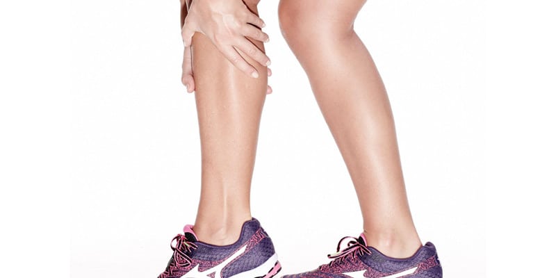
Twisting and turning down the slope can put a bit of a strain on your ankles and knees. Ankles get sprained when they are extended past the point they should be. Knees are devised to only bend in one direction and they are easily sprained when forced in an unnatural position.
Sprains are common among those who engage in sporting activities like skateboarding, snowboarding, and skiing and usually are nothing serious. If you suffer a sprain, you can expect pain, swelling, bruising, and a decrease in range of motion of the knee or ankle.
R.I.C.E.
The main formula for treating sprains is R.I.C.E. This stands for Rest, Ice, Compress, and Elevate.
- R: Rest. You should stop the activity as soon as it starts to hurt. Stay off the knee or ankle for a while and see if it gets better. If the discomfort stops after a brief rest, gently rotate and flex the area to see if there is any remaining discomfort. If not, then you are good to go. If it still hurts, it’s time to I.C.E. it, so read on.
- I: Ice. Do NOT apply it directly onto the skin! Instead, apply an ice pack wrapped in cloth for 20 minutes on the sprained knee or ankle. Ice is essential for minor turns and twists that don’t stop hurting after a brief rest period. For serious sprains, you should apply ice to your sprain immediately, pronto, or stat! Even if you are going to seek medical attention, you should ice the area down anyway. Apply an ice pack or just use a can of soda from the frig or a bag of frozen peas. Some people put ice in a Ziplock bag and cover it with a towel. Whatever works for you.
- C: Compression. This simply means you need to wrap your sprained ankle or knee with an ACE bandage. Be sure to wrap it firmly but not too tightly. The pressure encourages the stray fluid that is at the sprain to get back into the vessels where it belongs and circulate back to the heart. Do this for even minor strains to reduce the chance of swelling and support the injured area. Apply the ice on top of the bandage.
- E: Elevate. Simply raise the injured area above the heart right away. You should also do this at night. The purpose of this is so gravity will help drain the excess fluid from the tissues.
Frostbite and Hypothermia
Everybody has heard of Jack Frost. He’s that happy cold-weather fellow who likes to nip at your fingers, nose, and toes. If you feel that ‘pins and needles’ feeling, that’s your sign to get to a warm area. When frostbite occurs, your skin will be bright pink and then turn red and swollen at first. Serious frostbite makes the areas turn bluish-gray.
When hypothermia occurs, you will shiver and your heart beat and breathing will speed up as your body temperature goes down. You may feel clumsy, confused, and feel really sleepy. Both of these conditions are serious and you should seek medical help if they occur.
If your fingers, nose, or toes are turning bluish-gray, red, or dark pink from frostbite, get help immediately. Don’t use the frostbitten part of your body and don’t rub the skin. Warm up the area with warm water (not hot) and avoid heating lamps or campfires. Cover the chilled area if possible, and put your hands or fingers in your armpits for warmth. Remove any wet clothing and get to a warm place.
Sunburn
The sun’s rays are the hottest from 10 am to 4 pm. If you are out during this time, you are more likely to burn. If you do get sunburned, expect discomfort and pain, redness, swelling, and even blistering. A minor burn will make the skin be tender to touch. A more serious burn will involve blistering. If you have lots of blistering on your body, you will need to seek help immediately.
The main objective is to cool down the skin and limit the extent and discomfort of the burn. Take a shower with cool water or use aloe vera cooling gel on your skin. Drink plenty of fluids, too. Sunburns are dehydrating as they draw water from the body. If your skin is burned, you will need to stay out of the sun for a couple of days to let it heal.
Prevention of Skiing and Snowboarding Injuries
It is easier to prevent than to treat, so take this advice seriously and do what you can in the way of prevention. Here are some handy tips:
- Warm it up. Before starting a sports activity, jog in place, walk briskly, and stretch to warm up your muscles. Get into the sport gradually over a few minutes, giving your muscles a chance to get a good supply of blood.
- Cool it. Cool down afterwards by stretching or light walking to keep the muscles from shortening and trapping the by-products of exercise (lactic acid) in the tissue and resulting in stiffness.
- Cover up. For hypothermia and frostbite prevention, wear a hat, dress in layers, wear long underwear, and use a water-resistant coat and gloves. Another tip is to do buddy checks with a friend. Drink warm fluids before going out for your ski or snowboarding activity.
- Lather up. For sunburn prevention, use sunscreen with an SPF of at least 15. It is important to remember to put this on top of your head and in your part line if you aren’t wearing a hat.
- Dress up. For sunburn prevention, wear large shaded goggles or glasses and appropriate gear. Keep your head covered, as your part line and scalp is sensitive.


