
Patellar Tendon Tears

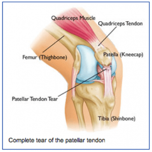
patellar1-300x300Anatomy
The patellar tendon attaches the lower pole of the kneecap (patella) to the lower leg bone (tibia). The quadriceps muscle attaches to the upper pole of the kneecap and pulls through the kneecap and patellar tendon to allow one to straighten out ones leg or to support ones weight while squatting.

When the patellar tendon is torn, one cannot support their weight when the knee is bent, such as getting up from a chair or going down the stairs.

Injury Mechanism
The patellar tendon is usually injured with a sudden high force across the tendon, such as landing from a jump. The patellar tendon can be injured by a direct blow or a sharp laceration across the tendon.

Symptoms
Patellar tendon injuries present as significant pain across the front of the knee and immediate weakness in supporting ones weight while squatting. Patients usually cannot straighten their knee out fully with a complete tear of the patellar tendon. Rapid swelling occurs. Often, one can feel a gap under their kneecap at the site of the rupture.

Diagnosis
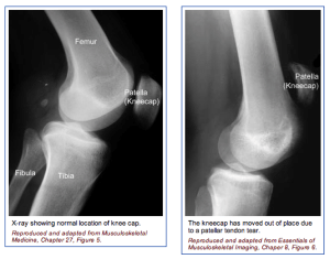
patellar2-300x235The physician’s work-up will start with a careful history and exam. The physician can often feel the defect in the patellar tendon and can appreciate the weakness on trying to straighten out the knee against resistance.

X-rays reveal a very high riding patella, since the pull of the quadriceps is no longer opposed by the tethering effect of an intact patellar tendon.

An MRI scan is often obtained to confirm the diagnosis, especially if the physician is concerned that the injury may be just a partial tear that may be able to heal without surgical repair. Plain x-rays show the bones of the knee, while MRI scans reveal the soft tissues around the knee including the ligaments, menisci, muscles and tendons.

Treatment
Your physician will discuss treatment options with you. Treatment decisions are based on whether the injury is a complete tear or a partial tear. Partial tears may be able to be treated with a brace. The brace is initially locked out completely straight to take all of the pressure off of the injured tendon.

As healing occurs, the hinges are unlocked and motion and strengthening are initiated. With complete tears, surgery is almost always recommended. Surgery involves sewing the two ends of the tendon together with strong suture material. If the tendon has pulled directly off of the bone, drill holes are made into the bone to repair the tendon directly to the bone. Risks and benefits of surgery are discussed thoroughly with the patient.

What to expect after surgery
Patellar tendon surgery is usually performed as an outpatient procedure. Patients are sent home with crutches and a knee brace with the knee fully straightened to protect the repair. Depending on the strength of the repair, patients start gentle range of motion and progressive weight bearing over 6-8 weeks.

Patients are using a stationary bike by 2 months, and an elliptical or stair climber shortly thereafter. Jogging is restricted until 4 months following surgery and full sports activities are not resumed until 6 months after surgery. Most patients can return to full activities, with no restrictions and no bracing at the 6th month point.
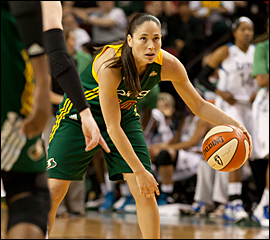
WNBA Star Sue Bird Recovering from Knee Surgery

Photo Via WNBA.com

As ESPN recently reported, Sue Bird, “The Seattle Storm star point guard is recovering from recent left knee surgery, which will sideline her for the entire summer season that began this weekend and runs through August.” Bird underwent knee surgery earlier this May to repair a cyst. She is hopeful that she will be able to play this fall when the team plays against Russia. In addition, she is optimistic that she will be back next spring.

In an interview, Bird said, “I’m doing well, I’m about almost two weeks post-op and had my follow-up appointment, everything’s good. It’s a waiting game now.” … read more

How to Prevent Achilles Tendon Injuries

Achilles Tendon

The tendon connecting your calf muscle to your heel bone is called your Achilles tendon. It is the largest tendon in your body. You use it every day when you jump, run, and walk. The Achilles tendon is prone to overuse (called tendinitis) because of the stresses it sees every day.

The Achilles tendon is one of the most commonly injured tendons, especially among athletes. Tendinitis is the most common form of injury, resulting in pain and swelling either within the tendon or where the tendon inserts into the heel bone (calcaneus). In worse cases, too much pressure on the Achilles tendon can cause it to partially tear or even rupture completely.

What can cause an Achilles tendon injury?

A rupture of your Achilles tendon often occurs when you abruptly start moving, such as while sprinting. Men age 30 years and up have more Achilles tendon injuries than other groups. We see these injuries a lot in so-called “weekend warriors.” There are a number of things that can cause or contribute to an Achilles tendon injury. Some of these might be:

  • Overuse
  • Inadequate stretching before a strenuous activity
  • Performing high-impact activities, such as jumping or plyometrics
  • Foot deformities, such as flat feet or high arches
  • Tight muscles and tendons in your legs

Athletes who participate in the following activities may experience Achilles tendon injuries:

  • Gymnastics & Dance
  • Running
  • Football
  • Baseball
  • Softball
  • Basketball
  • Volleyball
  • Tennis

How can I prevent an Achilles tendon injury?

There are many ways to prevent an Achilles tendon injury.

Maintain a Healthy Weight: Make sure you maintain a healthy weight range that is ideal for you. Avoid being underweight or overweight.

Eat Well: Make sure that you are getting the recommended amounts of vitamins and minerals. A diet lacking in calcium could cause Achilles tendon injuries. The U.S. Institute of Medicine’s Recommended Dietary Allowance (RDA) and Dietary Reference Intake (DRI) suggest that adults should get 1,000-1,200 mg of calcium daily and at least 700 mg of phosphorous.

Calcium is essential for your bones to grow and also helps with muscle contraction. Too little calcium or phosphorous intake will result in your body taking what is needed from other places, and this can lead to weaker bones.

Exercise Regularly: Injury prevention involves exercising often to keep your tendons, muscles, and bones strong. Make sure you only do exercises approved by your doctor. If you go to a gym, ask a trainer to help you use the equipment properly.

Pick the Right Shoes: Make sure that you are wearing the correct shoes for your feet. Shoes vary in the amount of cushioning and arch support they provide. Have your athletic shoes fitted by a specialist.

Avoid Hard Surfaces: Soft surfaces, such as cushioned floors, are better on your feet than hardwood. Too much time spent on hard surfaces can cause injuries to the Achilles tendon. Simple choices such as picking a grass court over a hard court, or running on a dirt trail instead of a sidewalk will help to reduce impact and may reduce your risk of injury.

Warm Up and Cool Down: To avoid injury to your Achilles tendon, you should always warm up and cool down before and after exercising or any physical activity.

Start Low and Go Slow: As is the case with other structures in the body, the Achilles tendon responds better to gradual increases in intensity. To avoid injury you should not suddenly increase your distance, speed, or steepness. Gradually increase your exercises rather than jumping right in and making big changes to your work out.

Get Strong: Calf muscles tend to be weak in many people, so it is important to strengthen them through exercise. You can do leg exercises with machines, elastic pull bands, and weights. Some good techniques to strengthen your leg muscles include lunges, leg presses, calf raises, squats, and leg curls.

Do Aerobics First: If you are planning on doing aerobics and strengthening on the same day, make sure that you do the aerobic exercise first. This includes running, stair climbing, and playing sports. These activities put strain on the Achilles tendon. If you perform your strengthening activities first, you put your Achilles tendon at risk for injury because it temporarily weakens the tendon.

Test Your Achilles Tendon: You should test your Achilles tendon for injuries after you finish working out. Use your thumb and forefinger to pinch different areas along your Achilles tendon looking for tenderness. If you experience any pain, this could indicate tendinitis.

What do I do if I have pain?

If you develop pain in your Achilles tendon, see an orthopedic foot and ankle surgeon as soon as possible. If the diagnosis of Achilles tendinitis is made early enough, several options such as immobilization, physical therapy, and stretching are available.

If you continue to exercise and do strenuous activity after you develop pain in the tendon, you risk rupturing the tendon. If this occurs, your surgeon may recommend surgical repair of the tendon.

Common Wrist Sports Injuries

As an orthopedic specialist, I see wrist injuries more commonly among people who participate in sporting activities, such as gymnastics, contact sports, skiing, skateboarding, snowboarding, and racquet sports. Below I will explain the four common mechanisms of injury, the common wrist sports injuries, and how these injuries are treated.

The wrist allows you to properly position your hand, representing arguably one the most complicated joints in the body. There are 15 bones and 27 articular surfaces in the wrist, not to mention its elaborate system of muscles, tendons and ligaments. Ligament injury is quite common among athletes, as the repetitive action of the wrist puts athletes at risk for injury. Wrist sprains result from a torn or partially torn ligament, and wrist strains are the result of a torn or partially torn tendon. The most common wrist fractures among athletes include: distal radius fractures and scaphoid fractures.

The Four Mechanisms of Wrist Injury

Throwing – With throwing injuries, there is an overuse of the wrist. These are common in baseball players, tennis athletes, and racquet ball participants.

Weight-bearing – I see many weight-bearing injuries among those who participate in gymnastics, weightlifting, and cheer-leading.

Twisting – With a twisting injury, the wrist suffers from a rapid rotation that disrupts the stability of the wrist. I see this type of injury a lot with radical skateboarders and snowboarders.

Impact – More common in football athletes, I treat impact injuries that result from either a direct impact or a fall onto an outstretched hand.

Wrist Sprains

The most common wrist injury among athletes is a sprain of the wrist. This often is an injury to one of the ligaments – the connective tissue that attaches one bone to another. Most sprains occur when the wrist is forcefully bent during a fall on an outstretched hand. Wrist sprains can be mild or severe, and I grade them based on the degree of injury. A grade 1 sprain indicates a stretched ligament without apparent tearing. A grade 2 sprain, however, involves partial tearing of a ligament. With a grade 3 sprain indicates ligaments are completely torn.

Distal Radius Fracture

The most common fracture is called a “distal radius fracture.” A distal radius fracture is a break that occurs at the wrist end of the radius bone. These breaks are common among athletes and can be mistaken for sprains. Wrist fractures often occur during a fall onto an outstretched hand. With fractures of the wrist, the break can occur in four ways: intra-articular, extra-articular, open, or comminuted (in many parts). Many can be treated with casting alone, though some require surgery.

Scaphoid Wrist Fracture

The scaphoid bone is one of the smaller bones of the wrist, but it is one that commonly breaks during sporting injuries. This bone is located on the thumb side of the wrist, and can be difficult to treat due to its tenuous blood supply. As with most wrist injuries, a break to the scaphoid bone typically occurs from falling onto an outstretched hand. Treatment usually requires casting if not displaced, or surgery if displaced.

Symptoms of Significant Wrist Injuries

  • Pain at the time of injury
  • Swelling
  • Bruising or discoloration
  • Difficulty moving the wrist
  • A “popping” or tearing sensation during the trauma
  • Warmth and tenderness of the skin

Treatment for Wrist Injuries

Treatment really depends on the type of injury you have. For mild sprains, I generally recommend the “RICE” method and over-the-counter pain relievers, like Tylenol or Motrin.

RICE

R – Rest the wrist for around 48 hours.
I – Ice the injured area to reduce swelling (use a pack wrapped in a towel).
C – Compress the wrist with an elastic ACE wrap.
E – Elevate the injury above heart level.

Nonsurgical Treatment

Simple Sprain –With mild to moderate wrist sprains, you will need to wear a splint for 1 to 3 weeks. This keeps the wrist immobilized while it heals. If you develop stiffness, I can teach you some stretching exercises to allow you to regain full range of motion of your wrist.

Simple Fracture –If your broken bone is in good position, I can treat it by applying a fiberglass or plaster cast. This is done so that the healing wrist bone remains protected from further injury while it heals. You may have to wear the cast for up to 6 weeks, depending on your injury.

Closed Reduction –If the alignment is out of place, I may need to “reduce” the bone and re-position the bone fragments. A “reduction” is the medical term for this process, and because I will not be operating on your wrist, the procedure is called a “closed reduction”. After I put the bone in proper position, I will apply a splint or cast for you to wear for 4 to 6 weeks. Depending on the nature of the injury, I will take X-rays at weekly intervals for around 3 to 6 weeks. After a 6 week period, I may recommend physical therapy for you to help improve your wrist strength and mobility.

Surgical Treatment

Complex Fracture –For those fractures that require surgery, I follow one simple rule – put the broken pieces back into position and prevent them from moving out of place while they heal. I offer several treatment procedures for distal radius fractures and scaphoid fractures, and the choice depends on your age, your athletic activity, and your injury. As with most wrist surgeries, I may order hand therapy and rehabilitation exercises following the repair. It may take as long as 6 to 8 weeks for a complex fracture to heal.

Open Reduction –To perform wrist surgery, I usually make an incision directly over the area of the broken bones and re-align them in a process called “open reduction”. It is considered “open” because I have to surgically correct the fracture. It may be necessary for me to insert pins, plate and screws to hold the bones in place. As with other surgical procedures, I may require you to undergo hand therapy after your cast or splint is removed. Keep in mind, and open reduction surgical procedure takes a while to heal, but with proper physical therapy and rehabilitation, you will regain strength and full function of the wrist.

Common Forearm Fractures in Children

Fractures of the forearm in children can occur near the wrist (at the distal bone end), in the middle of the bone, or near the elbow (at the proximal bone end). The two forearm bones are the radius and the ulna. A child’s bones are at risk for growth plate fractures because these sections are made of cartilage. Fortunately, children’s bones heal faster than adults’.

Forearm fractures make up around 50 percent of all childhood fractures, and the majority of these involve the wrist-end of the radius bone. Most forearm fractures in children occur from a fall onto an outstretched arm.

Types of Childhood Forearm Fractures

The main categories of fractures are non-displaced or displaced fractures and closed or open fractures. Displaced fractures involve the bone snapping into two or more parts and these parts do not line up. A non-displaced fracture is one in which the bones break but are still in anatomic position.

These fractures require a procedure to re-align the bones. A closed fracture is where the bone breaks but does not protrude through the skin. An open fracture, however, involves the bone breaking through the skin.

Torus Fracture

A torus fracture, is also called a “buckle” fracture, results in the top layer of the bone on one side compressing and buckling away from the growth plate. This is a stable fracture, with the broken segments not separated or displaced. These breaks hurt but do not cuase a deformity.

Metaphyseal Fracture

With a metaphyseal fracture, the break is across the upper or lower portion of the bone shaft. With this type of injury, the growth plate is not affected.

Greenstick Fracture

A greenstick fracture runs through a portion of the bone, causing it to bend on the other side. This term came about because doctors described this fracture like a green stick breaks. These types of injuries occur only in children, as adult bones won’t break in this manner.

Galeazzi Fracture

The Galeazzi fracture affects the radius and the ulna bones. These types of injuries are usually displaced with dislocation of the ulna at the wrist area.

Monteggia Fracture

With a Monteggia fracture, both forearm bones are affected. The ulna breaks and the top and the radius dislocates. This is a severe injury that requires immediate urgent care.

Growth Plate Fracture

Growth plate fractures, also called physeal fractures, occur at or across the growth plate, typically in the area where the radius attaches with the wrist. This area heals well, with less than 5 percent having complications.

Symptoms

Symptoms really depend on the type of fracture, but most fractures cause severe pain and numbness of the hand and forearm. Sometimes, the fracture causes a bent appearance of the forearm. Other symptoms include swelling, bruising, and inability to rotate or turn the arm.

Treatment

Treatment depends on the degree of displacement and the specific type of fracture. The severity of a fracture depends on the location and the amount of damage done to the tissue and bone. A minor fracture could heal within a few weeks, whereas a serious fracture could take months to heal.

Treatment also depends on the age of the child, the degree of deformity, and the stability of the break. The goal of treatment to a fracture involving the growth plate is restoration of normal alignment with minimal trauma.

Nonsurgical Treatment

Many fractures of the forearm in children can be treated without surgery. Casts and immobilizing devices protect the bones while they heal. Sometimes it is necessary for the orthopedic specialist to manipulate the bones into proper alignment, a process called reduction.

Surgical Treatment

Surgery to realign the bones and secure them in place is necessary if the skin is broken, if the fracture is unstable, if bone segments have been displaced, and if the bones cannot be manipulated to realign through reduction. Once the surgeon aligns the bone segments, he may use metal implants, pins, or a cast to hold these in place.

Long-Term Outcome

Once your child’s cast comes off, the wrist joint or elbow joint may be stiff for a few weeks. This will resolve without the need for physical therapy in most cases. Your child’s healing bones will be temporarily weak following immobilization, so you should not allow rough play, climbing, or contact sports for 3 to 4 weeks after the cast comes off.