Trigger Finger Causes and Treatment

One of the more recent national trends in the fitness industry is the rise of Crossfit, and with at least 12 of these fitness gyms located throughout the city, Seattle is no exception. This is a high intensity workout method that focuses on major body movements to train multiple muscle groups simultaneously. One of the movements performed regularly by avid Crossfitters is the pull up. Of course, a strong grip on the overhead bar is required to complete this exercise, especially the “kipping” variety, where the athlete rocks the body and uses the shifting body weight to assist in the lift. With its emphasis on pull-ups as a foundational exercise, Crossfitters have started to see their share of wrist and hand injuries, one of them being trigger finger.

The hand and wrist are often susceptible to overuse injuries, especially those who perform repetitive tasks related to sports activities or various occupations. One common condition that may arise as noted above is trigger finger, which is characterized by restricted movement in one or more fingers in the hand. With this repetitive use injury the affected finger typically becomes stuck in the bent, or flexed position.

Trigger finger, medically termed stenosing tenosynovitis is an inflammation of the finger tendon sheath located in the palm of the hand and can be present in any of the fingers or even the thumb. The tendons in the hand are encased in a layer of tissue called a sheath, which allows the tendon to glide easily as the finger is bent and straightened. The action is similar to a bicycle’s brake cable sliding within the outer casing as the brake lever is depressed and then released. However, at times the sheath may become inflamed, causing a restricted gliding action.

Generally the hand will be able to close and grasp objects without difficulty, but when the fingers are straightened, the tendon will become stuck within the sheath, leaving one bent finger. The person will then usually exert more force to straighten the finger, causing the tendon to snap through the sheath and rapidly straighten. The connotation is that of the hammer on a firearm snapping closed, and is where trigger finger gets its common name.

What causes Trigger Finger?

Trigger Finger is generally caused from repetitive use of the fingers. Those individuals whose occupations require repeated and forceful grasping of tools such as tradesmen may be susceptible to this injury. Others such as rock climbers, with their forceful use of the finger flexors, are also at risk.

Clinically, I generally see this condition in those between 40-60 years of age and it is more common in women than in men. Those individuals with diabetes or rheumatoid arthritis may be at increased risk for developing trigger finger. Because it is an inflammatory condition, trigger finger usually produces local pain over the affected area in addition to the hallmark trigger effect. You may also feel a small bump in the palm of the hand where the tendon is inflamed.

The first step in treating trigger finger is to see a physician who specializes in hand injuries. Your doctor will be able to diagnose the extent of the injury and guide you toward the most effective treatments, reducing recovery time.

How is Trigger Finger Treated?

Conservative steps such as anti-inflammatory medications and rest are usually advocated initially.  However, if the condition persists, I usually will treat with a steroid injection, which is a much more potent anti-inflammatory. This is a highly effective treatment modality for most patients and relief may last several months or longer. Should symptoms return after the injection wears off, surgical release of the tendon sheath may be indicated. This is a minimally invasive day surgery in which the sheath is widened, allowing the tendon to glide freely through the previously restricted area. This procedure has excellent long-term success and potential to return to 100% functional use of the hand.

There is usually a quick recovery time and post-operative physical therapy is not always needed. However, if you have had trigger finger for a long time, then there may be some underlying stiffness in the finger joints themselves. Should this be the case, I may refer you to a therapist who specializes in hand rehabilitation in order to expedite recovery and full range of motion in the hand.
Should you have any questions or concerns regarding trigger finger or any other issues concerning the hand, elbow, or shoulder, please feel free to contact my office for a consultation at (206) 633-8100 ext. 18133.

Wide-Bore MRI

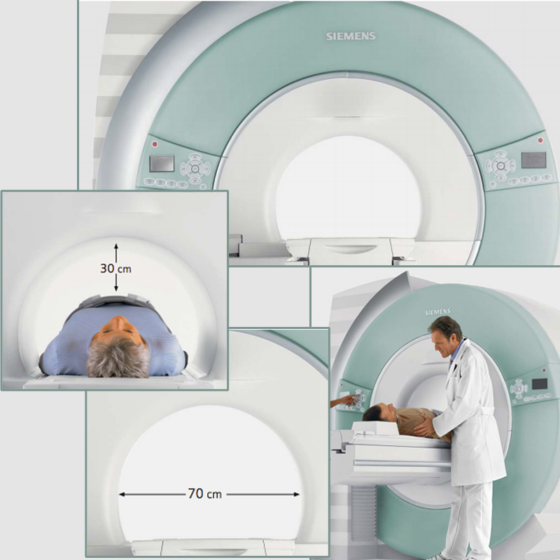
The benefits of a Wide Bore MRI, include a much more comfortable surrounding for you as the patient and features faster imaging and diagnosis. Other features of the Wide-Bore MRI include:

.70-cm open bore
.30 cm of face space
.Head-out exams
.Pain and mobility issues
.Respiratory problems
.Pediatric and elderly patients
.Claustrophobic patients
.Kyphosis
.Patients with special needs and conditions

Read more about all the features of the Wide-Bore MRI here.

New Open MRI at OSS


Patellar Tendon Tears

patellar1-300x300Anatomy
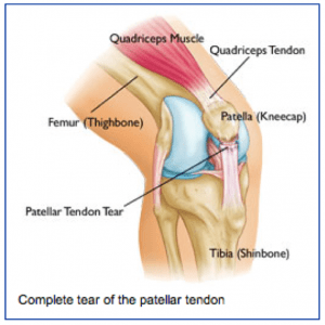
The patellar tendon attaches the lower pole of the kneecap (patella) to the lower leg bone (tibia). The quadriceps muscle attaches to the upper pole of the kneecap and pulls through the kneecap and patellar tendon to allow one to straighten out ones leg or to support ones weight while squatting.

When the patellar tendon is torn, one cannot support their weight when the knee is bent, such as getting up from a chair or going down the stairs.

Injury Mechanism
The patellar tendon is usually injured with a sudden high force across the tendon, such as landing from a jump. The patellar tendon can be injured by a direct blow or a sharp laceration across the tendon.

Symptoms
Patellar tendon injuries present as significant pain across the front of the knee and immediate weakness in supporting ones weight while squatting. Patients usually cannot straighten their knee out fully with a complete tear of the patellar tendon. Rapid swelling occurs. Often, one can feel a gap under their kneecap at the site of the rupture.

Diagnosis
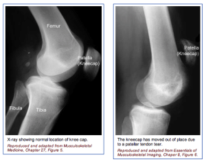
patellar2-300x235The physician’s work-up will start with a careful history and exam. The physician can often feel the defect in the patellar tendon and can appreciate the weakness on trying to straighten out the knee against resistance.

X-rays reveal a very high riding patella, since the pull of the quadriceps is no longer opposed by the tethering effect of an intact patellar tendon.

An MRI scan is often obtained to confirm the diagnosis, especially if the physician is concerned that the injury may be just a partial tear that may be able to heal without surgical repair. Plain x-rays show the bones of the knee, while MRI scans reveal the soft tissues around the knee including the ligaments, menisci, muscles and tendons.

Treatment
Your physician will discuss treatment options with you. Treatment decisions are based on whether the injury is a complete tear or a partial tear. Partial tears may be able to be treated with a brace. The brace is initially locked out completely straight to take all of the pressure off of the injured tendon.

As healing occurs, the hinges are unlocked and motion and strengthening are initiated. With complete tears, surgery is almost always recommended. Surgery involves sewing the two ends of the tendon together with strong suture material. If the tendon has pulled directly off of the bone, drill holes are made into the bone to repair the tendon directly to the bone. Risks and benefits of surgery are discussed thoroughly with the patient.

What to expect after surgery
Patellar tendon surgery is usually performed as an outpatient procedure. Patients are sent home with crutches and a knee brace with the knee fully straightened to protect the repair. Depending on the strength of the repair, patients start gentle range of motion and progressive weight bearing over 6-8 weeks.

Patients are using a stationary bike by 2 months, and an elliptical or stair climber shortly thereafter. Jogging is restricted until 4 months following surgery and full sports activities are not resumed until 6 months after surgery. Most patients can return to full activities, with no restrictions and no bracing at the 6th month point.

Gel-One Injections for Arthritis Treatment

Patients, who have been suffering from chronic arthritis throughout their lives, have a new treatment option worth investigating. The new treatment is one Dr. Peterson is proud to be offering to his patients. Patients, that could not find relief before, may find relief with Gel-One. Zimmer Corporation, the company that developed Gel-One, created a brand new single use injection that is unlike the older ones on the market. This product has no false injections into the knee.

Out with the Old – Hyaluronic Acid Injection Therapy

The current treatment method, when used in patients who have had their arthritis for over a decade, has only around a 50 percent chance of actually relieving the pain of arthritis of the knee for an extended period of time. Those who find success with it have relief that lasts approximately 6 months, on average.

A second aspect of the current method of treatment is that even if the hyaluronic acid injection is successful in providing relief, some methods of therapy require a patient undergo multiple injections weekly, typically three to five per week depending on the severity of the arthritic pain.

In with the New – Gel -One

The Gel-One product by the Zimmer Corporation is not the first single-use injection treatment to be offered on the market. The previously used injection often provided an amount to be injected that was not sufficient to properly reduce pain and inflammation in the knee, producing what is affectionately called a pseudosepsis (fake infection). The Zimmer Corporation has worked hard to counter this first injection option by working to produce one that is much more effective while being processed in a way that still allows for delivery in a single use injection system.

Gel-One however has been clinically tested in a controlled study group to confirm that it does not produce a pseudosepsis effect.  The study consisted of 379 randomized patients, which 248 of them receiving the Gel-One formula. The patients were compared to the control at the thirteenth week beyond the baseline and demonstrated a greater amount of pain relief, averaging out at approximately 40% reduced amount of pain.

Also in this study there were no unexpected side effects observed, lending to the confidence our clinic has in this product as a new therapy option for those with chronic arthritic pain.

Gel-One and Our Clinic

Dr. Peterson offers Gel-One to his patients, and early results seem promising. He is continuing to use other forms of hyaluronic acid for those patients who have done well with them, and prefer not to switch.

If you would like to discuss your arthritis treatment options and find out if Gel-One is  right for you, make an appointment with Dr. Peterson at Orthopedic Specialists of Seattle. OSS is a comprehensive orthopedic practice.

Direct Anterior Approach Total Hip Replacement

Over the last decade, direct anterior approach (also called anterior supine incision or ASI) total hip replacement has been gaining popularity in the United States.  The reasons for this surge in popularity can be traced to three factors.

More traditional posterior or anterolateral surgical approaches to total hip replacement have to cut muscles attached to the hip joint in order to expose the joint.  In ASI total hip replacement, no muscles are cut, which results in less initial postoperative pain and faster early recovery with a shorter hospital stay for many patients.  Most patients have a one-night stay after surgery, and are off crutches by about 2 weeks after surgery.

The ASI approach allows for the use of x-ray imaging during surgery to precisely position the components, and to ensure very accurate measurement of leg lengths.  Since component position is critical to optimal long-term function of the implant, and leg-length inequality is one of the most common reasons for repeat surgery after older methods of hip replacement, this use of precise x-ray control is very advantageous.

Dislocation of the prosthesis is one of the more frustrating and challenging complications of total hip replacement.  With other techniques, the reported rate of dislocation over the lifetime of the prosthesis is 3-5% (Medicare data).  To try and minimize this, many surgeons performing those older techniques will tell their patients never to bend more than a right angle at the waist, and not to scissor their legs.

Because no muscles are cut during the ASI approach, the dislocation rate is markedly less.  After more than 2000 total hips performed by surgeons at Orthopedic Specialists of Seattle, the dislocation rate is less than 0.1% (less than 1 in 1000).  We therefore put no restriction on patient motion after surgery.  Dr. Peterson’s only restriction for his patients is no running for exercise.

An excellent YouTube animation video of how the ASI approach is performed is available below. While this video was made in Omaha by Dr. Ajoy Jana, the technique used by Dr. Peterson is very similar in all regards.

The surgeons at OSS perform more ASI hip replacements than any other practice in the Northwest.  Dr. Peterson has been performing total hip replacements for over 20 years, and switched to the ASI approach for most cases several years ago after seeing the significant benefits for his patients.  He would be happy to discuss this remarkable new technique with you during your office visit.