Collateral Ligament Injuries

Anatomy
collateral-300x208
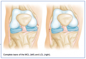
The medial collateral ligament (MCL) runs from the inner side (medial side) of the femur (thigh bone) to the inner (medial side) of the tibia (lower leg bone). It prevents the knee from opening on the inside when struck from the outside of the knee joint. The MCL lies on the outside of the joint capsule and has a good blood supply that contributes to its good healing potential.

The lateral collateral ligament (LCL) runs from the outer side (lateral side) of the femur (thigh bone) to the top of the fibula (the smaller of the two lower leg bones). It prevents the knee from opening on the outer side when struck from the inner side of the knee joint. The LCL is thinner and when completely disrupted often requires surgical repair.

Injury Mechanism
The MCL is often injured in sports when one is struck from the outer or lateral side of the knee, such as having an opponent fall against the outside of one’s knee in football. Another common mechanism of injury to the MCL is when the foot is forced out to the side away from the body, such as with a simultaneous kick of a soccer ball with the inside of the foot. LCL injuries are much more rare and usually occur when the knee is struck from the inside while the foot is planted, forcing a distraction force to the outside of the knee.

Symptoms
When patients sustain an injury to the collateral ligaments they often experience pain, localized swelling and bruising on the involved side of the knee. With partial tears, there is stiffness and pain when fully bending the knee, but no sense of instability. With a complete tear, the knee will feel unstable and will give way to the side with any lateral movements.

Diagnosis
The physician’s work-up will start with a careful history and exam. Often the description of a direct blow to either side of the knee can lead the physician to the suspected injury to the MCL or ACL. On examination, the physician can feel the instability when pulling the foot to one side or the other while stabilizing the knee. X-rays are often obtained to see that no fractures have occurred. Occasionally, a small avulsion fracture might hint that a collateral ligament injury has occurred. An MRI scan is often obtained to confirm the diagnosis and to evaluate any associated injuries to the menisci, other ligaments, and damage to the joint surfaces.

Treatment
Your physician will discuss treatment options with you. Treatment decisions are based on degree of instability. Minor tears (sprains) can be treated with rest, ice, elevation and compression. More significant tears in which many of the fibers of the ligament have been torn may require bracing for 6 weeks to keep the fibers from healing in a stretched out position. Occasionally physical therapy is needed to help regain full range of motion and strengthen the surrounding muscles after the period of bracing. Rarely is surgery recommended for an isolated MCL tear, but occasionally LCL injuries can benefit from surgical repair or reconstruction.

Knee Anatomy

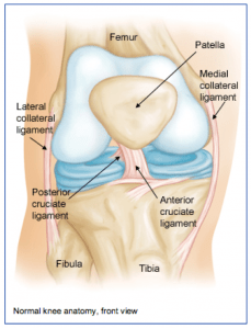
The knee joint is one of the largest joints in the body. It is a complex joint with four bones: the femur (thigh bone), the tibia (main lower leg bone), the fibula (smaller lower leg bone), and the patella (kneecap). The bones are connected with four main ligaments: ACL (anterior cruciate ligament), PCL (posterior cruciate ligament), MCL (medial collateral ligament), and the LCL (lateral collateral ligament).

The ACL and PCL control the forward/backwards movement of the knee joint and prevent pivoting of the knee. The MCL and LCL prevent giving away on either side of the knee. The quadriceps is a group of 4 muscles that converge on the front of the thigh and together allow one to straighten their knee by pulling through the kneecap and patellar tendon, which attaches to the front of the lower leg bone (tibia).

The hamstrings are the muscles on the back of the thigh that help bending the knee by crossing the joint in the back of the knee and attaching to the lower leg bones. Between the femur and tibia, sitting centrally in the knee joint, are two C-shaped pads (the medial and lateral menisci) that act as cushions or shock absorbers between the two bones. The meniscal pads are made of cartilage.

There is also about a quarter of an inch of cartilage on the distal end of the thighbone and on the proximal end of the lower leg bone. Arthritis occurs when that joint cartilage becomes damaged or thin.

Shoulder Anatomy

from orthoinfo.aaos.org

from orthoinfo.aaos.org

The shoulder joint is a complicated joint.

It is made up of three bones: the scapula (shoulder blade), the clavicle (collarbone) and the humerus (upper arm bone). The true shoulder joint is where the shoulder blade meets the upper arm bone.

A second joint exists where the collarbone meets the shoulder blade on the top and front of the shoulder and is called the AC or acromioclavicular joint. When people refer to a shoulder separation, it is really the AC joint that has been injured, not the true ball and socket (glenohumeral) joint of the shoulder. A third joint exists where the shoulder blade sits on the back of the ribcage called the scapulothoracic joint.

The shoulder joint is unique in that it can move in almost every direction, unlike the knee that just straightens and bends. The large ball on the relatively flat socket is what allows the shoulder such unrestricted motion.

What keeps the humeral head (the ball) on the flat surface of the glenoid (socket) is the muscles, tendons, and ligaments that surround the head plus a lip of cartilage (labrum) that surrounds and extends the socket to help contain the head.

The rotator cuff is a group of 4 muscles that converge on the outside of the shoulder to help elevate and rotate the arm. The tendinous attachment of these tendons to the upper arm bone is called the rotator cuff. The long head of the biceps tendon also runs over the top of the shoulder and attaches to the upper part of the socket through the upper labrum.

Between the top of the shoulder blade (the acromion) and the rotator cuff and biceps tendons is a large fluid filled sack called the bursa. It is normally quite thin and helps allow these tissues to slide under the acromion without rubbing or abrading.

Arthritis of the Knee

arthritisknee1-300x164
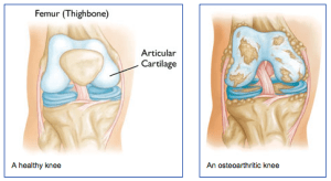
What is arthritis of the knee?
Arthritis of any joint is when there is inflammation and damage to the normal joint surface. The normal articular cartilage is smooth and firm. With arthritis, the surface of the joint becomes pitted, eroded, and irregular causing swelling and pain. There are many types of arthritis.

The most common type of arthritis is osteoarthritis, or wear and tear that is seen in older individuals. Another form of arthritis is rheumatoid arthritis. It is commonly involves multiple joints, especially in the hands and wrists.

It can occur at any age. One more common form of arthritis is post-traumatic arthritis. It follows an injury to the surfaces, the meniscus, or the ligaments, which makes the joint susceptible to arthritis often many years after the injury.

Symptoms
Patients with arthritis typically describe pain and stiffness, especially with the first few steps after getting up from a chair or first thing in the morning. In the early stages of arthritis, the knee is often swollen, and even warm to the touch. After years of having arthritis, the knee rarely swells, but it becomes more difficult to fully straighten or bend the knee. Arthritic knees are painful with activity.

Diagnosis
kneearthritis2-300x140The physician’s work-up will start with a careful history and exam. A history of prior injury, pain, stiffness and swelling may suggest arthritis. The exam often shows some swelling and decreased range of motion. Sometimes, patients develop deformities if one side of the knee wears out more than the other side.

The legs can then appear “knock-kneed” or “bow-legged.” X-rays are usually the key in determining the extent of the arthritis. Arthritic knees show loss of normal joint space, cysts and bone spurs on x-ray.

Treatment
Early treatment of arthritis may involve non-steroidal anti-inflammatories, ice, rest, injections and activity modification. If these methods are not satisfactory in keeping a patient from becoming sedentary, surgery is often discussed. Ideally for surgery, patients should be in their mid-60s (current knee replacements last about 20 years or more so are not ideal for those in their 40s or 50s), able to walk only a block or two (considered sedentary), and experience pain at night (effecting quality of life).

However, as techniques and materials become better, physicians are considering knee replacements in younger and younger patients.

Surgical options for arthritis range from arthroscopic debridement (cleaning out the loose pieces and smoothing the surfaces), osteotomies (cutting a wedge out of the bone above or below the knee to try to throw more of the weight onto the better half of the knee), cartilage replacement (transferring plugs of bone and cartilage to the involved areas), to partial or total knee replacements in which the arthritic surfaces are removed and replaced with metal and plastic surfaces.

Your orthopedist will have a thorough discussion with you about which surgical option is best for you, and discuss the risks and benefits of the surgery with you.

Osgood-Schlatter Disease

What is Osgood-Schlatter Disease?
Osgood-Schlatter Disease is a condition that occurs in adolescents that involves pain and inflammation that occurs where the patellar tendon attaches to the lower let bone (tibia). It is commonly seen during a sports season that involves running and jumping (basketball, soccer, track).

osgood-300x252Diagnosis
The diagnosis is usually made by identifying swelling and tenderness at the attachment of the patellar tendon to the front of the tibia. Often a bump is present at that location. Occasionally x-rays will reveal a bony prominence or sometimes an extra piece of bone where the patellar tendon attaches to the tibia.

MRI scans are not usually necessary to help make the diagnosis in these patients.

Treatment
Surgery is almost never needed, as most cases of Osgood-Schlatter Disease will resolve as one reaches the end of adolescence. Often, activity restrictions are necessary to help resolve the symptoms. Warming up and stretching before activities and icing after can be helpful. Non-steroidal anti-inflammatories are sometimes necessary to help with the discomfort.