
Dr. Grant Garcia talks about various treatment options for rotator cuff tears in all shapes and sizes.
Dr. Grant Garcia provides Stemless Shoulder Arthroplasty
Dr. Grant Garcia is one of a few surgeons in the Seattle area performing this surgery.
This system is designed to be a great bone-sparing option for total shoulder replacement.
It has faster surgery time, less blood loss, and decreased infection rate. Plus, it shows significant improvement in strength and range of motion in the shoulder.
Knee Bursitis
Spring is right around the corner, and with the warmer weather, many of us will be getting back to our gardens or home improvement projects we postponed over the winter. After weeding in the garden, laying down new flooring, or performing any other activity for which you spend time on your knees, it can be normal to develop some soreness. This may, however, develop into increased pain, swelling, and inflammation. These symptoms are commonly caused by a condition called knee bursitis.
What is knee bursitis?
In our body there are hundreds of fluid-filled sacks called bursa. They live in between various parts of our body such as between tendons and bones, muscles and other muscles, or skin and the underlying structures, to name a few.
Bursae act as a slippery surface that allow for the many parts of our body to easily move against one another.
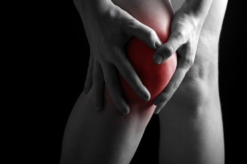
One of the places you have bursae is around your knee. The two main bursae are the prepatellar bursa, which lives in front of the patella (the small mobile bone on the front of the knee), and the infrapatellar bursa, which lives in front of the patellar tendon (the thick tendon that runs from the patella to the main bone of the lower leg).
Who gets knee bursitis?
Bursitis can affect individuals of any age; however, as we age the skin overlying the knee slowly begins to thin. With this thinning there is less cushion for the underlying bursae. As such, older individuals are at greater risk for knee bursitis. Additionally, patients on chronic glucocorticoid therapy are also at greater risk for knee bursitis.
Signs of Knee Bursitis
Bursitis typically feels like pain and swelling in the knee, and you may also see some redness. When the bursa becomes inflamed, it can fill with more fluid, leading to swelling in front of the knee. When the knee is bent, this fluid filled bursa becomes compressed against the front of the knee (imagine pushing against a water balloon). This causes the bursa to stretch, leading to pain. This is why pain associated with knee bursitis typically worsens with bending of the knee and improves when the knee remains straight. If the bursitis is left untreated, the fluid filled sack has the potential to rupture. This could then lead to an infection of the surrounding skin.
Knee Bursitis Diagnosis
The diagnosis of knee bursitis can be made by an experienced clinician, who will ask you about your knee pain and examine the affected knee. Additionally, the provider will likely take a sample of the fluid in the bursa using a needle and send this for further analysis. The provider may also order blood tests and x-rays to rule out a more serious infection of the joint.
Here at the Orthopedic Specialists of Seattle, we have two experienced surgeons, Dr. Jonathan Franklin and Dr. Charlie Peterson, who have a wealth of knowledge and experience in diagnosing and managing knee bursitis. At our clinic, we also have the laboratory and imaging resources to properly work-up your knee pain.
How do I treat knee bursitis?
Once the diagnosis of knee bursitis is made, the goal of treatment is to relieve the immediate symptoms and prevent it from getting worse. This is achieved in several different ways including a knee brace, pain medications, or applying heat or ice, and your clinician will decide which is best for you. Ultimately, the swelling and tenderness will resolve somewhere between a few weeks to a few months, depending on your condition.
To prevent knee bursitis, it is a good idea to avoid spending too much time on your knees and take breaks often. Or, if you will be working on your knees, it can be helpful to wear knee pads or use a kneeling pad. Here at Orthopedic Specialists of Seattle, our goal is to help you return to your beloved activities as soon as possible and to prevent further episodes of pain.
Magnetic Resonance Imaging (MRI) – Accredited Facility
Seattle Orthopedic Center has been awarded a three-year term of accreditation in magnetic resonance imaging (MRI) as the result of a recent review by the American College of Radiology (ACR).
The ACR gold seal of accreditation represents the highest level of image quality and patient safety. It is awarded only to facilities meeting ACR Practice Parameters and Technical Standards after a peer-review evaluation by board-certified physicians and medical physicists who are experts in the field. Image quality, personnel qualifications, adequacy of facility equipment, quality control procedures and quality assurance programs are assessed.

Broken Foot: Common Symptoms and Treatments
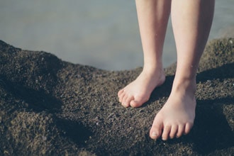
There are several medium and small bones that make up the three sections of the foot. The forefoot includes the slender bones that make up the 5 toes (phalanges) and the anterior portion of the arch of the foot (metatarsals).
The mid-foot is made up of more compact bones (navicular, cuboids, and cuneiforms) that make up the top of the arch of the foot. The hind-foot is made up of two bones, the talus that connects the foot to the lower leg and the calcaneus (the heel).
Foot injuries are common and can arise from a multitude of different mechanisms. Each of the bones of the foot are subject to fracture from externally applied forces. Fractures to the talus and calcaneus (hind foot) are typically caused by crushing forces, such as landing from a great height.
Fractures to the forefoot can be caused by crush, flexing, and twisting forces. In addition to trauma directly to the bone, there are ligaments and tendons attached to the bones of the foot. If enough force is applied to these ligaments and tendons, they can separate and take a small piece of bone with it.
In addition to fractures from a single traumatic blow, some fractures can result from repetitive impacts to a bone. This is called a stress fracture and commonly occurs in the metatarsals of the foot.
Common Symptoms
It can often be difficult to distinguish fractures from other types of injuries of the foot, such as sprains of the ligaments or bruises of the soft tissue. If a bone is fractured, there is typically pain localized to the fracture. There will also likely be soft tissue swelling around the affected area.
Treatment Options
To determine if there is a fracture radiology studies such as x-ray and CT may be necessary. Careful physical exam performed by an experienced surgeon can also help to elucidate whether or not a bone is fractured. Depending on the fracture type and location, there are multiple types of repair. These range from simple alignment and casting to surgical repair with hardware placement. Dr. Mark Reed of OSS is well versed in all of these repair techniques.