Pumpkin Carving Safety Tips

Every October, carved pumpkins peer out from porches and doorsteps in the United States and other parts of the world. Gourd-like orange fruits inscribed with ghoulish faces and illuminated by candles are a sure sign that Halloween and the holiday season is upon us. The practice of decorating “jack-o’-lanterns” – the name comes from an Irish folktale about a man named Stingy Jack – originated in Ireland, where large turnips and potatoes served as an early canvas.

Irish immigrants brought the tradition to America, home of the pumpkin, and it became an integral part of Halloween festivities. Click Here and find pumpkin carving safety tips for you and your loved ones!

Seahawks DE Michael Bennett taken off field on stretcher

On Sunday, September 29, seattlepi.com reported that Seahawks defensive end Michael Bennett, who has been a big story this season as Seattle’s sack leader, was taken off the field Sunday on a stretcher after he was injured on a play against the Texans in Houston. The article reported, “Late in the second quarter, Bennett was rushing Texans quarterback Matt Schaub when he was pushed from behind by a Houston defender into Schaub’s leg. Bennett’s head appeared to snap back, and his helmet flew off as he hit the ground. Bennett laid face-down on the turf for several minutes as trainers tended to him.”

According to the news article, “Bennett suffered a strained muscle in his back that was close to his vertebrae. The location of the injury was why medical personnel were extra-careful and carted Bennett off the field on a stretcher.” Head coach, Pete Carroll said that he was “fine” and a tweet was sent out the next day stating that Bennet was practicing and that he may be able to play in their upcoming game against Indianapolis.

Treatment of a lumbar muscle strain is important to understand. Once you know the cause of your symptoms, you can proceed with treatment. It is important that if you are not sure of the cause of low back pain, that you are evaluated by a physician. According to Dr. Charlie Peterson, “Back injuries can be painful, frustrating and even scary, but are also common. As such, the vast majority can be managed with a few simple techniques. However, if you have unusual symptoms or your pain persists, it’s time to seek advice from a specialist.”

If you are experiencing pain in your lower back or it has been injured as a result of physical activity, below is a list to help you treat your injury:

Step 1: Rest

The first step in the treatment of a lumbar muscle strain is to rest the back. This will allow the inflammation to subside and control the symptoms of muscle spasm. Bed rest should begin soon after injury, but should not continue beyond about 48 hours. While it is important to rest the injured muscles, it is just as important to not allow the muscle to become weak and stiff. Once the acute inflammation has subsided, some simple stretches and exercises should begin.

Step 2: Medications

Two groups of medications are especially helpful in treating the acute symptoms of a lumbar back strain. The first of these are anti-inflammatory medications. These medications help control the inflammation caused by the injury, and also help to reduce pain. There are many anti-inflammatory options, talk to your doctor about what medication is appropriate for you.

The second group of medications commonly prescribed for the treatment of lumbar strains is muscle relaxing medications. Again, there are several options that you may discuss with your doctor. These medications are often sedating, so they need to be used with care. For patients who have back spasm symptoms, these muscle relaxing mediations can be a very useful aspect of treatment.

Step 3: Physical Therapy/Exercises

Proper conditioning is important to both avoid this type of problem and recover from this injury. By stretching and strengthening the back muscles, you will help control the inflammation and better condition the lumbar back muscles. The exercises should not be painful. Without some simple exercises, the low back muscles can become “deconditioned,” or weak. When the low back muscles are “deconditioned”, it is very difficult to fully recover from low back injuries.

It is also important to understand that even if you are “in good shape,” you may have weak low back muscles. When you have a low back muscle injury, you should perform specific exercises that stretch and strengthen the muscles of the low back, hips and abdomen. These exercises are relatively simple, do not require special equipment, and can be performed at home.

Step 4: Further Evaluation

If your symptoms continue to persist despite treatment, it is appropriate to return to your doctor for further evaluation. Other causes of back pain should be considered, and perhaps x-rays or other studies (MRI, CT scan, bone scan, laboratory studies) may be needed to make an accurate diagnosis.

If you believe you are suffering from a back injury and need specialized orthopedic care, Orthopedic Specialists of Seattle has excellent treatment options available for you.

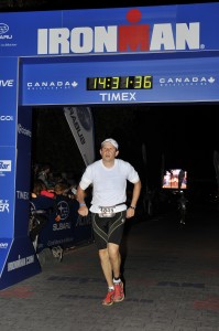
Congratulations Dr. Weil On Competing in the Ironman in Whistler, BC

Congratulations Dr. Weil for competing and completing in the Whistler Blackcomb Subaru Ironman Canada in Whistler, BC!

Dr-W-Weil-Ironman-picture-and-time-199x300

The race started with a two-loop swim in Alta Lake. Athletes then had a lakeside transition before starting a one-loop bike course via the Sea-to-Sky Highway, Callaghan Valley and Pemberton.

The two-loop run course utilized the Valley Trail, winding past Lost Lake and Green Lake before finishing adjacent to Whistler Olympic Plaza in the Village.

Dr. Weil’s official time was 14:31:36 for completing the Ironman! Way to go Dr. Weil!

Read full article here

Installation of Our New Open Bore MRI is Complete

Months of anticipation and planning were successful in the installation of our new Open Bore MRI.  We are happy to report that it is up and running.  Installing this sophisticated piece of equipment was an event in itself!  Thank you to everyone involved in the planning and installation of our new MRI!

Treatment of Cavus Foot Deformity

Cavus Foot Deformity

Now that it’s summer, we spend more time at the beach or swimming pool enjoying the warm weather. As an orthopedic surgeon that specializes in conditions of the foot and ankle, I tend to notice wet footprints across the pool deck or in the sand and cannot help but analyze the health of the individual’s feet. One of the conditions that I notice is the high arched foot, medically termed pes cavus. This is noticeable by a footprint that is pronounced in the heel and forefoot, with very minimal or even absent impression in the middle part of the foot.

Causes of Cavus Foot Deformity

Pes Cavus is caused by muscle imbalances in the lower leg and foot that draws the front of the foot, or sometimes draws the heel downward, making the arch higher than normal. It usually begins during childhood and may be associated with neurological conditions such as muscular dystrophy or spina bifida, but not always. In many cases the muscles of the feet become tighter or weaker for unknown reasons. As with many medical conditions, genetics play a role in who will become afflicted with high arches.

Symptoms of Cavus Foot Deformity

High arches can cause a number of symptoms, ranging from mild to severe. Pain in the forefoot is a common occurrence due to increased weight bearing in this area. Excessive callus buildup at the ball of the foot behind the great toe as well as just behind the fifth toe is common, as these become high-pressure areas during standing and walking. Tightness in the calf muscles is often present, and the individual may also suffer from recurring ankle sprains due to the inwardly rolled ankles associated with the deformity.

Diagnosis of Cavus Foot Deformity

Identifying pes cavus is a straightforward process. The high arched foot is noticeable to anyone, but an orthopedic surgeon should evaluate the individual in order to identify some of the nuances of the condition. Diagnosing which muscles are tight or weak and assessing their potential to be stretched or strengthened is important for initiating an effective treatment plan.

Also, the cavus foot causes increased body weight to be distributed through areas of the foot that are not designed for this purpose. Evaluation by the surgeon will aid in a proper prescription of orthotics, if deemed necessary.

Treatment of Cavus Foot Deformity

Conservative intervention is generally the rule when starting to treat high arches. Often times if the feet have become painful, orthotic inserts are prescribed. As opposed to pes planus (flat feet), which is often a flexible disorder of the foot that we try to correct with orthotics (i.e., push back into the correct position), pes cavus is usually a rigid deformity,meaning that the shape of the foot cannot be changed.

In this case, the goal of orthotics is to accommodate the shape of the foot and to redistribute body weight over a larger area. Because of the rigidity, the cavus foot is not able to flex and absorb impact as the individual walks. For this reason, orthotics are usually constructed of softer materials to act as a shock absorber.

Physical therapy may also be prescribed to stretch and strengthen the muscles of the lower leg. Tight calf muscles and weak muscles along the outside of the lower leg (peroneal muscles) are often present in pes cavus. While therapy cannot change the shape of the foot, it may be able to help with pain control and function. Because the foot is usually rolled inward along with the high arch, the individual is susceptible to chronic ankle sprains and some reactive muscle strengthening may be beneficial, along with ankle bracing.

If conservative treatment fails to achieve the desired result, then surgical correction maybe necessary. There are many types or surgeries that the physician can perform based on individual need:

  • Tendon lengthening: This procedure involves making precision cuts in the tight tendons of the lower leg to allow better alignment of the foot. Following surgery,there is a period of immobilization for several weeks to allow the tendons to heal.
  • Osteotomy (bone cut/realignment): If the condition has been present since childhood and the bony structure of the foot has grown abnormally, then small sections of bone may need to be removed in order to restore proper position of the foot. The first metatarsal, located in the midfoot behind the great toe, is often treated with an osteotomy. The metatarsal often is positioned at a downward angle that is greater than normal, which in turn rolls the ankle toward the outside of the foot as the person bears weight.

    The osteotomy procedure seeks to normalize that angle and place the foot in a neutral position that is perpendicular to the ground. Many times, this osteotomy is performed in conjunction with soft tissue surgery such as tendon lengthening. Also, the calcaneus (heel bone) tends to be oriented toward the midline in pes cavus, as opposed to away from the midline in the normal foot, and sometimes an osteotomy is required to correct this.

  • Arthrodesis: Also known as a joint fusion, this procedure permanently locks the affected joint into a fixed position. It is a last resort option, but sometimes necessary when the cavus foot deformity is severe or when arthritis is present.