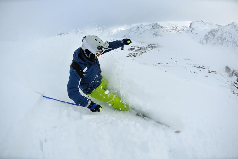
Preventing Ski Injuries through Conditioning

A busy ski resort in the United States may see dozens of injuries on the slopes each day. As an orthopedic surgeon, I also see many patients with ski-related injuries throughout the season. Most injuries are the result of poor conditioning, or equipment failure.

The most common injuries amongst downhill skiers are knee sprains, shoulder injuries, head/face injuries and wrist/thumb injuries. The knee is the most commonly injured joint, resulting in about one third of all ski injuries. Injury rates and type vary with uncontrollable factors such as weather and snow conditions. Proper equipment and conditioning, however, are factors that we can control.

Equipment

When skiers examine their equipment, it’s important to make sure that: Skis, poles, and boots are in good condition and properly sized for the individual’s weight,size and skill. Bindings are adjusted and tested prior to each ski season.Helmets are properly fitted and checked for damage prior to the ski season.Sunglasses, goggles and sunscreen are part of one’s safety equipment

Conditioning

Skiers can increase their safety and performance this winter by starting with a pre-conditioning program that includes four components: endurance, strength, flexibility, and balance. Aerobic fitness is the key to preventing the end of the day injuries (the last run).Cross training, which includes multiple sports and activities in the conditioning regimen, has become popular, especially with a seasonal sport such as skiing. Strength and flexibility focusing on the legs and trunk are vital in injury prevention specific for skiing. Balance training has been shown to be the single most important exercise for preventing ACL tears in women.
A typical conditioning program can include:

1. Aerobic fitness (5 days/week for at least 30 minutes)

  • Running
  • Cycling
  • Swimming
  • Elliptical or stair climber
  • Jumping rope
  • Treadmill

2. Strength (3 days/week, 2 sets of 60 seconds each)

  • Leg press
  • Wall squats
  • Hamstring curls
  • Toe raises
  • Lateral leg raises
  • Sit-ups

3. Flexibility (daily, 2 sets of 60 seconds each)

  • Hamstring stretches
  • Achilles stretches
  • Quad stretches

4. Balance Exercises (daily, 2 sets of 60 seconds)

  • Standing on one leg, perform mini squats
  • Single leg hop, holding landing for 5 seconds, repeat

In addition to a conditioning program, skiers need to adequately warm up – an activity that is often neglected with skiing. No one would think of running out on the football field or onto the basketball court without warming up first. But with skiing, one typically sits in the car for an hour or more to get to the slopes, and then stands in line for tickets and for the lift, before finally sitting on the chair for several minutes. By the time one has arrived on the top of the hill, he or she is often stiff and cold.

It’s important for skiers to remember to warm up and stretch before starting down the hill.Often an easy, predictable run is a good idea before heading to the more challenging terrain.The few minutes spent warming up will be well worthwhile in injury prevention.

More about knee injuries

Every ski season, I treat many knee injuries. In the 1970′s, ankle injuries were more common, resulting from soft, leather boots. The development of stiffer boots has transferred much of the force to the knee.

The most common knee injury from skiing is the MCL (medial collateral ligament) injury. It often results from catching an edge or having the skis diverge, so that the foot is forced away from the body. This creates a distraction force on the inside of the knee. Fortunately, the MCL has a good blood supply, and can be treated non-operatively, with a period of bracing for 4-8 weeks, depending on the severity of the injury.

ACL (anterior cruciate ligament) injuries are also common skiing injuries. They are thought to occur from the forces created by the long lever arm of the ski that are transmitted to the knee ligaments. Commonly, the ACL is injured with a hyperextension mechanism. In expert skiers,we see ACL injuries when saving a backwards fall by a strong quadriceps contraction, pulling the tibia (lower leg) forward with enough force to rupture the ACL. Recent boot and binding technology has reduced the rate of ACL injuries. In young, active individuals, the ACL injuries often require surgical reconstruction. Success rates from surgery are excellent, but require aggressive rehabilitation and six months of recovery time before one can return to skiing or other twisting or pivoting sports.

No one wants to go down the path of surgery and recovery. But too many people wait to think about preparing for skiing until half way through the season, when snow has already accumulated and they are on their way to the top of the mountain. Many times, this is to late. Although injury is a risk we all take when participating in any sport, a conscientious approach to skiing – including equipment inspection and conditioning – will minimize the occurrence. Not only will these precautions reduce injury rate, but they will also enhance performance, decrease fatigue, and ultimately, increase one’s enjoyment of the sport.

We are fortunate in the Pacific Northwest to have great skiing terrain so close. Be safe and prepared so you can enjoy a great skiing season this year.
ski article

Seattle Seahawks WR, Percy Harvin and His Hip Labral Tear

Percy Harvin, Seattle Seahawk’s Wide Receiver is waiting out another week before he can return to the football playing field. NFL Spinzone reports, “Harvin was able to return to practice a couple of weeks ago, but the Seahawks have prudently decided to rest him. He has recovered quickly and well from off-season hip surgery, but Pete Carroll is 100% correct in being as cautious as possible with their new star receiver. The last thing they need is for the oft-injured Harvin to have a setback, especially since the 8-1 Seahawks clearly have their eyes set on a Super Bowl-winning season.”

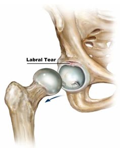
The labrum is a unique structure found in the hip. The labrum is a specialized structure that lines the edge or rim of the hip socket. The shape and function of the labrum can be most easily understood as being similar to a bumper cushion on a pool table. The shape of the labrum is triangular in cross-section. The labrum acts as a cushion between the ball and socket of the hip joint during flexion of the hip.

The labrum is filled with nerve fibers that make tears in the labrum very painful.

What causes a labral tear?hip-labral-tear

Labral tears typically occur from:

  • Repetitive motion. Physical activity that requires one to repeatedly pivot the body can lead to joint wear and tear. This is common in athletes such as golfers and baseball players, who must repeatedly twist their bodies to hit a ball.
  • Osteoarthritis. Increased friction from osteoarthritis symptoms inside the hip joint may lead to labral tear.
  • Trauma. Dislocation or violent, direct impact to the hip joint may cause labral tears. This is common in athletes and victims of car accidents.
  • Birth defects. Some people are born with abnormal hip structures, which increase the progression of wear and tear on the hip joint. These problems may lead to labral tears.

What does it feel like?

Some labral tears cause no signs or symptoms; however, when symptoms are present, they include:

  • Radiating pain in the hip and groin
  • A catching or locking sensation in the hip joint
  • Stiffness in the joint
  • A limited range of motion

How do I know if I have a labral tear?

A physical examination by your doctor will help diagnose the cause of your symptoms. X-rays will allow your physician to take a closer look at the bone and rule out other possible underlying conditions. Magnetic resonance imaging (MRI) may also be used to further examine the soft tissues.

How are labral tears treated?

The level of treatment depends upon the severity of the condition. Some patients recover in only a few weeks using conservative treatments. However, patients with severe labral tearing may require surgery. One or a combination of these treatments may be used:

  • Rest and activity modification. By resting and allowing the inflammation of the joint to settle down, symptoms may be alleviated.
  • Medication. Anti-inflammatory medicines may help alleviate the pain and inflammation associated with labral tears. Your doctor may also recommend cortisone injections.
  • Physical therapy. Learning new exercises can help maximize hip range of motion, strength, and stability.
  • Surgery. In general, labral tears can be either repaired or trimmed during an arthroscopic procedure. Depending on the severity of the tear, the surgeon may cut out and remove the torn piece of labrum or repair the torn tissue by sewing it back together.

According to Dr. Watt, “Hopefully Percy will continue his recovery and return to the field soon. If you think you have labral tear or hip condition that is not getting better, I would be happy to evaluate your hip and devise an appropriate treatment plan.”


In the words of Russell Wilson, “Go Hawks!”


5 Common Hockey Injuries

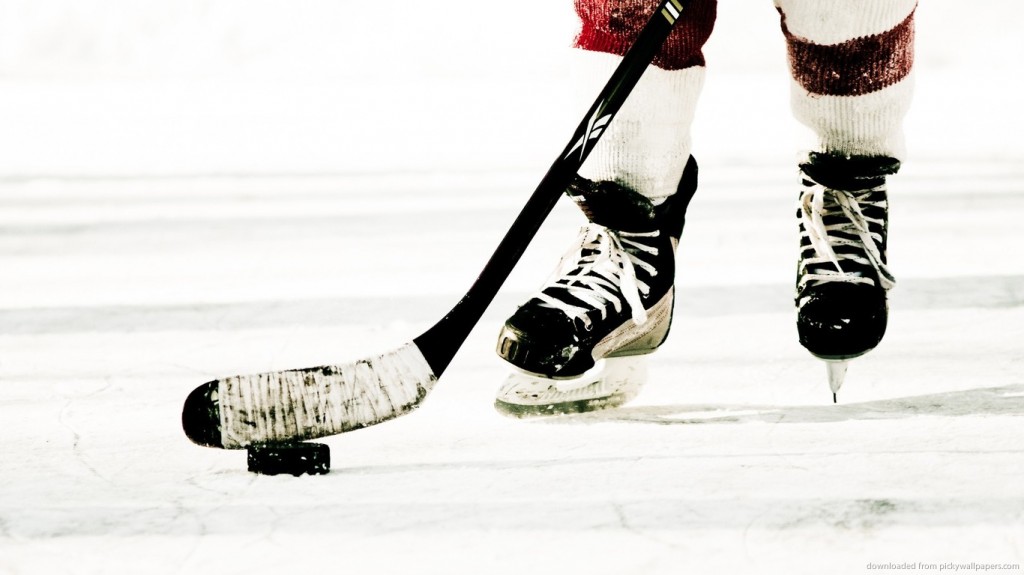
Hockey photo

The regular NHL season is well underway and the Seattle Thunderbirds are respectively improving their stats with their recent win over the Vancouver Giants at ShoWare Center.

In the regular season as well as the off season, players experience a variety of sports-related injuries as a result. Ice hockey is a contact sport where the players and the puck move at high speeds, so when players run into each other or objects, great force is used. This is why hockey is considered a collision sport. Injuries are fairly common, but efforts can be made to avoid them with training and proper equipment. Listed below are five common injuries that can occur while playing hockey.

AC Joint Injury

The acromioclavicular joint, or AC joint, is one of the joints in the shoulder responsible for motion and stability. The ligaments that hold the AC joint together can be torn through sudden impact to the shoulder, which can cause separation to occur in the AC joint. This sudden impact can happen in hockey when players skating at high speeds collide with one another or into a rigid surface. Swelling, bruising, pain, and motion range loss are all symptoms of AC joint separation. There may also be visible bumps on the shoulder if the bones separate.

Shoulder Dislocation

Shoulder dislocation generally refers to a dislocation in the glenohumeral joint in the shoulder. This happens when the top of the humerus, or upper arm bone, is forced out of the glenoid, the socket in the shoulder joint it usually nestles in. If a player falls or receives a heavy blow or sudden impact on the shoulder, it can cause dislocation if the upper arm is forced to move in an abnormal way. Symptoms of shoulder dislocation include pain, weakness, and mobility issues. The arm may also appear to hang incorrectly off the shoulder.

Muscle Strain

Muscle strain occurs when a muscle is pushed past its limit. This can happen if a player’s muscle is suddenly presented with a heavy load or stretched beyond its normal ability. If a player’s muscles are tight but not warmed up or not conditioned well, tearing or straining is a risk. Symptoms of a muscle tear include pain at rest or when the muscle is used, and weakness or inability to use the muscle.

Meniscus Injury

The meniscus is a C-shaped piece of cartilage in the knee. There are two menisci in each knee joint, and if they are torn they can affect stability in the knee. This tearing can happen if the cartilage is worn down or through the quick movements and stress put on the knees by ice skating. Symptoms vary depending on how and where the meniscus is torn, but symptoms can include pain, instability or feeling the knee “giving,” stiffness, swelling, and an impaired range of motion. Sliding, popping, or locking may occur if the tear is left untreated because loose fragments from the meniscus tear will drift into the joint.

Gamekeeper’s Thumb

The ulnar collateral ligament, or UCL, connects the bones at the base of the thumb, which prevents the thumb from moving too far from the hand. When an acute sprain or tear of the UCL occurs, it is called a UCL injury. When the injury is chronic and develops over time from repeated UCL stretching, it’s called gamekeeper’s thumb. UCL injuries are commonly caused by injury or trauma in which the thumb is bent away from the hand at the MCP joint. This can happen in sports hockey, or in any situation in which a fall is landed on an outstretched hand.

This injury might also be sustained when a person is gripping something that is suddenly moving, like a hockey stick during a fall. Swelling, pain, and tenderness on the ulnar side of the thumb are all symptoms of UCL injury. You may also have difficulty pinching and gripping with the thumb, and you may have limitations in your range of movement. In severe cases, a bump under the skin, called a Stener lesion, may form due to the ends of the torn ligament being held apart by a nearby tendon.

If you believe you are suffering from a sports injury and need specialized orthopedic care, Orthopedic Specialists of Seattle has excellent treatment options available for you. Please feel free to contact Orthopedic Specialists of Seattle at (206) 633-8100 to schedule an appointment.

MCL Tears and Repairs By Dr. Charlie Peterson, MD

MCL Tears and Repairs

The medial collateral ligament (MCL), located on the inside portion of the knee, is one of the more common sporting injuries to the lower extremity. It is usually an “acute” injury, meaning that it happens suddenly due to trauma. In sports, the athlete may take a sudden blow to the outside of the knee, creating excessive tensile force to the MCL, such as being tackled in football. This injury also occurs commonly in sports where the ankle is immobilized such as hockey and downhill skiing, where the ankle is stabilized in a skate or boot. This immobilization leaves the knee to absorb the full impact of a collision or fall and increases the risk of knee injury.

Functional Anatomy

The skeletal anatomy of the knee consists of three bones. The thigh bone, medically termed the femur connects with the shin bone, called the tibia. In the front of the knee is the knee cap, or the patella. Holding these bones together are the four major knee ligaments. Two are located deep within the joint and are called cruciate ligaments. They prevent excessive forward and backward motion, as well as rotation. The remaining two are the collateral ligaments, and are located on the sides of the knee. Their job is to prevent lateral, or sideways, motion of the knee. The MCL is located on the inside of the knee joint and prevents the knee from collapsing inward. In addition to the bones and ligaments, the knee has two cartilage pads called the medial meniscus and lateral meniscus. These pads act as shock absorbers within the knee.

Types of MCL Injury

Tears to the MCL are usually a result of direct trauma, either from a blow to the outside of the knee, such as with a football tackle, or a fall that pushes the lower leg sideways. Partial tears will cause varying degrees of instability within the knee, and are often treated successfully with conservative interventions including bracing and physical therapy to strengthen the surrounding musculature. Complete tears may cause significant instability in the knee, especially if in combination with other ligament injuries such as the ACL.

If isolated, even high-grade MCL tears can still often be treated with bracing alone. However, such tears often occur in conjunction with other structures such as the medial meniscus or the anterior cruciate ligament (ACL). The medial meniscus has a direct connection to the MCL, making it particularly susceptible to injury during an acute MCL sprain. Should this be the case, surgical intervention may be required to restore full function due to the degree of instability caused by multiple injuries.

Non-operative Treatment

MCL tears are most often treated successfully without surgery. With significant tears there may be an initial degree if instability following injury. A hinged knee brace may be prescribed to limit control of this aberrant movement. As the ligament heals, your orthopedic surgeon may refer you to physical therapy to strengthen the leg musculature surrounding the knee, and also to restore normal movement patterns that may have been disrupted following injury and immobilization. Patients are able to perform most of their normal daily activities during this process, with the possible exception of high intensity athletics, and generally have very good outcomes following four to eight weeks of rehabilitation.

Surgical Treatment

In cases where non-operative treatment has failed or in some multiple ligament injuries, the surgeon will recommend repair or reconstruction surgery. This means that the damaged MCL will be repaired with sutures if possible. If that is not possible, then a new ligament can be fashioned from a soft-tissue “graft,” a piece of tendon taken from either the patient or a cadaver. A small incision is made to gain access to the area, and the repair made, or the tendon graft is anchored in place with surgical screws.

Following surgery, there will be a period of immobilization, followed by physical therapy. The duration and intensity of the rehabilitation process is dependent on the type of MCL repair or reconstruction, and the other injuries present. In most cases, patients can return to full function including athletics at the conclusion of treatment.