mm

About Orthopedic Specialists

Orthopedic Specialists of Seattle provides new and advanced procedures including endoscopic carpel tunnel release surgery for carpal tunnel syrome, complex joint restoration procedures, anterior approach hip replacement surgery, and more.

Reverse Total Shoulder Replacement

Dr. Peterson and Dr. Shapiro have been performing a relatively new procedure called reverse total shoulder replacement for the last several years.

This particular procedure is designed for people who have rotator cuff arthropathy or a large, irreparable rotator cuff tear. The rotator cuff is a group of muscles and tendons that surround the shoulder joint and allow you to lift your arm over your head. When this structure is severely torn, shoulder arthritis can set in and mobility is limited.

During this procedure, the surgeon removes damaged bone joint tissue.  A smooth,  polished, spherical alloy metal “glenosphere” is then fixed to the old bony “cup” of the shoulder, and a stemmed alloy and polymer cup to the shaft of the upper-arm bone.

Why is Reverse Total Shoulder Replacement Done?

This surgery was developed because traditional shoulder surgeries do not work well when patients also have a severe rotator cuff tear with arthritis. With reverse total shoulder replacement, the deltoid muscle powers the new prosthesis, allowing pain free motion overhead in many patients.

Who is a Candidate for Surgery?

Reverse total shoulder replacement may be recommended if you have:

  • A completely torn rotator cuff that cannot be repaired.
  • Cuff tear arthropathy (arthritis with a severe cuff tear).
  • A previous should replacement that was unsuccessful.
  • Severe shoulder pain and difficulty lifting your arm.
  • Tried other treatments that have not relieved your shoulder pain.

Reverse shoulder replacement may not be recommended for people who have:

  • Poor general health and may not tolerate anesthesia and surgery well.
  • An active infection or are at risk for infection.
  • Severe weakness of or damage to the deltoid muscle of the shoulder.
  • A shoulder problem deemed appropriate for more traditional replacement procedures.

How do I Prepare for this Procedure?

Anesthesia – This procedure can be performed under general or regional anesthesia, depending on what your orthopedic surgeon prefers.

Antibiotics – You will probably be prescribed antibiotics to take before and after the surgery to prevent infection.

Medications – Be sure you tell your orthopedic specialists about all the medications you are taking. He may advise you to stop certain medications before the procedure.

Home Planning – There are some things you should be aware of that will make your recovery period much easier. First of all, you will need to take several weeks off from work following the surgery. When you come home, you will need help for a few weeks with dressing, bathing, and simple household chores. Also, you may not be permitted to drive following the surgery and for a few weeks.

What Happens During the Surgery?

A reverse total shoulder replacement usually takes about 1.5 hours. The surgeon will make an incision at the top or front of your shoulder and remove the damaged bone. Then he will position the new components to restore function to your shoulder joint. The incision will then be closed with sutures.What Should I Expect After the Procedure?

After your procedure, the healthcare professionals will give you pain medication to keep you comfortable and several doses of antibiotics. Most patients are allowed to eat solid food and get out of bed the day after the surgery. You will go home on the first or second day following your procedure.

When you leave the surgical center, your arm will be in a sling to provide support. Your orthopedic specialist will instruct you on exercises to increase your mobility and endurance and plan a physical therapy program to strengthen your shoulder and improve your flexibility. Full recovery from this surgery usually occurs in 4-6 months.

Arthritis of the Thumb

Arthritis is a condition that irritates or destroys a joint. Although there are several types of arthritis, the one that most often affects the joint at the base of the thumb (the basal joint) is osteoarthritis (degenerative or “wear-and-tear” arthritis).

Osteoarthritis occurs when the smooth cartilage that covers the ends of the bones begins to wear away. Cartilage enables the bones to glide easily in the joint; without it, bones rub against each other, causing friction and damage to the bones and the joint.

The joint at the base of the thumb, near the wrist and at the fleshy part of the thumb, enables the thumb to swivel, pivot, and pinch so that you can grip things in your hand. Arthritis of the base of the thumb is more common in women than in men, and usually occurs after age 40.

Prior fractures or other injuries to the joint may increase the likelihood of developing this condition.

Symptoms

  • Pain with activities that involve gripping or pinching, such as turning a key, opening a door, or snapping your fingers.
  • Swelling and tenderness at the base of the thumb.
  • An aching discomfort after prolonged use.
  • Loss of strength in gripping or pinching activities.
  • An enlarged, “out-of-joint” appearance.
  • Development of a bony prominence or bump over the joint.
  • Limited motion.

Diagnosis
Your physician will ask you about your symptoms, any prior injury, pain patterns, or activities that aggravate the condition. The physical examination may show tenderness or swelling at the base of the thumb.

One of the tests used during the examination involves holding the joint firmly while moving the thumb. If pain or a gritty feeling results, or if a grinding sound (crepitus) can be heard, the bones are rubbing directly against each other.

An X-ray may show deterioration of the joint as well as any bone spurs or calcium deposits that have developed.
Many people with arthritis at the base of the thumb also have symptoms of carpal tunnel syndrome, so your physician may check for that as well.

Treatment
In its early stages, arthritis at the base of the thumb will respond to nonsurgical treatment.

  • Ice the joint for five to fifteen minutes several times a day.
  • Take an anti-inflammatory medication such as aspirin or ibuprofen to help reduce inflammation and swelling
  • Wear a supportive splint to limit the movement of the thumb, and allow the joint to rest and heal. The splint may protect both the wrist and the thumb.
  • It may be worn overnight or intermittently during the day.

Because arthritis is a progressive, degenerative disease, the condition may worsen over time. The next phase in treatment involves a steroid solution injection into the joint. This will usually provide relief for several months. However, these injections cannot be repeated indefinitely.

Surgical Options
When conservative treatment is no longer effective, surgery is an option. The operation can be performed on an outpatient basis, and several different procedures can be used. One option involves fusing the bones of the joint together.

This, however, will limit movement. Another option is to remove part of the joint and reconstruct it using either a tendon graft or an artificial substance. You and your physician will discuss the options and select the one that is best for you.

After surgery, you will have to wear a cast for several weeks. A rehabilitation program, often involving a physical therapist, helps you regain movement and strength in the hand. You may feel some discomfort during the initial stages of the rehabilitation program, but this will diminish over time.

Full recovery from surgery takes several months. Most patients are able to resume normal activities and are quite satisfied with the results.

Open MRI Available at OSS

Our OPEN MRI (Magnetic Resonance Imaging) system utilizes a completely safe magnetic field and an advanced computer system to produce exceptional quality images of any body part in any desired direction.
The result of our OPEN MRI is a sophisticated diagnostic picture of the area your orthopedic specialist wishes to view. What’s more, there is no pain, no known side effects, and no radiation used with our OPEN MRI.

This high-tech machine is ideal for patients who are claustrophobic. Patients who have had both a closed MRI and an open MRI have commented that the open MRI is much quieter and ear plugs are not necessary. … read more

Collateral Ligament Injuries

Anatomy
collateral-300x208
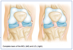
The medial collateral ligament (MCL) runs from the inner side (medial side) of the femur (thigh bone) to the inner (medial side) of the tibia (lower leg bone). It prevents the knee from opening on the inside when struck from the outside of the knee joint. The MCL lies on the outside of the joint capsule and has a good blood supply that contributes to its good healing potential.

The lateral collateral ligament (LCL) runs from the outer side (lateral side) of the femur (thigh bone) to the top of the fibula (the smaller of the two lower leg bones). It prevents the knee from opening on the outer side when struck from the inner side of the knee joint. The LCL is thinner and when completely disrupted often requires surgical repair.

Injury Mechanism
The MCL is often injured in sports when one is struck from the outer or lateral side of the knee, such as having an opponent fall against the outside of one’s knee in football. Another common mechanism of injury to the MCL is when the foot is forced out to the side away from the body, such as with a simultaneous kick of a soccer ball with the inside of the foot. LCL injuries are much more rare and usually occur when the knee is struck from the inside while the foot is planted, forcing a distraction force to the outside of the knee.

Symptoms
When patients sustain an injury to the collateral ligaments they often experience pain, localized swelling and bruising on the involved side of the knee. With partial tears, there is stiffness and pain when fully bending the knee, but no sense of instability. With a complete tear, the knee will feel unstable and will give way to the side with any lateral movements.

Diagnosis
The physician’s work-up will start with a careful history and exam. Often the description of a direct blow to either side of the knee can lead the physician to the suspected injury to the MCL or ACL. On examination, the physician can feel the instability when pulling the foot to one side or the other while stabilizing the knee. X-rays are often obtained to see that no fractures have occurred. Occasionally, a small avulsion fracture might hint that a collateral ligament injury has occurred. An MRI scan is often obtained to confirm the diagnosis and to evaluate any associated injuries to the menisci, other ligaments, and damage to the joint surfaces.

Treatment
Your physician will discuss treatment options with you. Treatment decisions are based on degree of instability. Minor tears (sprains) can be treated with rest, ice, elevation and compression. More significant tears in which many of the fibers of the ligament have been torn may require bracing for 6 weeks to keep the fibers from healing in a stretched out position. Occasionally physical therapy is needed to help regain full range of motion and strengthen the surrounding muscles after the period of bracing. Rarely is surgery recommended for an isolated MCL tear, but occasionally LCL injuries can benefit from surgical repair or reconstruction.

Knee Anatomy

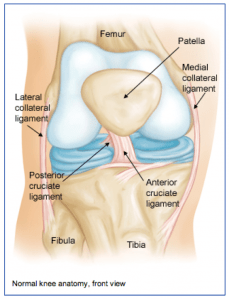
The knee joint is one of the largest joints in the body. It is a complex joint with four bones: the femur (thigh bone), the tibia (main lower leg bone), the fibula (smaller lower leg bone), and the patella (kneecap). The bones are connected with four main ligaments: ACL (anterior cruciate ligament), PCL (posterior cruciate ligament), MCL (medial collateral ligament), and the LCL (lateral collateral ligament).

The ACL and PCL control the forward/backwards movement of the knee joint and prevent pivoting of the knee. The MCL and LCL prevent giving away on either side of the knee. The quadriceps is a group of 4 muscles that converge on the front of the thigh and together allow one to straighten their knee by pulling through the kneecap and patellar tendon, which attaches to the front of the lower leg bone (tibia).

The hamstrings are the muscles on the back of the thigh that help bending the knee by crossing the joint in the back of the knee and attaching to the lower leg bones. Between the femur and tibia, sitting centrally in the knee joint, are two C-shaped pads (the medial and lateral menisci) that act as cushions or shock absorbers between the two bones. The meniscal pads are made of cartilage.

There is also about a quarter of an inch of cartilage on the distal end of the thighbone and on the proximal end of the lower leg bone. Arthritis occurs when that joint cartilage becomes damaged or thin.