Tennis Elbow (Lateral Epicondylitis)

master_71Description
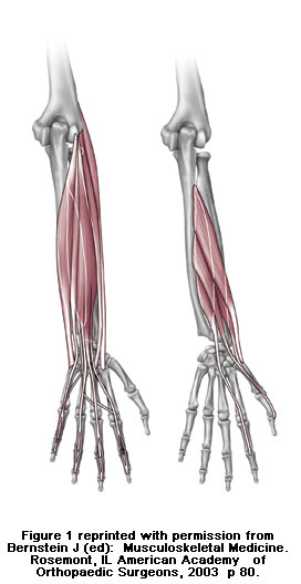
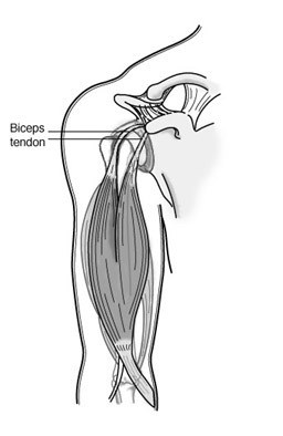
Tennis elbow is a degenerative condition of the tendon fibers that attach on the bony prominence (epicondyle) on the outside (lateral side) of the elbow. The tendons involved are responsible for anchoring the muscles that extend or lift the wrist and hand (see Figure 1).

Risk Factors/Prevention
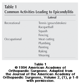
Tennis elbow happens mostly in patients between the ages of 30 years to 50 years. It can occur in any age group. Tennis elbow can affect as many as half of athletes in racquet sports. However, most patients with tennis elbow are not active in racquet sports. Most of the time, there is not a specific traumatic injury before symptoms start. Many individuals with tennis elbow are involved in work or recreational activities that require repetitive and vigorous use of the forearm muscles (see Table 1). Some patients develop tennis elbow without any specific recognizable activity leading to symptoms.

Symptoms
Patients often complain of severe, burning pain on the outside part of the elbow. In most cases, the pain starts in a mild and slow fashion. It gradually worsens over weeks or months. The pain can be made worse by pressing on the outside part of the elbow or by gripping or lifting objects. Lifting even very light objects (such as a small book or a cup of coffee) can lead to significant discomfort. In more severe cases, pain can occur with simple motion of the elbow joint. Pain can radiate to the forearm.
To diagnose tennis elbow, tell the doctor your complete medical history. He or she will perform a physical examination.

  • The doctor may press directly on the bony prominence on the outside part of the elbow to see if it causes pain.
  • The doctor may also ask you to lift the wrist or fingers against pressure to see if that causes pain.

cons1_304_132X-rays are not necessary. Rarely, MRI (magnetic resonance imaging) scans may be used to show changes in the tendon at the site of attachment onto the bone.

Treatment Options
In most cases, nonoperative treatment should be tried before surgery. Pain relief is the main goal in the first phase of treatment. The doctor may tell you to stop any activities that cause symptoms. You may need to apply ice to the outside part of the elbow. You may need to take acetaminophen or an anti-inflammatory medication for pain relief.
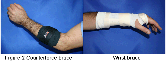
Orthotics can help diminish symptoms of tennis elbow. The doctor may want you to use counterforce braces and wrist splints. These can reduce symptoms by resting the muscles and tendons (see Figure 2).

cons1_305_132

Symptoms should improve significantly within four weeks to six weeks. If not, the next step is a corticosteroid injection around the outside of the elbow. This can be very helpful in reducing pain. Corticosteroids are relatively safe medications. Most of their side effects (i.e., further degeneration of the tendon and wasting of the fatty tissue below the skin) occur after multiple injections. Avoid repeated injections (more than two or three in a specific site).

cons1_306_132
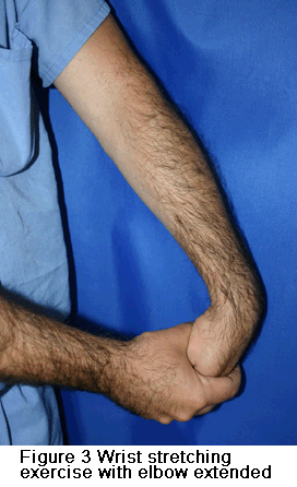
After pain is relieved, the next phase of treatment starts. Modifying activities can help make sure that symptoms do not come back. The doctor may want you to do physical therapy. This may include stretching and range of motion exercises and gradual strengthening of the affected muscles and tendons (see Figure 3). Physical therapy can help complete recovery and give you back a painless and normally functioning elbow. Nonoperative treatment is successful in approximately 85 percent to 90 percent of patients with tennis elbow.

Treatment Options: Surgical
Surgery is considered only in patients who have incapacitating pain that does not get better after at least six months of nonoperative treatment.
The surgical procedure involves removing diseased tendon tissue and reattaching normal tendon tissue to bone (see Figure 4). The procedure is an outpatient surgery; you do not need to stay in the hospital overnight. It can be performed under regional or general anesthesia.

Technique for surgical treatment of lateral epicondylitis. A, Skin incision over the lateral epicondyle. B, Distal reflection of the extensor mechanism exposing the lateral compartment of the elbow. C, Excision of pathologic tissue from the underside of the extensor mechanism. D, Decortication of the lateral epicondyle. E, Drilling of two V-shaped tunnels within the lateral epicondyle. F, Reattachment of the extensor mechanism to the lateral epicondyle. G, Side-to-side repair of the extensor tendon mechanism.

cons1_307_132

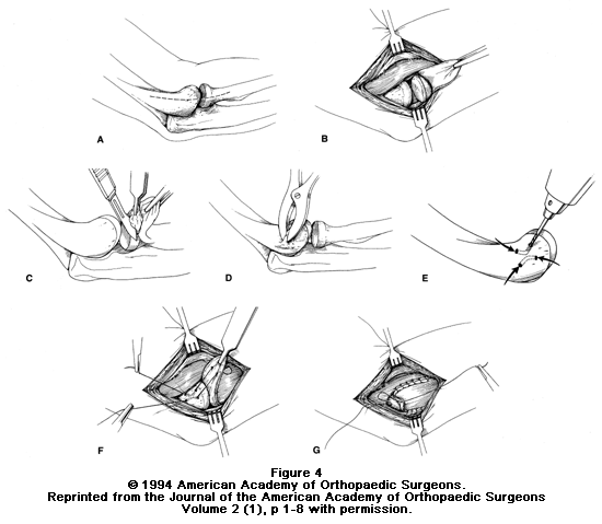
Most commonly, the surgery is performed through a small incision over the bony prominence on the outside of the elbow. Recently, an arthroscopic surgery method has been developed.

So far, no significant benefits have been found to using the arthroscopic method over the more traditional open incision.

After surgery, the elbow is placed in a small brace and the patient is sent home. About one week later, the sutures and splint are removed. Then exercises are started to stretch the elbow and restore range of motion. Light, gradual strengthening exercises are started two months after surgery. The doctor will tell you when you can return to athletic activity. This is usually approximately four months to six months after surgery. Tennis elbow surgery is considered successful in approximately 90 percent of patients.

Osteoarthritis of the Elbow

Description
Osteoarthritis occurs when the cartilage surface of the elbow is damaged or becomes worn. This can happen because of a previous injury such as elbow dislocation or fracture. It may occur due to degeneration of the joint cartilage from age. Osteoarthritis usually affects the weightbearing joints, such as the hip and knee. The elbow is one of the least affected joints due to its well matched joint surfaces and strong stabilizing ligaments. This makes the joint able to tolerate large forces across it without becoming unstable.

A doctor can usually diagnose elbow arthritis based upon a patient’s symptoms and standard X-rays (Figure 1). X-rays show the arthritic changes. Most of the time, advanced imaging studies such as CT (computed tomography) or MRI (magnetic resonance imaging) scans are not needed. Elbow osteoarthritis that occurs without previous injury is more common in men than women. It usually begins after age 50, although some patients can have symptoms earlier.

master_72Risk Factors/Prevention
Most patients who are diagnosed with elbow osteoarthritis have a history of injury to the elbow, such as a fracture that involved the surface of the joint, or an elbow dislocation.

The risk for elbow arthritis increases if:

  • The patient needed surgery to repair the injury or reconstruct the joint
  • There is loss of joint cartilage
  • The joint surface cannot be repaired or reconstructed to its pre-injury level

Injury to the ligaments resulting in an unstable elbow can also lead to arthritis, even if the elbow surface is not damaged. That’s because the normal forces across the elbow are altered, causing the joint to wear out more rapidly.

Sometimes there is no single injury. Work or outside activities may also lead to elbow arthritis if the patient places more demands on the joint than it can bear.

For example, professional baseball pitchers place unusually high demands on their throwing elbows. This can lead to failure of the stabilizing ligaments. It usually needs surgical reconstruction. High shear forces placed across the joint can lead to cartilage breakdown over a period of years.

The best way to prevent elbow arthritis is to avoid injury to the joint. When injury does happen, it is important to recognize it right away and get treatment. Individuals involved in heavy work or sports activities should maintain muscular strength around the elbow. Always use proper conditioning and technique.

Symptoms

The most common symptoms of elbow arthritis are:

  • Pain
  • Loss of range of motion

You might not have both symptoms at once. Patients usually complain of a “grating” or “locking” sensation in the elbow. The “grating” is due to loss of the normal smooth joint surface. This is caused by cartilage damage or wear. The “locking” is caused by loose pieces of cartilage or bone. These can dislodge from the joint and become trapped between the moving joint surfaces, blocking motion.

Joint swelling may also occur. But this does not usually happen at first. Swelling occurs later, as the disease progresses.

In later stages, patients might also notice numbness in their ring finger and small finger. This can be caused by elbow swelling or limited range of motion in the joint. The “funny bone” (ulnar nerve) is located in a tight tunnel behind the inner (medial) side of the elbow. Swelling in the elbow joint can put increased pressure on the nerve. This causes tingling. If the elbow cannot be moved through its normal range of motion, it may stiffen into a position where it is bent (flexion). This can also cause pressure around the nerve to increase.

Treatment Options
Treatment options depend on the stage of the disease, prior history, what the patient desires, overall medical condition, and the results of X-rays.

For the early stages, the most common treatment is non-surgical. This includes oral medications such as Tylenol® or Advil®, physical therapy, activity modification and joint injections.
Sometimes corticosteroid injections are used to treat arthritis symptoms. Steroid medication has typically been used with good results. The affects are temporary. But injections may give significant relief until symptoms progress enough to need additional treatment. An alternative to steroids has been the injection of hyaluronic acid in various forms. This attempts to increase the fluid in a joint, a process called viscosupplementation. It surrounds the diseased cartilage with a thicker and more “cushioned” environment.

This treatment has been recently studied in people with osteoarthritis of the knee. While there was enthusiasm for this treatment, research has not shown it to be better than traditional steroid injections. Additionally, the hyaluronic injections were significantly more expensive. The results of these “viscosupplementation” injections in the elbow or other joints have not been investigated.

When nonsurgical interventions are not enough to control symptoms, surgery may be needed.

Treatment Options: Surgical
By the time arthritis can be seen on X-rays, there has been significant wear or damage to the joint surfaces. If the wear or damage is limited, arthroscopy can offer a minimally invasive surgical treatment. It may be an option for patients with earlier stages of arthritis.

Arthroscopy has been shown to provide symptom improvement at least in the short term. It involves removing any loose bodies or inflammatory/degenerative tissue in the joint. It also attempts to smooth out irregular surfaces. Multiple small incisions are used to complete the surgery. It can be performed as an outpatient procedure. The recovery is reasonably rapid.

If the joint surface has worn away completely it is unlikely that anything other than a joint replacement would bring about relief. There are several different types of joint replacement available (Figure 2).

cons1_328_239In appropriately selected patients, the improvement in pain and function can be dramatic. With an experienced surgeon, the results of elbow replacement are the same as the results of hip replacement and knee replacement. For patients who are too young or who are too active to have prosthetic joint replacement, there are other reasonably good options.

If loss of motion is the primary symptom, the surgeon can release the contracture and smooth out the joint surface. At times, a new surface made from the patient’s own body tissues can be made. These procedures can give years of symptom improvement.

Research on the Horizon/What’s New?
Recently, joint supplementation has been used as an alternative to traditional oral and injectable medication. For oral medication, this involves a glucosamine/chondroitin supplement. These “nutraceuticals” attempt to give the body more of the basic elements that make up cartilage. Then the body may attempt to maintain or “build back” cartilage. There have been few well-controlled research studies on glucosamine/chondroitin. They have not included patients with elbow arthritis. So the short and long term effects are not yet known. Anecdotal reports have been favorable.

In cases where there has been loss or damage to areas of the joint, a cartilage/bone graft can be considered. This procedure attempts to return the joint to its prior smooth appearance and form in an attempt to prevent further deterioration of the joint. As our understanding of cartilage growth and regeneration improves, this may allow replacement of larger areas of joint damage or degeneration. Newer elbow replacements have also been designed with the goals of greater longevity and easier insertion compared with prior designs.

Olecranon (Elbow) Fractures

When you bend your elbow, you can easily feel its “tip,” a bony prominence that extends from one of the lower arm bones (the ulna). That tip is called the olecranon (oh-lek’-rah-nun). It is positioned directly under the skin of the elbow, without much protection from muscles or other soft tissues. So it can easily break if you experience a direct blow to the elbow or fall on a bent elbow.

Signs and symptoms

  • Sudden, intense pain.
  • Bruising around the elbow.
  • Rupture or abrasion of the overlying skin.
  • Possible deformity, if there is also a dislocation of the bone.
  • Tenderness and swelling over the bone site.
  • Numbness in one or more fingers.
  • Pain with movement of the joint.

Evaluation and classification
It is important to see a physician and verify that there is no associated damage to nerves or blood vessels. Your physician will use X-rays to confirm the diagnosis and classify the type of fracture. Fractures are generally divided into three types, depending on the stability of the joint and the amount of separation among the broken pieces of bone. (Note: Some fractures can have characteristics of more than one category.)

  • Type I fractures are generally stable with little displacement. These fractures can generally be treated nonsurgically.
  • Type II fractures are the most common. They are relatively stable, although there is displacement of the bone pieces.
  • Type III fractures are displaced and involve more than 50 percent of the joint surface, resulting in joint instability.

Treatment
Treatment depends on the type of fracture.

  • Type I fracture can usually be treated with a splint or sling to hold the elbow at a 90 degree angle. The physician will request a second set of X-rays after 10 days to make sure that the broken pieces have not become displaced. Gentle motion is permitted, and hand and wrist exercises should be done daily.
  • Type II fracture is best treated surgically. The orthopaedic surgeon will use a plate or a combination of wires and pins or screws to hold the bones in place. Physical therapy to maintain range of motion will start a day or two after the operation, and continue for at least six weeks.
  • Type III fractures are also treated surgically, usually with a plate that fits under the ulna and around the tip of the elbow. Screws hold the plate in place. You will have to wear a splint for a couple of days, then physical therapy to maintain range of motion will begin.

Fractures of the tip of the olecranon that do not involve the joint are may be treated by removing the small fragment and repairing the tendon that has pulled off. Elderly people who experience a type II or type III fracture may be treated with a sling and early range of motion instead of surgery. Athletes who have stress fractures of the olecranon are treated with activity restriction, stretching and range of motion exercises, and substitution activities for 8 to 12 weeks, although complete recovery may take three to six months.

Elbow (Olecranon) Bursitis

Description
The bursa is a slippery sac between the loose skin and the bones of your elbow. The bursa allows the skin to move freely over the underlying bone. It is located at the tip of the elbow. Normally, the bursa is flat and it’s hard to tell it is even there. If the bursa becomes irritated or inflamed, a condition known as elbow bursitis develops.

Risk Factors/Prevention
Common causes of elbow bursitis include:

  • Trauma: A hard blow to the tip of the elbow could cause the bursa to produce excess fluid and swell.
  • Prolonged pressure: Leaning on the tip of the elbow for long periods of time on hard surfaces such as a tabletop may cause the bursa to swell. Typically, this type of bursitis would develop over several months.
  • Infection: If the tip of the elbow has an injury that breaks the skin, such as an insect bite or a scrape, bacteria may get inside the bursa and cause an infection. The infected bursa produces fluid, redness and swelling. If the infection goes untreated, the fluid may turn to pus.
  • Medical conditions: Certain conditions such as rheumatoid arthritis and gout are associated with development of elbow bursitis.

Symptoms
Swelling is often the first symptom. The skin on the back of the elbow is loose, so you may not notice small amounts of swelling right away. As the swelling continues, the bursa gets larger. This causes pain as the bursa is stretched, since the bursa contains nerve endings. The swelling may get large enough to restrict motion in the elbow.

If the bursitis is infected, the skin becomes red and warm. If the infection is not treated right away, it may spread to other parts of the arm or move into the bloodstream. This can cause serious illness.

cons1_414_166

See your doctor to diagnose elbow bursitis. You may need an X-ray so the doctor can look for a foreign body or a bone spur. Bone spurs are often found on the tip of the bone in the elbow in patients who have recurrent problems with elbow bursitis.

Treatment Options: Nonsurgical
First, the doctor must determine whether the bursitis is due to an infection. If the doctor suspects this, fluid removal (aspiration) of the swollen area may be recommended. This is commonly performed as an office procedure. Fluid removal helps relieve symptoms and gives the doctor a sample that can be looked at in a laboratory to identify if any bacteria are growing.

This also lets the doctor know if a specific antibiotic is needed to fight the infection.

Often, the doctor may start you on antibiotics before the exact bacteria can be identified. This is done to prevent the infection from progressing. The antibiotic that the doctor recommends in this case will cover a number of possible infections.

If the bursitis is not from an infection, it is treated with elevation, ice and other nonoperative treatments such as an elbow pad and avoidance of direct pressure on the swollen elbow. Oral medications such as ibuprofen or other anti-inflammatories may also be used.

If the swelling and pain do not respond to these measures, your doctor may recommend removing fluid from the bursa and injecting a corticosteroid medication into the bursa. The steroid medication is an anti-inflammatory that is stronger than the medication that can be taken by mouth.

Treatment Options: Surgical
Infected bursa that do not improve with antibiotics and/or removing fluid from the elbow may require surgery. Patients who have surgery for elbow bursitis may need to stay in the hospital for a period of time.

If elbow bursitis is not a result of infection, surgery may be needed if nonoperative treatments don’t work. Surgery to remove the bursa is usually performed as an outpatient procedure. The surgery does not disturb any muscle, ligament or joint structure. Physical therapy after surgery is not always needed. Postoperative casting or prolonged immobilization is not typically required.

Biceps Tendinitis

The biceps muscle, in the front of the upper arm, helps stabilize the upper arm bone (humerus) in the shoulder socket. It also helps accelerate and decelerate the arm during overhead movement in activities like tennis or pitching. Strong, cord-like structures called tendons connect one end of the biceps muscle to the shoulder in two places.

At the other end of the muscle, tendons connect the biceps muscle to the smaller bone (radius) in the lower arm. If the tendons become inflamed or irritated, the condition is called tendinitis.

Signs and symptoms
Injuries to the biceps tendons are commonly caused by repetitive overhead activity. Symptoms include:

  • Pain when the arm is overhead or bent.
  • Localized tenderness as the tendon passes over the groove in the upper arm bone.
  • Occasionally, a snapping sound or sensation in the shoulder area.

cons1_139_209Diagnosis and treatment
During the physical examination, the doctor will assess the shoulder area for range of motion, tenderness and signs of shoulder instability. He or she may ask you to raise or rotate the arm.

X-rays may be requested to uncover associated conditions that might cause irritation. The doctor may also request an MRI that can show any damage to the tendons. Overuse, aging and stress can cause the tendon to deteriorate, even if there is no inflammation present.

Initial treatment is conservative. The first step is to rest the arm and shoulder. Switch to another sport or activity for awhile. Ice applications and nonsteroidal anti-inflammatory medications such as ibuprofen can help reduce inflammation.

Your physician can also recommend stretching and progressive strengthening exercises to build muscle endurance and restore range of motion. Then you can gradually return to overhead activity.

Surgical options
If the pain results from shoulder instability or from pressure on the tendon from the shoulder bones, your orthopaedist may recommend arthroscopic surgery. Using fiber optic technology and miniature instruments inserted through a small incision, the surgeon can examine the shoulder joint and anchor the tendon properly.

After surgery, your orthopaedist will prescribe a rehabilitation program that includes stretching and strengthening exercises. Early movement is important, but you should wait for your physician’s approval before doing any heavy lifting or returning to sports.