Biceps Tendon Tears

From orthoinfo.aaos.org

From orthoinfo.aaos.org

Anatomy: The biceps is a large muscle in the front of the shoulder. The upper end of the biceps has to heads (attachment sites). The long head of the biceps or its attachment to the labrum (cartilage lip at the top of the shoulder socket) is commonly injured.

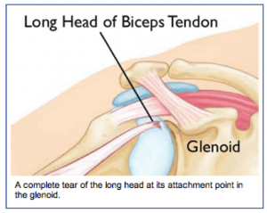
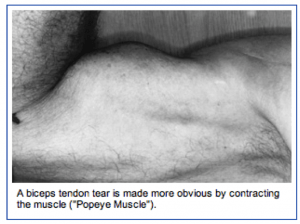
When it is torn at the upper end or at the labral attachment, there is often pain in the front of the shoulder. When the long head of the biceps is completely torn, the biceps muscle has a typical bulging appearance, but oftentimes does not cause much discomfort of weakness.

Injury Mechanism: Acute injuries to the long head of the biceps occur with heavy lifting or by grabbing the railing to support one’s body weight during a fall. Tears of the biceps can also occur with repetitive use of the shoulder in sports or work.

Symptoms: Acute complete tears of the long head of the biceps usually present as a sharp pain in the front of the shoulder, followed by bruising and the “Popeye muscle” deformity of the biceps (bulging in the middle of the upper arm where the muscle has retracted). Chronic tears or fraying of the biceps usually presents as aching in the front of the shoulder with use of the biceps.

From orthoinfo.aaos.orgDiagnosis: The physician’s work-up will start with a careful history and exam. X-rays will be normal as they show the bones and joints, not the muscles and tendons. An MRI scan can be obtained to confirm the diagnosis, especially if the diagnosis is not obvious by history and exam.

Treatment: Your physician will discuss treatment options with you. Most complete tears of the rotator cuff can be treated without surgery. Rarely patients notice weakness or cramping in the biceps. Stretching, strengthening, anti-inflammatories, and time will usually settle down symptoms to a tolerable level.

The partial tears are usually more painful. Injections can sometimes manage the discomfort, but occasionally surgery is necessary to help alleviate symptoms.

From orthoinfo.aaos.orgSurgery is usually accomplished through the arthroscope to remove the frayed tissue.

If the partial tear is nearly complete, the tendon is released from its attachment to the socket (labrum) and attached further down on the upper arm bone (humerus). This procedure is called a biceps tenodesis. Sometimes a small incision is necessary to re-attach the tendon to the bone.

What to expect after surgery: If the frayed tendon is just removed through the arthroscope, the recovery is rapid. Usually, after a few days of rest, gentle exercise is started. A full recovery is expected after 4 to 6 weeks. If the tendon is re-attached to bone, a sling is used with the elbow bent for 3 or 4 weeks to protect the repair. No resisted use of the biceps is started until 6 to 8 weeks after the repair.

Bursitis and Tendinitis

From orthoinfo.aaos.org

From orthoinfo.aaos.org

Both bursitis and tendinitis are forms of impingement syndrome. Bursitis occurs when the bursa (normally thin fluid filled sack that allows the rotator cuff to glide smoothly under the acromion) becomes swollen or inflamed.

Tendinitis occurs when the tendons of the rotator cuff or the long head of the biceps becomes swollen or inflamed (normally thin fluid filled sack that allows the rotator cuff to glide smoothly under the acromion) becomes swollen or inflamed.

Injury Mechanism: Bursitis and tendinitis are commonly seen in throwing athletes, those doing overhead lifting, or with repetitive motions and overuse of the shoulder. Often, one does not recall a specific injury, but awakens with pain.

Symptoms: When patients have bursitis, they describe pain over the lateral aspect of the shoulder with overhead reaching or laying on that shoulder at night. With tendinitis, pain occurs when using the rotator cuff or biceps in addition to the pain with overhead reaching or laying on that shoulder at night.

Diagnosis: The physician’s work-up will start with a careful history and exam. With tendinitis, the physician can isolate the tendons involved by eliciting pain with use of those tendons plus the impingement test is positive. With bursitis, pain cannot be elicited with use of specific tendons, but the impingement test is still positive.

X-rays are often normal with bursitis or tendinitis, but occasionally a spur is identified on a special “outlet view” that can be obtained in the physician’s office. An MRI scan is sometimes obtained to confirm that the rotator cuff is not torn, especially when patients do not respond to initial treatment.

Treatment: The mainstay of treatment for bursitis and tendinitis is non-surgical. Most patients respond to a short course of anti-inflammatories, stretching, and gentle strengthening of the external rotators of the shoulder. This can be accomplished at home with rubber tubing. If symptoms persist, one can inject the bursa with cortisone to cause the swollen tissues to shrink so they will no longer rub, or impinge.

If the symptoms respond temporarily to the injections, but keep recurring, one can consider surgery to shave down the undersurface of the acromion (acromioplasty) to make more clearance for the rotator cuff and bursa to slide underneath without rubbing. This type of surgery is performed through the arthroscope, using 2 or 3 small incisions, and looking inside the shoulder with a small lens and camera.”

What to expect after surgery: Most shoulder surgery is now performed as an outpatient procedure. In the rare event that an acromioplasty is needed to stop the impingement process, most patients find that they do best by resting their shoulder in a sling for 3 or 4 days following surgery.

They are then encouraged to come out of the sling for gentle stretching exercises. Once comfortable (usually 1 or 2 weeks following surgery), gentle strengthening is initiated with rubber tubing. Once the patient is off of his/her medication, they can resume driving and returning to office work. Most patients can return to full activities in 6 to 8 weeks.

Reverse Total Shoulder Replacement

Dr. Peterson and Dr. Shapiro have been performing a relatively new procedure called reverse total shoulder replacement for the last several years.

This particular procedure is designed for people who have rotator cuff arthropathy or a large, irreparable rotator cuff tear. The rotator cuff is a group of muscles and tendons that surround the shoulder joint and allow you to lift your arm over your head. When this structure is severely torn, shoulder arthritis can set in and mobility is limited.

During this procedure, the surgeon removes damaged bone joint tissue.  A smooth,  polished, spherical alloy metal “glenosphere” is then fixed to the old bony “cup” of the shoulder, and a stemmed alloy and polymer cup to the shaft of the upper-arm bone.

Why is Reverse Total Shoulder Replacement Done?

This surgery was developed because traditional shoulder surgeries do not work well when patients also have a severe rotator cuff tear with arthritis. With reverse total shoulder replacement, the deltoid muscle powers the new prosthesis, allowing pain free motion overhead in many patients.

Who is a Candidate for Surgery?

Reverse total shoulder replacement may be recommended if you have:

  • A completely torn rotator cuff that cannot be repaired.
  • Cuff tear arthropathy (arthritis with a severe cuff tear).
  • A previous should replacement that was unsuccessful.
  • Severe shoulder pain and difficulty lifting your arm.
  • Tried other treatments that have not relieved your shoulder pain.

Reverse shoulder replacement may not be recommended for people who have:

  • Poor general health and may not tolerate anesthesia and surgery well.
  • An active infection or are at risk for infection.
  • Severe weakness of or damage to the deltoid muscle of the shoulder.
  • A shoulder problem deemed appropriate for more traditional replacement procedures.

How do I Prepare for this Procedure?

Anesthesia – This procedure can be performed under general or regional anesthesia, depending on what your orthopedic surgeon prefers.

Antibiotics – You will probably be prescribed antibiotics to take before and after the surgery to prevent infection.

Medications – Be sure you tell your orthopedic specialists about all the medications you are taking. He may advise you to stop certain medications before the procedure.

Home Planning – There are some things you should be aware of that will make your recovery period much easier. First of all, you will need to take several weeks off from work following the surgery. When you come home, you will need help for a few weeks with dressing, bathing, and simple household chores. Also, you may not be permitted to drive following the surgery and for a few weeks.

What Happens During the Surgery?

A reverse total shoulder replacement usually takes about 1.5 hours. The surgeon will make an incision at the top or front of your shoulder and remove the damaged bone. Then he will position the new components to restore function to your shoulder joint. The incision will then be closed with sutures.What Should I Expect After the Procedure?

After your procedure, the healthcare professionals will give you pain medication to keep you comfortable and several doses of antibiotics. Most patients are allowed to eat solid food and get out of bed the day after the surgery. You will go home on the first or second day following your procedure.

When you leave the surgical center, your arm will be in a sling to provide support. Your orthopedic specialist will instruct you on exercises to increase your mobility and endurance and plan a physical therapy program to strengthen your shoulder and improve your flexibility. Full recovery from this surgery usually occurs in 4-6 months.

Arthritis of the Thumb

Arthritis is a condition that irritates or destroys a joint. Although there are several types of arthritis, the one that most often affects the joint at the base of the thumb (the basal joint) is osteoarthritis (degenerative or “wear-and-tear” arthritis).

Osteoarthritis occurs when the smooth cartilage that covers the ends of the bones begins to wear away. Cartilage enables the bones to glide easily in the joint; without it, bones rub against each other, causing friction and damage to the bones and the joint.

The joint at the base of the thumb, near the wrist and at the fleshy part of the thumb, enables the thumb to swivel, pivot, and pinch so that you can grip things in your hand. Arthritis of the base of the thumb is more common in women than in men, and usually occurs after age 40.

Prior fractures or other injuries to the joint may increase the likelihood of developing this condition.

Symptoms

  • Pain with activities that involve gripping or pinching, such as turning a key, opening a door, or snapping your fingers.
  • Swelling and tenderness at the base of the thumb.
  • An aching discomfort after prolonged use.
  • Loss of strength in gripping or pinching activities.
  • An enlarged, “out-of-joint” appearance.
  • Development of a bony prominence or bump over the joint.
  • Limited motion.

Diagnosis
Your physician will ask you about your symptoms, any prior injury, pain patterns, or activities that aggravate the condition. The physical examination may show tenderness or swelling at the base of the thumb.

One of the tests used during the examination involves holding the joint firmly while moving the thumb. If pain or a gritty feeling results, or if a grinding sound (crepitus) can be heard, the bones are rubbing directly against each other.

An X-ray may show deterioration of the joint as well as any bone spurs or calcium deposits that have developed.
Many people with arthritis at the base of the thumb also have symptoms of carpal tunnel syndrome, so your physician may check for that as well.

Treatment
In its early stages, arthritis at the base of the thumb will respond to nonsurgical treatment.

  • Ice the joint for five to fifteen minutes several times a day.
  • Take an anti-inflammatory medication such as aspirin or ibuprofen to help reduce inflammation and swelling
  • Wear a supportive splint to limit the movement of the thumb, and allow the joint to rest and heal. The splint may protect both the wrist and the thumb.
  • It may be worn overnight or intermittently during the day.

Because arthritis is a progressive, degenerative disease, the condition may worsen over time. The next phase in treatment involves a steroid solution injection into the joint. This will usually provide relief for several months. However, these injections cannot be repeated indefinitely.

Surgical Options
When conservative treatment is no longer effective, surgery is an option. The operation can be performed on an outpatient basis, and several different procedures can be used. One option involves fusing the bones of the joint together.

This, however, will limit movement. Another option is to remove part of the joint and reconstruct it using either a tendon graft or an artificial substance. You and your physician will discuss the options and select the one that is best for you.

After surgery, you will have to wear a cast for several weeks. A rehabilitation program, often involving a physical therapist, helps you regain movement and strength in the hand. You may feel some discomfort during the initial stages of the rehabilitation program, but this will diminish over time.

Full recovery from surgery takes several months. Most patients are able to resume normal activities and are quite satisfied with the results.

Open MRI Available at OSS

Our OPEN MRI (Magnetic Resonance Imaging) system utilizes a completely safe magnetic field and an advanced computer system to produce exceptional quality images of any body part in any desired direction.
The result of our OPEN MRI is a sophisticated diagnostic picture of the area your orthopedic specialist wishes to view. What’s more, there is no pain, no known side effects, and no radiation used with our OPEN MRI.

This high-tech machine is ideal for patients who are claustrophobic. Patients who have had both a closed MRI and an open MRI have commented that the open MRI is much quieter and ear plugs are not necessary. … read more